Collection of materials relating to neuro-ophthalmology as part of the Neuro-Ophthalmology Virtual Education Library.
NOVEL: https://novel.utah.edu/
TO
- NOVEL492
| Title | Creator | Description | Subject | ||
|---|---|---|---|---|---|
| 1 |
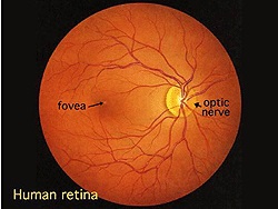 |
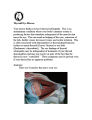
Simple Anatomy of the Retina (Webvision) | Helga Kolb, MD | Description of the anatomy of the retina with diagrams. | Retina Anatomy |
| 2 |
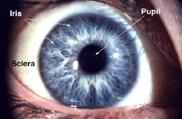 |
Gross Anatomy of the Eye (Webvision) | Helga Kolb, MD | Description of the gross anatomy of the eye, with diagrams. | Gross Anatomy Eye |
| 3 |
 |
Retinal Causes of a Neurologic-Type Visual Field Defect | Omar Ozgur, MD; Rudrani Banik, MD | Power point of case presentation of 47 year old female with history of breast cancer with new onset temporal visual field defect and photopsias. Differential diagnosis of homonymous hemianopia discussed; retinal causes of neurologic-type visual field defects reviewed including: white dot syndrome (m... | Homonymous Hemianopia; Neurologic Visual Field Defect; Temporal Visual Field Defect; White Dot Syndrome; Multiple Evanescent White Dot Syndrome (MEWDS); Cancer-Associated Retinopathy; Tamoxifen Retinopathy; Autoimmune Retinopathy |
| 4 |
 |
Pseudotumor cerebri and Chiari Malformation | Nicole Scripsema, MD; Rudrani Banik, MD | Power point of case presentation of pseudotumor cerebri with co-existing Chiari malformation. Management of severe visual loss associated with chronic papilledema discussed, as well as possible relationship between raised intracranial pressure from pseudotumor cerebri and Chiari malformation. | Pseudotumor Cerebri; Papilledema; Chiari Malformation |
| 5 |
 |
Part II: Anatomy and Physiology of the retina | Olaf Strauss, MD; Ralph Nelson; Dustin M. Graham | Section of Webvision covering the anatomy and physiology of the retina: The retinal pigment epithelium by Olaf Strauss Photoreceptors by Helga Kolb Outer Plexiform Layer by Helga Kolb Inner Plexiform Layer by Helga Kolb Morphology and Circuitry of Ganglion Cells by Helga Kolb Ganglion Cell Physiolog... | Anatomy; Physiology; Retina |
| 6 |
 |
Lemierre Syndrome - A Neuroophthalmological Approach | Vinzenz A. C. Vadasz, MD; Christina Gerth-Kahlert, MD | Case report of a twenty-two year old woman with double vision after tonsillitis, caused through multiples thrombosis by an infection with fusobacterium necrophorum known as the Lemierre-Syndrome. Fig. 1: Ocular motility at ICU (lying position) Fig. 2: white arrows show thrombosis of the right opht... | Lemierre-Syndrome; Fusobacterium Necrophorum; Septic Thrombosis |
| 7 |
 |
Oculopharyngeal Muscular Dystrophy (OPMD) | Natasha Nayak, MD; Rudrani Banik, MD | Power point of case presentation of chronic, progressive ophthalmoplegia and bilateral ptosis in adult male with positive family history of similar ocular findings. Differential diagnosis with associated findings reviewed. Work up done: EMG testing consistent with myopathy. Genetic testing positiv... | Ophthalmoplegia;, Ptosis; Oculopharygneal Muscular Dystrophy; Genetic Disorder |
| 8 |
 |
Non-Organic Visual Loss | Omar Ozgur, MD; Rudrani Banik, MD | Power point of case presentation of 12 year old girl with recurrent monocular visual loss. Examination is normal. Differential diagnosis discussed, including non-organic visual loss. Diagnostic testing for non-organic visual loss reviewed. Slide 4: Figure 1: Table of exam findings Slide 5: Figure 2... | Non-organic Visual Loss; Monocular Visual Loss |
| 9 |
 |
Montreal Cognitive Assessment (MOCA) | Ziad Nasreddine, MD | The MoCA© is a cognitive screening test designed to assist Health Professionals in detection of mild cognitive impairment. For more information, contact: info@moca.org | Cognitive Disorders; Cognitive Assessment; MOCA Test |
| 10 |
 |
Neuromyelitis Optica | Omar Ozgur, MD; Rudrani Banik, MD | Power point of case presentation of female with bilateral, sequential atypical optic neuritis. MRI Brain normal with no demyelination; MRI Spine shows enhancement at multiple levels and NMO antibody positive, confirming diagnosis of neuromyelitis optica (NMO). History of NMO discussed, diagnostic c... | Neuromyelitis Optica; Atypical Optic Neuritis; MRI; Plasmapheresis |
| 11 |
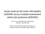 |
Acute Zonal Occult Outer Retinopathy (AZOOR) versus Multiple Evanescent White Dot Syndrome (MEWDS) | Asim V. Farooq, MD; Michael T. Andreoli, MD; Heather E. Moss, MD | PPT case report on acute zonal occult outer retinopathy (AZOOR) versus multiple evanescent white dot syndrome (MEWDS). | AZOOR; MEWDS; Paracentral Scotoma; Goldmann Visual Field; Photoreceptor Loss |
| 12 |
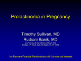 |
Prolactinoma in Pregnancy | Timothy Sullivan, MD; Rudrani Banik, MD | Power point of case of prolactinoma which became symptomatic during pregnancy with visual field loss. Discussion of prolactinomas and their management. Patient underwent observation only. Post-partum examination revealed resolution of bitemporal field defect with reduction in size of prolactinoma ... | Prolactinoma; Pregnancy; Bitemporal Defect |
| 13 |
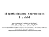 |
Idiopathic Bilateral Neuroretinitis in a Child | Asim V. Farooq, MD; Michael T. Andreoli, MD; Molly Gilbert, MD; Heather E. Moss, MD | PPT case describing idiopathic bilateral neuroretinitis in a child. | Neuroretinitis; Pediatric; Idiopathic; Optic Atrophy |
| 14 |
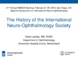 |
The History of the International Neuro-Ophthalmology Society | Klara Landau, MD, FEBO | This presentation provides an ovreview of hte hisotry of the International Neuro-ophthalmology Society (INOS), with maps and photos. | International Neuro-Ophthalmology Society: INOS |
| 15 |
 |
Palinopsia: Some Visions Never Fade | Amrita-Amanda D. Lakraj, MD; Ryan D. Walsh, MD | This is a PowerPoint presentation, which teaches the symptom of palinopsia through a video of a patient's chief complaint in which he describes the symptom almost according to a textbook. This video is followed by a brief explanation of the etiology, management, and importance of diagnosing this sym... | Palinopsia; Visual Disturbance; Ghosting |
| 16 |
 |
Pituitary Apoplexy and Hemifield Slide Phenomenon | Helen H. Yeung, MD; Rudrani Banik, MD | PowerPoint of case presentation of pituitary apoplexy. Patient presented with bilateral severe visual loss and bilateral ophthalmoplegia from partial third nerve palsies (pupil-sparing with no ptosis) from midbrain compression. After transsphenoidal surgery with decompression of mass and steroids, ... | Pituitary Apoplexy; Hemifield Slide; Bitemporal Defect; Partial Third Nerve Palsy |
| 17 |
 |
Wallenberg Syndrome and Skew Deviation | Lauren Schneider, MD; Rudrani Banik, MD | Power point of case presentation of acute Wallenberg Syndrome associated with vertical diplopia, found by 3 step and supine testing to be consistent with skew deviation. | Wallenberg Syndrome; Skew Deviation; Vertical Diplopia |
| 18 |
 |
Cone Dystrophy | Gregory P. Van Stavern, MD | PowerPoint discussing Cone Dystrophy: Early loss of central and color vision; Color impairment often out of proportion to loss of VA; Hemeralopia ("day blindness") prominent; Light sensitivity and photophobia; Macular changes variable, and may occur late- may "Bull's Eye" pattern; Abnormal Photost... | Cone Dystrophy; Occult Macular Dystrophy; Central Cone Dystrophy |
| 19 |
 |
Pupillary Light Reflex | Wade Crow, MD | Illustration of the Pupillary Light Reflex. | Pupillary Light Reflex |
| 20 |
 |
Acute Multifocal Pigment Epithelium Epitheliopathy (AMPEE) | Gregory P. Van Stavern, MD | Images providing example of Acute Multifocal Pigment Epithelium Epitheliopathy (AMPEE) | Acute Multifocal Pigment Epithelium Epitheliopathy (AMPEE) |
| 21 |
 |
Histoplasmosis | Gregory P. Van Stavern, MD | Histoplasmosis, a fungus, can present acutely as a systemic condition. This image shows signs of Histoplasmosis. | Histoplasmosis |
| 22 |
 |
Multifocal Choroiditis | Gregory P. Van Stavern, MD | Multi-focal choroiditis is usually a bilateral choroidopathy seen more frequently in women associated with punched out appearing lesions occasionally with pigment around the edges. Image provides example. | Multi-Focal Choroiditis Panuveitis |
| 23 |
 |
Retinitis Pigmentosa | Gregory P. Van Stavern, MD | Retinitis pigmentosa is a retinal/choroidal degeneration caused by various genetic defects. The term retinitis pigmentosa is really a misnomer since it is not inflammation (retinitis) and it is not a disease of the pigmentary system (pigmentosa). | Retinitis Pigmentosa |
| 24 |
 |
Serpiginous Choroidopathy | Gregory P. Van Stavern, MD | Serpiginous choroidopathy (also known as Geographic choroidopathy) usually affects the choroid, the choriocapillaris and the retinal pigment epithelium in both eyes. | Serpiginous Choroidopathy |
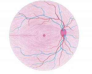
| 25 |
 |
Birdshot | Gregory P. Van Stavern, MD | Birdshot Retinochoroidopathy is a posterior uveitis seen in women 30-60 years of age who present with floaters, changes in color vision, and difficulty with night vision. | Birdshot Choroidopathy |
| 26 |
 |
Pars Planitis | Gregory P. Van Stavern, MD | Pars planitis is an inflammatory condition seen in children and young adults. It is associated with inflammation of the pars plana--at the far periphery of the retina. | Pars Planitis |
| 27 |
 |
Vogt Koyanagi-Harada (VKH) Syndrome | Gregory P. Van Stavern, MD | Vogt-Koyanagi disease causes bilateral uveitis, along with alopecia, vitiligo, and hearing loss. | Vogt Koyanagi-Harada Syndrome (VKH) |
| 28 |
 |
Stargardt's Disease | Gregory P. Van Stavern, MD | Stargardt's disease is an inherited maculopathy which frequently presents with a loss of central vision. | Stargardt's Disease |
| 29 |
 |
Bardet-Biedl Syndrome | Gregory P. Van Stavern, MD | PowerPoint discussing Bardet-Biedl Syndrome, a hereditary condition characterized by rod-cone dystrophy (RP), truncal obesity, polydactyly, hypogonadotropic hypogonadism (males), GU abnormalities (females), and cognitive impairment | Bardet-Biedl Syndrome; Genetics |
| 30 |
 |
Usher Syndrome | Gregory P. Van Stavern, MD | Powerpoint describing Usher Syndrome, a hereditary condition characterized by congenital, bilateral, and profound sensorineural hearing loss, adolescent onset Retinitis Pigmentosa (RP) and vestibular areflexia | Usher Syndrome; Retinal Dystrophy; Retinitis Pigmentosa; Hearing Loss |
| 31 |
 |
Vision and Alzheimer's Disease | Victoria S. Pelak, MD | Slideshow describing condition. | Alzheimer's Disease |
| 32 |
 |
Visual Evoked Responses (Webvision) | Donnell J. Creel, MD, University of Utah | WebVision: The terms visually evoked potential (VEP), visually evoked response (VER) and visually evoked cortical potential (VECP) are equivalent. They refer to electrical potentials, initiated by brief visual stimuli, which are recorded from the scalp overlying visual cortex, VEP waveforms are ext... | Electrophysiology; Visual Evoked Responses |
| 33 |
 |
What is White? Normal White Structures | Gregory P. Van Stavern, MD | The only inherently "white" element in the normal eye is the sclera. | White in the Retina |
| 34 |
 |
Aberrant Regeneration of Third Nerve | Gregory P. Van Stavern, MD | 48 year old woman S/P rupture and repair of right sided posterior communicating artery aneurysm Video shows residual partial right third nerve palsy, with aberrant regeneration, causing a pseudo Von Graefe's sign (elevation of the right upper eyelid with attempted infraduction of the right eye) Se... | Aberrant Regeneration of Third Nerve; Third Nerve Palsy |
| 35 |
 |
Aberrant Regeneration Third Nerve | Gregory P. Van Stavern, MD | 48 year old woman S/P rupture and repair of right sided posterior communicating artery aneurysm Video shows residual partial right third nerve palsy, with aberrant regeneration, causing a pseudo Von Graefe's sign (elevation of the right upper eyelid with attempted infraduction of the right eye) | Aberrant Regeneration Third Nerve; Third Nerve Palsy |
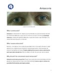
| 36 |
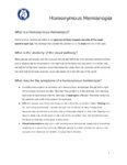
 |
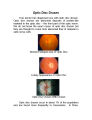
Acute Retinal Necrosis (ARN) | Gregory P. Van Stavern, MD | Acute Retinal Necrosis causes inflammation and subsequent retinal detachment. This powerpoint provides images depicting ARN. | Acute Retinal Necrosis (ARN) |
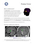
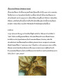
| 37 |
 |
Congenital and Secondary Syphilis | Gregory P. Van Stavern, MD | Images showing evideince of Congenital and Secondary Syphilis | Syphilis |
| 38 |
|
Vision & Alzheimer's Disease | Victoria S. Pelak, MD | Alzheimer's Disease (AD) is an age-related neurodegenerative disorder with progressive loss of cognitive function over time. A clinical diagnosis for Probable AD Dementia requires the following: a loss of cognitive function in two or more cognitive domains (or in one cognitive domain along with a ch... | Vision; Alzheimer's Disease |
| 39 |
 |
White Dot Syndromes: MEWDS, AZOOR, AIBSE | Gregory P. Van Stavern, MD | Some have lumped Multiple Evanescent White Dot Syndrome (MEWDS), Acute Idiopathic Blind Spot Enlargement (AIBSE) with acute macular neuroretinopathy, and pseudo-presumed ocular histoplasmosis syndrome together with AZOOR (Acute Zonal Occult Outer Retinopathy). These conditions all present with visua... | White Dot Syndromes: MEWDS, AZOOR, AIBSE |
| 40 |
 |
Superonasal Transconjunctival Optic Nerve Sheath Decompression: A Modified Surgical Technique Without Extraocular Muscle Disinsertion | Kevin E. Lai, MD; Kenneth C. Lao, MD; Peter L. Hildebrand, MD; Bradley K. Farris, MD | Report on the surgical technique and outcomes of a modified medial transconjunctival approach to optic nerve sheath decompression (ONSD) in 15 patients. Supplemental Digital Content : Video that demonstrates the stONSD procedure. m4v: http://content.lib.utah.edu/cdm/ref/collection/EHSL-NOVEL/id/22... | Superonasal Transconjunctival Optic Nerve Sheath Decompression (ONSD); Surgical Technique |
| 41 |
 |
Superonasal Transconjunctival Optic Nerve Sheath Decompression (stONSD) | Kevin E. Lai, MD; Kenneth C. Lao, MD; Peter L. Hildebrand, MD; Bradley K. Farris, MD | This video demonstrates the surgical technique and outcomes of a modified medial transconjunctival approach to optic nerve sheath decompression (ONSD). Disease/Diagnosis: Papilledema; Idiopathic Intracranial Hypertension (IIH). | Superonasal Transconjunctival Optic Nerve Sheath Decompression (ONSD); Surgical Technique |
| 42 |
 |
Disability Evaluation Under Social Security | John Pula, MD | A. How do we evaluate visual disorders? 1. What are visual disorders? Visual disorders are abnormalities of the eye, the optic nerve, the optic tracts, or the brain that may cause a loss of visual acuity or visual fields. A loss of visual acuity limits your ability to distinguish detail, read, or do... | Visual Impairment; Visual Disorders; Legal Blindness |
| 43 |
 |
Retinitis Pigmentosa - Rod Dystrophy | Gregory P. Van Stavern, MD | PowerPoint discussing retinitis pigmentosa, rod dystrophy. Retinitis Pigmentosa is a generalized retinal dystrophy with peripheral rather than central onset Primarily rod-cone dystrophy. Provides images. | Rod Dystrophy; Rod Dystrophy; Retinitis Pigmentosa; Night Dlindness |
| 44 |
 |
Tonic Pupil | Adesina, Ore-Ofe, MD | PowerPoint presentation covering tonic pupil, which is damage to ciliary ganglion or short posterior ciliary nerves. It causes denervation of the ciliary body and iris sphincter muscle. | Tonic Pupil |
| 45 |
 |
Horner's Carotid Dissection | Gregory P. Van Stavern, MD | PowerPoint describing Horner's Syndrome and Carotid Dissection. | Horner's Syndrome; Carotid Dissection; Dark Adaptation; Rod Dystrophy |
| 46 |
 |
2013 William F Hoyt Lecture: Neuro-Ophthalmology in Review: Around the Brain with 50 Fellows | Nancy J. Newman, MD | No matter what their ultimate specialty, every ophthalmologist needs to master the basics of neuroophthalmology. To that end, we must ensure that we continue to train effective teachers of neuro-ophthalmology. This is William F. Hoyt's most important lasting legacy and charge. In this same spirit, E... | History |
| 47 |
 |
Diffusion Weighted Imaging (DWI) | John Pula, MD | Diffusion weighted imaging sequences are often included as part of a routine brain MRI protocol. Imaging provides examples of DWI. | Diffusion Weighted Imaging; DWI |
| 48 |
 |
Photophobia for Patients - Large Print | Kathleen B. Digre, MD | The symptoms of light sensitivity are: an uncomfortable sense of brightness, squinting, frequent blinking, and redness of the eye (especially if the eye is dry). Involuntary eye closure and excessive blinking is seen with blepharospasm. Individuals will tend to seclude themselves in darkness. | Photophobia |
| 49 |
 |
Neuromyelitis Optica (NMO) | John Pula, MD | Slideshow describing condition. | Neuromyelitis Optica; NMO |
| 50 |
 |
Radiological Examination of the Visual System | John Pula, MD | An explanation of imaging types. | Visual System; Radiology; Imaging |
| 51 |
 |
Photophobia for Patients | Kathleen B. Digre, MD | The symptoms of light sensitivity are: an uncomfortable sense of brightness, squinting, frequent blinking, and redness of the eye (especially if the eye is dry). Involuntary eye closure and excessive blinking is seen with blepharospasm. Individuals will tend to seclude themselves in darkness. | Photophobia |
| 52 |
 |
Diffusion Tensor Imaging (DTI) | John Pula, MD | Diffusion tensor (DT) MRI applies the direction of water diffusion through tissues to map out neural pathways in the brain, such as white matter tracts. | Diffusion Tensor Imaging; DTI |
| 53 |
 |
Multiple Sclerosis Treatment Strategies | John Pula, MD | Slideshow exploring current treatment of multiple sclerosis. | Multiple Sclerosis; Multiple Sclerosis Treatment |
| 54 |
 |
Facts About Ambulatory Care Accreditation | Joint Commission on Accreditation of Healthcare Organizations (JCAHO) | The Joint Commission's Ambulatory Care Accreditation Program was established in 1975, and today more than 2,000 freestanding ambulatory care organizations are Joint Commission-accredited. These organizations generally fall into the broad categories of surgical, medical/dental and diagnostic/therapeu... | Ambulatory Care Accreditation |
| 55 |
 |
Branch Retinal Artery Occlusion with Multiple Retinal Emboli | Kathleen B. Digre, MD; James J. Corbett, MD | Slideshow describing condition. | Retinal Emboli; Emboli |
| 56 |
 |
Branch Retinal Vein Occlusion (BRVO) | Kathleen B. Digre, MD; James J. Corbett, MD | Slideshow describing condition. | Occlusion |
| 57 |
 |
Branch Retinal Artery Occlusion | Kathleen B. Digre, MD; James J. Corbett, MD | Slideshow describing condition. | Occlusion |
| 58 |
 |
Branch Retinal Emboli | Kathleen B. Digre, MD; James J. Corbett, MD | Slideshow describing condition. | Emboli |
| 59 |
 |
Calcific Emboli | Kathleen B. Digre, MD; James J. Corbett, MD | Slideshow describing condition. | Emboli |
| 60 |
 |
Central Retinal Vein Occlusion | Kathleen B. Digre, MD | Slideshow describing condition. | Occlusion |
| 61 |
 |
Craniopharyngioma and Optic Atrophy | Kathleen B. Digre, MD; James J. Corbett, MD | Slideshow describing condition. | Craniopharayngioma; Otpic Atrophy |
| 62 |
 |
CRAO with Ciliary Artery Sparing | Kathleen B. Digre, MD; James J. Corbett, MD | Slideshow describing condition. | CRAO |
| 63 |
 |
Central Cone Dystrophy Occult Macular Dystrophy | Gregory Van Stavern, MD | Slideshow describing condition of Central Cone Dystrophy Occult Macular Dystrophy | Central Cone Distrophy; Macular Dystrophy; Occult Macular Dystrophy |
| 64 |
 |
Pupillary reflex and the APD | Wade Crow, MD | Illustrations describing pupillary reflex. | Pupillary Reflex, APD |
| 65 |
 |
Vasospastic Amaurosis Fugax | Kathleen B. Digre, MD; James J. Corbett, MD | Slideshow describing condition. | Vasospastic Amaurosis Fugax |
| 66 |
 |
Acquired Hyperopia | AAO/NANOS - American Academy of Ophthalmology / North American Neuro-Ophthalmology Society | Choroidal folds may result from choroidal tumors, compression on the eye wall from thyroid ophthalmopathy, orbital pseudotumor, orbital tumor, posterior scleritis, hypotony, scleral laceration, retinal detachment, marked hyperopia, or secondary to papilledema. Intraocular pressure measurements, refr... | Acquired Hyperopia |
| 67 |
 |
Fat Emboli | Kathleen B. Digre, MD; James J. Corbett, MD | Slideshow describing condition. | Emboli |
| 68 |
 |
Fluorescein Angiography | Kathleen B. Digre, MD; James J. Corbett, MD | Fluorescein angiography in neuro-ophthalmology. | Fluorescein Angiography; History |
| 69 |
 |
Fibrin-Platelet Emboli | Kathleen B. Digre, MD; James J. Corbett, MD | Slideshow describing condition. | Emboli; Platelet Emboli |
| 70 |
 |
High Myopia | Kathleen B. Digre, MD; James J. Corbett, MD | Slideshow describing condition. | Myopia |
| 71 |
 |
Hollenhorst Plaque | Kathleen B. Digre, MD; James J. Corbett, MD | Slideshow describing condition. | Hollenhorst Plaque |
| 72 |
 |
Ischemic Ocular Syndrome | Kathleen B. Digre, MD; James J. Corbett, MD | Slideshow describing condition. | Ischemic Ocular Syndrome |
| 73 |
 |
Best's Vittelform Maculopathy | Gregory P. Van Stavern, MD | This 14 year old presented with decreased vision, headaches and central scotomas. She was found to have bilateral papilledema related to IIH and also Best's vitilliform maculopathy. The maculas are commonly described as having a "fried egg" sunny side up appearance. | Best Macular Dystrophy |
| 74 |
 |
Septo-Optic Dysplasia | Kathleen B. Digre, MD; James J. Corbett, MD | Slideshow describing the condition. | Septo-optic Dysplasia |
| 75 |
 |
Retino-choroidal Collateral Vessels | Kathleen B. Digre, MD; James J. Corbett, MD | Slideshow describing condition. | Collateral Vessels |
| 76 |
 |
Viewing the Red Reflex | Kathleen B. Digre, MD; James J. Corbett, MD | Slideshow describing eye examination of children. | Eye Examination |
| 77 |
 |
Papilledema 2013 | Kathleen B. Digre, MD | Objectives: What types of disc findings can be confused for papilledema List the features of true disc swelling Describe the tests you would order to evaluate and w/u papilledema List differential diagnosis of papilledema Describe possible treatments for papilledema (medical and surgical) | Papilledema |
| 78 |
 |
Optic Disc Anatomy, Variants, and Usual Discs | Kathleen B. Digre, MD | Examination of optic disc, disc anatomy, disc variation. | Optic Disc; Normal Disc Anatomy |
| 79 |
 |
Retino-choroidal Collaterals Due to Meningioma | Kathleen B. Digre, MD; James J. Corbett, MD | Slideshow describing condition. | Meningioma; Menigioma Treatment |
| 80 |
 |
Scleritis (Posterior Scleritis) | Kathleen B. Digre, MD; James J. Corbett, MD | Slideshow describing condition. | Scleritis; Posterior Scleritis |
| 81 |
 |
Pit Tumor Chiasm Compression | Kathleen B. Digre, MD; James J. Corbett, MD | Slideshow describing condition. | Pituitary Tumor; Pituitary Mass; Chiasm; Chiasm Compression |
| 82 |
 |
Hermann Ludwig Ferdinand von Helmholtz | Kathleen B. Digre, MD; James J. Corbett, MD | Biography of Hermann Ludwig Ferdinand von Helmholtz. | Hermann Ludwig Ferdinand von Helmholtz; History |
| 83 |
 |
Chiasmal Neuritis in Multiple Sclerosis | Kathleen B. Digre, MD; James J. Corbett, MD | Slideshow describing condition. | Multiple Sclerosis |
| 84 |
 |
Occlusion of the Central Retinal Artery | Kathleen B. Digre, MD; James J. Corbett, MD | Slideshow describing occlusion of the central retinal artery. | Occlusion; Retinal Artery |
| 85 |
 |
Ophthalmic Artery Occlusion | Kathleen B. Digre, MD; James J. Corbett, MD | Slideshow describing condition. | Occlusion; Artery |
| 86 |
 |
Cilioretinal Artery Occlusion | Kathleen B. Digre, MD; James J. Corbett, MD | Slideshow describing cilioretinal artery occlusion. | Occlusion; Artery Occlusion |
| 87 |
 |
Vascular Supply of the Eye | Kathleen B. Digre, MD; James J. Corbett, MD | Slideshow describing vascular supply of the eye. | Eye Anatomy; Vascular Supply |
| 88 |
 |
Venous Drainage of the Eye | Kathleen B. Digre, MD; James J. Corbett, MD | Slideshow describing condition. | Eye Anatomy; Venous Anatomy |
| 89 |
 |
Jean Martin Charcot | Kathleen B. Digre, MD; James J. Corbett, MD | Biography of Jean Martin Charcot | Jean Martin Charcot; History |
| 90 |
 |
William Gowers' Textbook | Kathleen B. Digre, MD; James J. Corbett, MD | Presentation on William Gowers' contribution to neuro ophthalmology. | William Gowers; History of Neuro-ophthalmology |
| 91 |
 |
William Gowers | Kathleen B. Digre, MD; James J. Corbett, MD | Biography of William Gowers. | William Gowers; History; Biography |
| 92 |
 |
Early Reflecting Ophthalmoscope | Kathleen B. Digre, MD; James J. Corbett, MD | The use of the early reflecting ophthalmoscope. | Reflecting Ophthalmoscope; History |
| 93 |
 |
The May Ophthalmoscope | Kathleen B. Digre, MD; James J. Corbett, MD | History of the ophthalmoscope developed by Charles May | Ophthalmosocope; History |
| 94 |
 |
Albrecht von Graefe | Kathleen B. Digre, MD; James J. Corbett, MD | Biography of Albrecht von Graefe. | Albrecht von Graefe; History |
| 95 |
 |
Johannes E. Purkinjé | Kathleen B. Digre, MD; James J. Corbett, MD | Biography of Johannes E. Purkinjé. | Johannes E. Purkinjé; History |
| 96 |
 |
Hughlings Jackson | Kathleen B. Digre, MD; James J. Corbett, MD | Biography of Hughlings Jackson. | Hughlings Jackson; History |
| 97 |
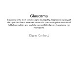 |
Glaucoma | Kathleen B. Digre, MD; James J. Corbett, MD | Slideshow describing condition. | Glaucoma |
| 98 |
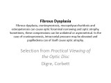 |
Fibrous Dysplasia | Kathleen B. Digre, MD; James J. Corbett, MD | Slideshow describing condition. | Dysplasia; Fibrous Dysplasia |
| 99 |
 |
Shaken Baby Syndrome | Kathleen B. Digre, MD; James J. Corbett, MD | Neuro-ophthalmologic implications of shaken baby syndrome. | Shaken Baby Syndrome; Child Abuse; Abuse |
| 100 |
 |
Direct Carotid Cavernous Fistula | Emory Eye Center | Slideshow describing condition. | Fistula |
| 101 |
 |
Terson's Syndrome | Kathleen B. Digre, MD; James J. Corbett, MD | Slideshow describing condition. | Terson's Syndrome; Hemhorrhage |
| 102 |
 |
Susac's Syndrome | Kathleen B. Digre, MD; James J. Corbett, MD | Slideshow describing condition. | Susac's Syndrome |
| 103 |
 |
Lessons From Bench Bedside | Shirley H. Wray, MD, PhD, FRCP | See also: http://content.lib.utah.edu/cdm/ref/collection/ehsl-shw/id/69, http://content.lib.utah.edu/cdm/ref/collection/ehsl-shw/id/282, http://content.lib.utah.edu/cdm/ref/collection/ehsl-shw/id/94, and http://content.lib.utah.edu/cdm/ref/collection/ehsl-shw/id/103 | Bilateral Internuclear Ophthalmoplegia; Pendular Horizontal Oscillations; Lid Nystagmus; Upbeat Nystagmus; Botulinum Toxin Therapy; Multiple Sclerosis; Horizontal Pendular Nystagmus; Gaze Evoked Upbeat Nystagmus; Abducting Nystagmus; Normal Convergence; Gaze Evoked Downbeat Nystagmus; Sac... |
| 104 |
 |
Ocular Myasthenia Gravis: Past, Present, Future | Victoria S. Pelak, MD | Slideshow describing condition. | Bilateral Myasthenia Gravis; Bilateral Ptosis; Bilateral Weakness of Adduction; Decompensated Phoria; External Ophthalmoplegia; Familial Myasthenia Gravis; Myasthenic Lid Twitch; Ocular Myasthenia Gravis; Positive Tensilon Test; Pseudo-internuclear Ophthalmoplegia; Tensilon Test; Unilateral Myasthen... |
| 105 |
 |
Multiple Sclerosis | Shirley H. Wray, MD, PhD, FRCP | Slideshow describing condition. | Bilateral Lid Nystagmus; Horizontal Saccadic Dysmetria; Jerk Oscillations; Lid Nystagmus; Multiple Sclerosis; Primary Position Upbeat Nystagmus; Rotary Nystagmus; Saccadic Dysmetria; Saccadic Pursuit; Square Wave Jerks; Torsional Nystagmus; Upbeat Nystagmus |
| 106 |
 |
Migraine Visual Aura | Shirley H. Wray, MD, PhD, FRCP | Slideshow describing condition. | Alice in Wonderland Syndrome; Macropsia - Hemi-macropsia; Metamorphopsia; Migraine Visual Aura Without Headache; Occipital Lobe; Visual Phenomena |
| 107 |
 |
Midbrain Hemorrhage | Shirley H. Wray, MD, PhD, FRCP | Slideshow describing condition. | Cavernous Angioma; Convergence Retraction Nystagmus; Light/Near Dissociation of the Pupils; Midbrain Hemorrhage; Normal Convergence; Pretectal Syndrome; Skew Deviation; Supranuclear Paralysis of Upgaze Hemorrhage; Vertical Oculocephalic Reflex Normal |
| 108 |
 |
Miller Fisher Syndrome: A Variant of Guillan Barré Syndrome | Sarah I. Sheikh, BM, BCh, MRCP | Presentation covering the Miller Fisher Syndrome, a variant of Guillan Barre Syndrome. | Acute Inflammatory Demyelinating Neuropathy; Areflexia; Bilateral Ptosis; Bilateral Sixth Nerve Palsy; Esotropia; Facial Diplegia; Facial Weakness; Guillian Barre Syndrome - Miller Fisher Syndrome; Normal Pupils; Paresis of Conjugate Upgaze; Total External Ophthalmoplegia; Voluntary Ptosis |
| 109 |
 |
Dry Eye Syndrome (Spanish) | NANOS | People with abnormalities of the tear film are diagnosed with "dry eyes", but some patients with "dry eyes" may not feel that their eyes are "dry". Itching, burning, a scratchy sensation, a sensation that there is sand or grit in the eye, or intermittent blurring of the vision can all be symptoms of... | Dry Eye Syndrome; Patient Brochure |
| 110 |
 |
Dry Eye Syndrome | NANOS | People with abnormalities of the tear film are diagnosed with "dry eyes", but some patients with "dry eyes" may not feel that their eyes are "dry". Itching, burning, a scratchy sensation, a sensation that there is sand or grit in the eye, or intermittent blurring of the vision can all be symptoms of... | Dry Eye Syndrome; Patient Brochure |
| 111 |
 |
Anisocoria | NANOS | Anisocoria is a medical term for unequal pupil size. Normally our pupils are relatively the same size. While small differences in pupil size are normal and can even come and go ( physiologic anisocoria ), constant and significant differences in pupil sizes may be a sign of damage to the nerves that ... | Anisocoria; Patient Brochure |
| 112 |
 |
Homonymous Hemianopia | NANOS | This refers to an absence of vision towards one side of the visual world in each eye. The damage that caused this problem is in the brain and not in the eyes. Updated April 2020. | Homonymous Hemianopia; Patient Brochure |
| 113 |
 |
Optic Disc Drusen | NANOS | Optic disc drusen are abnormal deposits of protein-like material in the optic disc - the front part of the optic nerve. Updated April 2020. | Optic Disc Drusen; Patient Brochure |
| 114 |
 |
Pituitary Tumor | NANOS | Pituitary tumors are benign (non-cancerous) overgrowth of cells that make up the pituitary gland (the master gland that regulates other glands in the body). Updated April 2020. | Pituitary Tumor; Patient Brochure |
| 115 |
 |
Hemifacial Spasm - Large Print | NANOS | Involuntary contractions, called "spasms," of the muscles on one side of the face. The affected side of the face seems to "scrunch up" while the other side of the face remains normal. | Hemifacial Spasm; Patient Brochure |
| 116 |
 |
Hemifacial Spasm | NANOS | Involuntary contractions, called "spasms," of the muscles on one side of the face. The affected side of the face seems to "scrunch up" while the other side of the face remains normal. | Hemifacial Spasm; Patient Brochure |
| 117 |
 |
Hemifacial Spasm (French) | NANOS | Involuntary contractions, called "spasms," of the muscles on one side of the face. The affected side of the face seems to "scrunch up" while the other side of the face remains normal. | Hemifacial Spasm; Patient Brochure |
| 118 |
 |
Optic Disc Drusen - Large Print | NANOS | Optic disc drusen are abnormal deposits of protein-like material in the optic disc - the front part of the optic nerve. | Optic Disc Drusen; Patient Brochure |
| 119 |
 |
Optic Disc Drusen (Hebrew) | NANOS | Optic disc drusen are abnormal deposits of protein-like material in the optic disc - the front part of the optic nerve. | Optic Disc Drusen; Patient Brochure |
| 120 |
 |
Optic Disc Drusen (French) | NANOS | Optic disc drusen are abnormal deposits of protein-like material in the optic disc - the front part of the optic nerve. | Optic Disc Drusen; Patient Brochure |
| 121 |
 |
Hemifacial Spasm (Spanish) | NANOS | Involuntary contractions, called "spasms," of the muscles on one side of the face. The affected side of the face seems to "scrunch up" while the other side of the face remains normal. | Hemifacial Spasm; Patient Brochure |
| 122 |
 |
Hemifacial Spasm (Hebrew) | NANOS | Involuntary contractions, called "spasms," of the muscles on one side of the face. The affected side of the face seems to "scrunch up" while the other side of the face remains normal. | Hemifacial Spasm; Patient Brochure |
| 123 |
 |
Migraine (Large Print) | NANOS | Headache on one or both sides of the brain, and may include symptoms of nausea, vomiting, and sensitivity to light. | Migraine; Patient Brochure |
| 124 |
 |
Migraine (Spanish) | NANOS | Headache on one or both sides of the brain, and may include symptoms of nausea, vomiting, and sensitivity to light. | Migraine; Patient Brochure |
| 125 |
 |
Migraine (Hebrew) | NANOS | Headache on one or both sides of the brain, and may include symptoms of nausea, vomiting, and sensitivity to light. | Migraine; Patient Brochure |
| 126 |
 |
Migraine (German) | NANOS | Headache on one or both sides of the brain, and may include symptoms of nausea, vomiting, and sensitivity to light. | Migraine; Patient Brochure |
| 127 |
 |
Migraine (French) | NANOS | Headache on one or both sides of the brain, and may include symptoms of nausea, vomiting, and sensitivity to light. | Migraine; Patient Brochure |
| 128 |
 |
Migraine | NANOS | Headache on one or both sides of the brain, and may include symptoms of nausea, vomiting, and sensitivity to light. | Migraine; Patient Brochure |
| 129 |
 |
Dry Eye Syndrome (Hebrew) | NANOS | People with abnormalities of the tear film are diagnosed with "dry eyes", but some patients with "dry eyes" may not feel that their eyes are "dry". Itching, burning, a scratchy sensation, a sensation that there is sand or grit in the eye, or intermittent blurring of the vision can all be symptoms of... | Dry Eye Syndrome; Patient Brochure |
| 130 |
 |
Dry Eye Syndrome (German) | NANOS | People with abnormalities of the tear film are diagnosed with "dry eyes", but some patients with "dry eyes" may not feel that their eyes are "dry". Itching, burning, a scratchy sensation, a sensation that there is sand or grit in the eye, or intermittent blurring of the vision can all be symptoms of... | Dry Eye Syndrome; Patient Brochure |
| 131 |
 |
Thyroid Eye Disease - Large Print | NANOS | This is an autoimmune condition where your body's immune system is producing factors that stimulate enlargement of the muscles that move the eye. | Thyroid Eye Disease; Thyroid Orbitopathy; Patient Brochure |
| 132 |
 |
Thyroid Eye Disease (Spanish) | NANOS | This is an autoimmune condition where your body's immune system is producing factors that stimulate enlargement of the muscles that move the eye. | Thyroid Eye Disease; Thyroid Orbitopathy; Patient Brochure |
| 133 |
 |
Thyroid Eye Disease (French) | NANOS | This is an autoimmune condition where your body's immune system is producing factors that stimulate enlargement of the muscles that move the eye. | Thyroid Eye Disease; Thyroid Orbitopathy; Patient Brochure |
| 134 |
 |
Myasthenia Gravis (Spanish) | NANOS | This is an autoimmune condition where the body's immune system has damaged receptors on your muscles and can result in double vision or drooping lid. | Myasthenia Gravis; Patient Brochure |
| 135 |
 |
Myasthenia Gravis (German) | NANOS | This is an autoimmune condition where the body's immune system has damaged receptors on your muscles and can result in double vision or drooping lid. | Myasthenia Gravis; Patient Brochure |
| 136 |
 |
Myasthenia Gravis (French) | NANOS | This is an autoimmune condition where the body's immune system has damaged receptors on your muscles and can result in double vision or drooping lid. | Myasthenia Gravis; Patient Brochure |
| 137 |
 |
Anterior Ischemic Optic Neuropathy - Large Print | NANOS | Loss of blood supply to the optic nerve results in diminished visual acuity. | Anterior Ischemic Optic Neuropathy; Patient Brochure |
| 138 |
 |
Myasthenia Gravis - Large Print | NANOS | This is an autoimmune condition where the body's immune system has damaged receptors on your muscles and can result in double vision or drooping lid. | Myasthenia Gravis; Patient Brochure |
| 139 |
 |
Thyroid Eye Disease (German) | NANOS | This is an autoimmune condition where your body's immune system is producing factors that stimulate enlargement of the muscles that move the eye. | Thyroid Eye Disease; Thyroid Orbitopathy; Patient Brochure |
| 140 |
 |
Thyroid Eye Disease (Hebrew) | NANOS | This is an autoimmune condition where your body's immune system is producing factors that stimulate enlargement of the muscles that move the eye. | Thyroid Eye Disease; Thyroid Orbitopathy; Patient Brochure |
| 141 |
 |
Myasthenia Gravis (Hebrew) | NANOS | This is an autoimmune condition where the body's immune system has damaged receptors on your muscles and can result in double vision or drooping lid. | Myasthenia Gravis; Patient Brochure |
| 142 |
 |
Optic Neuritis (German) | NANOS | In the most common form of optic neuritis, the optic nerve has been attacked by the body's overactive immune system and results in decreased vision. | Optic Neuritis; Patient Brochure |
| 143 |
 |
Dry Eye Syndrome (Danish) | NANOS | People with abnormalities of the tear film are diagnosed with "dry eyes", but some patients with "dry eyes" may not feel that their eyes are "dry". Itching, burning, a scratchy sensation, a sensation that there is sand or grit in the eye, or intermittent blurring of the vision can all be symptoms of... | Dry Eye Syndrome; Patient Brochure |
| 144 |
 |
Central Retinal Artery Occlusion | Natasha Nayak, MD; Rudrani Banik, MD | Power point of case presentation of acute central retinal artery occlusion (CRAO) treated with tPA. Risk factors for stroke and results of EAGLE study reviewed. Imaging: Number of Figures and legend for each: 12 Slide 3: Figure 1: Table 1: Exam Findings Slide 3: Figure 2: Table 2: Exam Findings Cont... | Central Retinal Artery Occlusion; Stroke; Tissue Plasminogen Activator; EAGLE Study |
| 145 |
 |
Pseudotumor Cerebri - Large Print | NANOS | This is a condition in which high pressure inside your head can cause problems with vision and headache. | Pseudotumor Cerebri; Patient Brochure |
| 146 |
 |
Pseudotumor Cerebri (Spanish) | NANOS | This is a condition in which high pressure inside your head can cause problems with vision and headache. | Pseudotumor Cerebri; Patient Brochure |
| 147 |
 |
Pseudotumor Cerebri (French) | NANOS | This is a condition in which high pressure inside your head can cause problems with vision and headache. | Pseudotumor Cerebri; Patient Brochure |
| 148 |
 |
Pseudotumor Cerebri (Hebrew) | NANOS | This is a condition in which high pressure inside your head can cause problems with vision and headache. | Pseudotumor Cerebri; Patient Brochure |
| 149 |
 |
Pseudotumor Cerebri (German) | NANOS | This is a condition in which high pressure inside your head can cause problems with vision and headache. | Pseudotumor Cerebri; Patient Brochure |
| 150 |
 |
Pseudotumor Cerebri | NANOS | This is a condition in which high pressure inside your head can cause problems with vision and headache. | Pseudotumor Cerebri; Patient Brochure |
| 151 |
 |
Thyroid Eye Disease (Thai) | NANOS | This is an autoimmune condition where your body's immune system is producing factors that stimulate enlargement of the muscles that move the eye. | Thyroid Eye Disease; Patient Brochure |
| 152 |
 |
Thyroid Eye Disease-Tagalog | NANOS | This is an autoimmune condition where your body's immune system is producing factors that stimulate enlargement of the muscles that move the eye. | Thyroid Eye Disease; Patient Brochure |
| 153 |
 |
Thyroid Eye Disease (Korean) | NANOS | This is an autoimmune condition where your body's immune system is producing factors that stimulate enlargement of the muscles that move the eye. | Thyroid Eye Disease; Patient Brochure |
| 154 |
 |
Thyroid Eye Disease-Tamil | NANOS | This is an autoimmune condition where your body's immune system is producing factors that stimulate enlargement of the muscles that move the eye. | Thyroid Eye Disease; Patient Brochure |
| 155 |
 |
Dry Eye syndrome (Korean) | NANOS | People with abnormalities of the tear film are diagnosed with "dry eyes", but some patients with "dry eyes" may not feel that their eyes are "dry". Itching, burning, a scratchy sensation, a sensation that there is sand or grit in the eye, or intermittent blurring of the vision can all be symptoms of... | Dry Eye Syndrome; Patient Brochure |
| 156 |
 |
Microvascular CNP (Korean) | NANOS | Microvascular cranial nerve palsy is one of the most common causes of double vision in the older poulation. They are often referred to as "diabetic" palsies. They will resolve without leaving any double vision. | Microvascular CNP; Patient Brochure |
| 157 |
 |
Migraine (Tagalog) | NANOS | Headache on one or both sides of the brain, and may include symptoms of nausea, vomiting, and sensitivity to light. | Migraine; Patient Brochure |
| 158 |
|
Migraine (Korean) | NANOS | Headache on one or both sides of the brain, and may include symptoms of nausea, vomiting, and sensitivity to light. | Migraine; Patient Brochure |
| 159 |
|
Migraine (Tamil) | NANOS | Headache on one or both sides of the brain, and may include symptoms of nausea, vomiting, and sensitivity to light. | Migraine; Patient Brochure |
| 160 |
 |
Dry Eye Syndrome (Tagalog) | NANOS | People with abnormalities of the tear film are diagnosed with "dry eyes", but some patients with "dry eyes" may not feel that their eyes are "dry". Itching, burning, a scratchy sensation, a sensation that there is sand or grit in the eye, or intermittent blurring of the vision can all be symptoms of... | Dry Eye Syndrome; Patient Brochure |
| 161 |
 |
Hemifacial Spasm (Tagalog) | NANOS | Involuntary contractions, called "spasms," of the muscles on one side of the face. The affected side of the face seems to "scrunch up" while the other side of the face remains normal. | Hemifacial Spasm; Patient Brochure |
| 162 |
 |
Migraine (Danish) | NANOS | Headache on one or both sides of the brain, and may include symptoms of nausea, vomiting, and sensitivity to light. | Migraine; Patient Brochure |
| 163 |
 |
Pituitary Tumor (Hebrew) | NANOS | Pituitary tumors are benign (non-cancerous) overgrowth of cells that make up the pituitary gland (the master gland that regulates other glands in the body). | Pituitary Tumor; Patient Brochure |
| 164 |
 |
Pituitary Tumor (Thai) | NANOS | Pituitary tumors are benign (non-cancerous) overgrowth of cells that make up the pituitary gland (the master gland that regulates other glands in the body). | Pituitary Tumor; Patient Brochure |
| 165 |
 |
Pituitary tumor (Korean) | NANOS | Pituitary tumors are benign (non-cancerous) overgrowth of cells that make up the pituitary gland (the master gland that regulates other glands in the body). | Pituitary Tumor; Patient Brochure |
| 166 |
 |
Pituitary Tumour-Tamil | NANOS | Pituitary tumors are benign (non-cancerous) overgrowth of cells that make up the pituitary gland (the master gland that regulates other glands in the body). | Pituitary Tumor; Patient Brochure |
| 167 |
 |
Pituitary Tumor-Danish | NANOS | Pituitary tumors are benign (non-cancerous) overgrowth of cells that make up the pituitary gland (the master gland that regulates other glands in the body). | Pituitary Tumor; Patient Brochure |
| 168 |
 |
Pseudotumor cerebri (Korean) | NANOS | This is a condition in which high pressure inside your head can cause problems with vision and headache. | Pseudotumor Cerebri; Patient Brochure |
| 169 |
 |
Optic Disc Drusen (Danish) | NANOS | Optic disc drusen are abnormal deposits of protein-like material in the optic disc - the front part of the optic nerve. | Optic Disc Drusen; Patient Brochure |
| 170 |
 |
Microvascular Cranial Nerve Palsy (Spanish) | NANOS | Microvascular cranial nerve palsy is one of the most common causes of double vision in the older poulation. They are often referred to as "diabetic" palsies. They will resolve without leaving any double vision. | Microvascular Cranial Nerve Palsy; Patient Brochure |
| 171 |
 |
Microvascular Cranial Nerve Palsy (French) | NANOS | Microvascular cranial nerve palsy is one of the most common causes of double vision in the older poulation. They are often referred to as "diabetic" palsies. They will resolve without leaving any double vision. | Microvascular Cranial Nerve Palsy; Patient Brochure |
| 172 |
 |
Microvascular Cranial Nerve Palsy (German) | NANOS | Microvascular cranial nerve palsy is one of the most common causes of double vision in the older poulation. They are often referred to as "diabetic" palsies. They will resolve without leaving any double vision. | Microvascular Cranial Nerve Palsy; Patient Brochure |
| 173 |
 |
Microvascular Cranial Nerve Palsy (Hebrew) | NANOS | Microvascular cranial nerve palsy is one of the most common causes of double vision in the older poulation. They are often referred to as "diabetic" palsies. They will resolve without leaving any double vision. | Microvascular Cranial Nerve Palsy; Patient Brochure |
| 174 |
 |
Dry Eye Syndrome (Traditional Chinese) | NANOS | People with abnormalities of the tear film are diagnosed with "dry eyes", but some patients with "dry eyes" may not feel that their eyes are "dry". Itching, burning, a scratchy sensation, a sensation that there is sand or grit in the eye, or intermittent blurring of the vision can all be symptoms of... | Dry Eye Syndrome; Patient Brochure |
| 175 |
 |
Benign Essential Blepharospasm (Korean) | NANOS | Uncontrolled blinking, squeezing, and eyelid closure that occurs in both eyes without an apparent environmental cause. | Blepharospasm; Patient Brochure |
| 176 |
 |
Anterior Ischemic Optic Neuropathy (Korean) | NANOS | Loss of blood supply to the optic nerve results in diminished visual acuity. | Anterior Ischemic Optic Neuropathy; Patient Brochure |
| 177 |
 |
Anisocoria (Tagalog) | NANOS | The pupil in the right eye and left eye are not the same size. | Anisocoria; Patient Brochure |
| 178 |
|
Anisocoria (Korean) | NANOS | The pupil in the right eye and left eye are not the same size. | Anisocoria; Patient Brochure |
| 179 |
 |
Dry Eye Syndrome (Simplified Chinese) | NANOS | People with abnormalities of the tear film are diagnosed with "dry eyes", but some patients with "dry eyes" may not feel that their eyes are "dry". Itching, burning, a scratchy sensation, a sensation that there is sand or grit in the eye, or intermittent blurring of the vision can all be symptoms of... | Dry Eye Syndrome; Patient Brochure |
| 180 |
 |
Benign Essential Blepharospasm (Danish) | NANOS | Uncontrolled blinking, squeezing, and eyelid closure that occurs in both eyes without an apparent environmental cause. | Blepharospasm; Patient Brochure |
| 181 |
 |
Benign Essential Blepharospasm (Tamil) | NANOS | Uncontrolled blinking, squeezing, and eyelid closure that occurs in both eyes without an apparent environmental cause. | Blepharospasm; Patient Brochure |
| 182 |
|
Anterior Ischemic Optic Neuropathy (Tagalog) | NANOS | Loss of blood supply to the optic nerve results in diminished visual acuity. | Anterior Ischemic Optic Neuropathy; Patient Brochure |
| 183 |
 |
Anterior Ischemic Optic Neuropathy (Tamil) | NANOS | Loss of blood supply to the optic nerve results in diminished visual acuity. | Anterior Ischemic Optic Neuropathy; Patient Brochure |
| 184 |
 |
Anisocoria (Tamil) | NANOS | The pupil in the right eye and left eye are not the same size. | Anisocoria; Patient Brochure |
| 185 |
 |
Anterior Ischemic Optic Neuropathy (Danish) | NANOS | Loss of blood supply to the optic nerve results in diminished visual acuity. | Anterior Ischemic Optic Neuropathy; Patient Brochure |
| 186 |
 |
Microvascular Cranial Nerve Palsy (Tamil) | NANOS | Microvascular cranial nerve palsy is one of the most common causes of double vision in the older poulation. They are often referred to as "diabetic" palsies. They will resolve without leaving any double vision. | Microvascular CNP; Patient Brochure |
| 187 |
 |
Homonymous Hemianopia (Tamil) | NANOS | This refers to an absence of vision towards one side of the visual world in each eye. The damage that caused this problem is in the brain and not in the eyes. | Homonymous Hemianopsia; Patient Brochure |
| 188 |
 |
Hemifacial Spasme - Hemifacial Spasm (Danish) | NANOS | Involuntary contractions, called "spasms," of the muscles on one side of the face. The affected side of the face seems to "scrunch up" while the other side of the face remains normal. | Hemifacial Spasm; Patient Brochure |
| 189 |
|
Hemifacial Spasm (Tamil) | NANOS | Involuntary contractions, called "spasms," of the muscles on one side of the face. The affected side of the face seems to "scrunch up" while the other side of the face remains normal. | Hemifacial Spasm; Patient Brochure |
| 190 |
 |
Homonym Hemianopsi - HH (Danish) | NANOS | This refers to an absence of vision towards one side of the visual world in each eye. The damage that caused this problem is in the brain and not in the eyes. | Homonymous Hemianopsia; Patient Brochure |
| 191 |
 |
Homonymous Hemianopia (Tagalog) | NANOS | This refers to an absence of vision towards one side of the visual world in each eye. The damage that caused this problem is in the brain and not in the eyes. | Homonymous Hemianopsia; Patient Brochure |
| 192 |
|
Miscrovasc CN Palsy (Tagalog) | NANOS | Microvascular cranial nerve palsy is one of the most common causes of double vision in the older poulation. They are often referred to as "diabetic" palsies. They will resolve without leaving any double vision. | Microvascular CNP; Patient Brochure |
| 193 |
 |
Myasthenia Gravis (Tagalog) | NANOS | This is an autoimmune condition where the body's immune system has damaged receptors on your muscles and can result in double vision or drooping lid. | Myasthenia Gravis; Patient Brochure |
| 194 |
 |
Myasthenia Gravis (Danish) | NANOS | This is an autoimmune condition where the body's immune system has damaged receptors on your muscles and can result in double vision or drooping lid. | Myasthenia Gravis; Patient Brochure |
| 195 |
 |
Optic Neuritis (Danish) | NANOS | In the most common form of optic neuritis, the optic nerve has been attacked by the body's overactive immune system and results in decreased vision. | Optic Neuritis; Patient Brochure |
| 196 |
 |
Optic Neuritis (Korean) | NANOS | In the most common form of optic neuritis, the optic nerve has been attacked by the body's overactive immune system and results in decreased vision. | Optic Neuritis; Patient Brochure |
| 197 |
 |
Optic Neuritis (Tamil) | NANOS | In the most common form of optic neuritis, the optic nerve has been attacked by the body's overactive immune system and results in decreased vision. | Optic Neuritis; Patient Brochure |
| 198 |
 |
Optic Disc Drusen (Tagalog) | NANOS | Optic disc drusen are abnormal deposits of protein-like material in the optic disc - the front part of the optic nerve. | Optic Disc Drusen; Patient Brochure |
| 199 |
|
Optic Disc Drusen (Korean) | NANOS | Optic disc drusen are abnormal deposits of protein-like material in the optic disc - the front part of the optic nerve. | Optic Disc Drusen; Patient Brochure |
| 200 |
|
Optic Disc Drusen (Tamil) | NANOS | Optic disc drusen are abnormal deposits of protein-like material in the optic disc - the front part of the optic nerve. | Optic Disc Drusen; Patient Brochure |