Collection of materials relating to neuro-ophthalmology as part of the Neuro-Ophthalmology Virtual Education Library.
NOVEL: https://novel.utah.edu/
TO
Filters: Collection: "ehsl_novel_novel"
1 - 25 of 17
| Title | Creator | Description | Subject | ||
|---|---|---|---|---|---|
| 1 |
 |
Myotonic Dystrophy | Brian Villafuerte, MD, Ezequiel Piccione, MD | Presentation covering an overview of myotonic dystrophy. | Myotonic Dystrophy |
| 2 |
 |
Anaesthesia for Eye Surgery and Associated Complications Slides | Julie Smith, MBBS, FANZCA | Lecture covering commonly performed eye surgery and anaesthetic techniques. | Eye Surgery; Anesthesia |
| 3 |
 |
Ocular Neuromyotonia Video | Bashaer Aldhahwani, MD; Joshua Pasol, MD | A video demonstrates ocular neuromyotonia in the left eye of a patient with a history of cranial radiation of parasellar mass. Ocular neuromyotonia (ONM) is a rare ocular motor disorder characterized by intermittent, tonic spasms of one or more of the extraocular muscles, resulting in strabismus and... | Ocular Neuromyotonia |
| 4 |
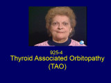 |
Thyroid Associated Orbitopathy (TAO) (PPT) | Shirley H. Wray, MD, PhD, FRCP | This 71 year old woman (Wray case 925-4) was referred with bilateral optic neuropathy and thyroid associated ophthalmopathy (TAO) of Graves' Disease. She had been treated for primary hyperthyroidism on three occasions with radioactive iodine and was taking Tapazole 5 mg daily. Neuro-ophthalmological... | Bilateral Lid Retraction; Lid Lag; Bilateral Exophthalmus; Restrictive Orbitopathy of Graves' Disease; Lid Retraction; Thyroid Orbitopathy; Restriction Syndromes; Thyroid Eye Disease; Thyroid-Associated Ophthalmopathy; Blow-Out Fracture |
| 5 |
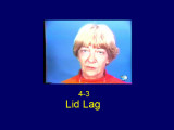 |
Lid Lag (PPT) | Shirley H. Wray, MD, PhD, FRCP | The classical eye signs of thyroid associated ophthalmopathy (TAO) of Graves' Disease is illustrated by case ID925-4. This 50 year old woman with TAO is included in the collection because she illustrates very well lid lag (persistent elevation of the upper eyelid in downgaze) - von Graefe sign Eyeli... | Lid Lag; Bilateral Exophthalmos; Restrictive Orbitopathy of Graves' Disease |
| 6 |
 |
Thyroid Eye Disease | Helen Jiang MD; Rudrani Banik MD | Power point of case presentation of 50 year old male with newly diagnosed hyperthyroidism who presents with ocular hypertension, acute onset proptosis and visual loss. Diagnosed with thyroid eye disease and compressive optic neuropathy. Treatment options discussed for visual loss in thyroid eye dis... | Thyroid Eye Disease; Compressive Optic Neuropathy; Proptosis; Ocular Hypertension |
| 7 |
 |
Ocular Neuromyotonia | Raed Behbehani, MD | Ocular Neuromytonia is a characterised by by paroxysmal tonic contraction of the extraocular muscles supplied by the oculomotor nerve. It is has been reported after cranial radiation therapy, especially to the sellar-parasellar region and from compressive lesions such tumours or aneurysms. The patho... | Ocular Neuromyotania |
| 8 |
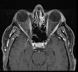 |
Curtain Sign (Enhanced Ptosis) - Associated Image 1 | Bashaer Aldhahwani, MD; Hong Jiang, MD, PhD | This is a 78-year-old male patient who presented with diplopia, right eyelid ptosis, and ophthalmoplegia. He had severe ptosis OD and pseudo-proptosis (lid retraction) OS at baseline, but when the right eyelid was manually elevated, there was marked enhanced ptosis of the left eyelid (Video). He was... | Myasthenia GravIs; Clinical Signs |
| 9 |
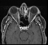 |
Curtain Sign (Enhanced Ptosis) - Associated Image 2 | Bashaer Aldhahwani, MD; Hong Jiang, MD, PhD | This is a 78-year-old male patient who presented with diplopia, right eyelid ptosis, and ophthalmoplegia. He had severe ptosis OD and pseudo-proptosis (lid retraction) OS at baseline, but when the right eyelid was manually elevated, there was marked enhanced ptosis of the left eyelid (Video). He was... | Myasthenia GravIs; Clinical Signs |
| 10 |
 |
Curtain Sign (Enhanced Ptosis) | Bashaer Aldhahwani, MD; Hong Jiang, MD, PhD | This is a 78-year-old male patient who presented with diplopia, right eyelid ptosis, and ophthalmoplegia. He had severe ptosis OD and pseudo-proptosis (lid retraction) OS at baseline, but when the right eyelid was manually elevated, there was marked enhanced ptosis of the left eyelid (Video). He was... | Myasthenia GravIs; Clinical Signs |
| 11 |
 |
Radiological Examination of the Visual System | John Pula, MD | An explanation of imaging types. | Visual System; Radiology; Imaging |
| 12 |
 |
The Anatomic Course of Cranial Nerve IV | Divya Chauhan, MD | Overview of the intracranial course of the trochlear nerve. | Cranial Nerve IV; Trochlear Nerve; Anatomy |
| 13 |
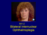 |
Bilateral Internuclear Ophthalmoplegia | Shirley H. Wray, MD, PhD, FRCP | This patient was seen at the Yale Eye Center at the age of 37. She had a long history of multiple sclerosis. At age 22, she had an acute attack of optic neuritis in the left eye which recovered fully within three weeks. Some months later she had a recurrent episode in the same eye, which also recove... | Bilateral Internuclear Ophthalmoplegia; Pendular Horizontal Oscillations; Lid Nystagmus; Upbeat Nystagmus; Botulinum Toxin Therapy; Multiple Sclerosis; Horizontal Pendular Nystagmus; Gaze Evoked Upbeat Nystagmus; Abducting Nystagmus |
| 14 |
 |
Tolosa Hunt Syndrome | Sahil Aggarwal, MD; Jason Liss, MD | Presentation covering an overview of Tolosa Hunt Syndrome. | Tolosa Hunt Syndrome |
| 15 |
 |
Neuro-Ophthalmic Manifestations of Mitochondrial Disease | Abhimanyu S. Ahuja, BS; Sidney M. Gospe III, MD, PhD | Classically, mitochondrial diseases exhibit a maternal inheritance pattern because pathogenic mutations in mitochondrial DNA (mtDNA) are transmitted exclusively via the maternal lineage due to rapid degradation of sperm-derived mitochondria early in embryogenesis. However, mutations in mtDNA may occ... | Mitochondrial Disease; Pathophysiology |
| 16 |
 |
Amyotrophic Lateral Sclerosis (ALS) | Natali V. Baner, MD; Ali G. Hamedani, MD, MHS | PowerPoint providing an overview of the definition, clinical presentation and treatment of amyotrophic lateral sclerosis (ALS). | Amyotrophic Lateral Sclerosis (ALS) |
| 17 |
 |
Non-accidental Eye Injuries in Pediatric Patients for the Neuro-ophthalmologist | Rachelle Srinivas; Melanie Truong-Le | Physical abuse, or non-accidental trauma (NAT), remains a leading cause of injury and death among young children, with abusive head trauma (AHT) being a major contributor. Common associated ocular findings include retinal hemorrhages (RH), subconjunctival hemorrhage, and optic nerve abnormalities, w... | Abusive Head Trauma; Child Abuse; Eye Injury; Retinal Hemorrhage |
1 - 25 of 17
