Collection of materials relating to neuro-ophthalmology as part of the Neuro-Ophthalmology Virtual Education Library.
NOVEL: https://novel.utah.edu/
TO
Filters: Collection: "ehsl_novel_novel"
| Title | Creator | Description | Subject | ||
|---|---|---|---|---|---|
| 1 |
 |
Acquired Hyperopia | AAO/NANOS - American Academy of Ophthalmology / North American Neuro-Ophthalmology Society | Choroidal folds may result from choroidal tumors, compression on the eye wall from thyroid ophthalmopathy, orbital pseudotumor, orbital tumor, posterior scleritis, hypotony, scleral laceration, retinal detachment, marked hyperopia, or secondary to papilledema. Intraocular pressure measurements, refr... | Acquired Hyperopia |
| 2 |
 |
Albrecht von Graefe | Kathleen B. Digre, MD; James J. Corbett, MD | Biography of Albrecht von Graefe. | Albrecht von Graefe; History |
| 3 |
 |
Best's Vittelform Maculopathy | Gregory P. Van Stavern, MD | This 14 year old presented with decreased vision, headaches and central scotomas. She was found to have bilateral papilledema related to IIH and also Best's vitilliform maculopathy. The maculas are commonly described as having a "fried egg" sunny side up appearance. | Best Macular Dystrophy |
| 4 |
 |
Branch Retinal Artery Occlusion | Kathleen B. Digre, MD; James J. Corbett, MD | Slideshow describing condition. | Occlusion |
| 5 |
 |
Branch Retinal Artery Occlusion with Multiple Retinal Emboli | Kathleen B. Digre, MD; James J. Corbett, MD | Slideshow describing condition. | Retinal Emboli; Emboli |
| 6 |
 |
Branch Retinal Emboli | Kathleen B. Digre, MD; James J. Corbett, MD | Slideshow describing condition. | Emboli |
| 7 |
 |
Branch Retinal Vein Occlusion (BRVO) | Kathleen B. Digre, MD; James J. Corbett, MD | Slideshow describing condition. | Occlusion |
| 8 |
 |
Calcific Emboli | Kathleen B. Digre, MD; James J. Corbett, MD | Slideshow describing condition. | Emboli |
| 9 |
 |
Central Cone Dystrophy Occult Macular Dystrophy | Gregory Van Stavern, MD | Slideshow describing condition of Central Cone Dystrophy Occult Macular Dystrophy | Central Cone Distrophy; Macular Dystrophy; Occult Macular Dystrophy |
| 10 |
 |
Central Retinal Vein Occlusion | Kathleen B. Digre, MD | Slideshow describing condition. | Occlusion |
| 11 |
 |
Chiasmal Neuritis in Multiple Sclerosis | Kathleen B. Digre, MD; James J. Corbett, MD | Slideshow describing condition. | Multiple Sclerosis |
| 12 |
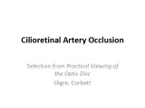 |
Cilioretinal Artery Occlusion | Kathleen B. Digre, MD; James J. Corbett, MD | Slideshow describing cilioretinal artery occlusion. | Occlusion; Artery Occlusion |
| 13 |
 |
Craniopharyngioma and Optic Atrophy | Kathleen B. Digre, MD; James J. Corbett, MD | Slideshow describing condition. | Craniopharayngioma; Otpic Atrophy |
| 14 |
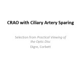 |
CRAO with Ciliary Artery Sparing | Kathleen B. Digre, MD; James J. Corbett, MD | Slideshow describing condition. | CRAO |
| 15 |
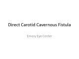 |
Direct Carotid Cavernous Fistula | Emory Eye Center | Slideshow describing condition. | Fistula |
| 16 |
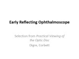 |
Early Reflecting Ophthalmoscope | Kathleen B. Digre, MD; James J. Corbett, MD | The use of the early reflecting ophthalmoscope. | Reflecting Ophthalmoscope; History |
| 17 |
 |
Fat Emboli | Kathleen B. Digre, MD; James J. Corbett, MD | Slideshow describing condition. | Emboli |
| 18 |
 |
Fibrin-Platelet Emboli | Kathleen B. Digre, MD; James J. Corbett, MD | Slideshow describing condition. | Emboli; Platelet Emboli |
| 19 |
 |
Fibrous Dysplasia | Kathleen B. Digre, MD; James J. Corbett, MD | Slideshow describing condition. | Dysplasia; Fibrous Dysplasia |
| 20 |
 |
Fluorescein Angiography | Kathleen B. Digre, MD; James J. Corbett, MD | Fluorescein angiography in neuro-ophthalmology. | Fluorescein Angiography; History |
| 21 |
 |
Glaucoma | Kathleen B. Digre, MD; James J. Corbett, MD | Slideshow describing condition. | Glaucoma |
| 22 |
 |
Hermann Ludwig Ferdinand von Helmholtz | Kathleen B. Digre, MD; James J. Corbett, MD | Biography of Hermann Ludwig Ferdinand von Helmholtz. | Hermann Ludwig Ferdinand von Helmholtz; History |
| 23 |
 |
High Myopia | Kathleen B. Digre, MD; James J. Corbett, MD | Slideshow describing condition. | Myopia |
| 24 |
 |
Hollenhorst Plaque | Kathleen B. Digre, MD; James J. Corbett, MD | Slideshow describing condition. | Hollenhorst Plaque |
| 25 |
 |
Hughlings Jackson | Kathleen B. Digre, MD; James J. Corbett, MD | Biography of Hughlings Jackson. | Hughlings Jackson; History |