Collection of materials relating to neuro-ophthalmology as part of the Neuro-Ophthalmology Virtual Education Library.
NOVEL: https://novel.utah.edu/
TO
Filters: Collection: "ehsl_novel_novel"
1 - 25 of 21
| Title | Creator | Description | Subject | ||
|---|---|---|---|---|---|
| 1 |
 |
Optic Chiasm | Yesha Shah, BSA, BBA; Amanda Henderson, MD | Overview of the anatomy of the optic chiasm. | Optic Chiasm; Anatomy |
| 2 |
 |
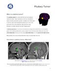
Pit Tumor Chiasm Compression | Kathleen B. Digre, MD; James J. Corbett, MD | Slideshow describing condition. | Pituitary Tumor; Pituitary Mass; Chiasm; Chiasm Compression |
| 3 |
 |
Metastatic Glioblastoma to Intracranial Optic Nerves, Optic Chiasm and Optic Tracts | Bashaer Aldhahwani, MD; Mariam S. Vilá-Delgado, MD | The patient with pathology confirmed glioblastoma after resectioning the superior frontal lobe tumor followed by 6 weeks of radiation therapy with concurrent temozolomide. The patient started bevacizumab to treat steroid-refractory vasogenic cerebral edema/radiation necrosis. 8 months after radiatio... | Metastatic Glioblastoma; Infiltrative Chiasmal Lesion |
| 4 |
 |
Neuro-Ophthalmic Manifestations of Sellar and Parasellar Masses | Rudrani Banik, MD | Neuroanatomy of the Chiasm. | Parasellar Masses |
| 5 |
 |
Illustrations of the Afferent Visual Pathway and Concepts Surrounding Trans-Synaptic Neuroaxonal Degeneration in the Visual Pathway in Multiple Sclerosis | Olwen C. Murphy; Peter A. Calabresi; Shiv Saidha | Image 1 title: Functionally-eloquent organization of the afferent visual pathway; Image 1 description: The afferent visual pathway is a sensory pathway comprised of 3 neurons. The 1st order neurons are the shortest neurons in the pathway and are entirely unmyelinated. The cell bodies of the 1st orde... | Optic Neuritis; Multiple Sclerosis; Neuroaxonal Degeneration; Trans-synaptic Degeneration; Visual Pathway; Functional Eloquence |
| 6 |
 |
Anatomy of the Oculomotor Nerve (CN III) | Lucas E. Morgan, MS4; Nicholas A. Koontz, MD; Devin D. Mackay, MD | A detailed overview of the anatomic course of CN III, including a detailed pathway description and labeled MRI images, gross anatomy pictures, and structural models. | CN III; Third Cranial Nerve; Oculomotor Nerve; Anatomy; MRI |
| 7 |
 |
Radiological Examination of the Visual System | John Pula, MD | An explanation of imaging types. | Visual System; Radiology; Imaging |
| 8 |
 |
Common Patterns of Visual Field Defects | Sean Gratton, MD; Sarah Lam, 6th year BA/MD | Lecture covering common visual field defects, including those of the retina, optic nerve, chiasm, and retrochiasmal. | Visual Field Defects |
| 9 |
 |
The Visual Pathway: Neuroanatomy Video Lab - Brain Dissections | Suzanne S. Stensaas, PhD | A brief review of the anatomy of the eye and the photic stimulation of the receptors is followed by a gross exploration of the visual pathway from the optic nerve, chiasm, and tract to the thalamus stressing how the left part of the visual world reaches the right hemisphere. Visual fields are relate... | Visual Pathway; Brain; Dissections |
| 10 |
 |
Ethambutol Optic Neuropathy | Hailey Mair, BS; Padmaja Sudhakar, MD | This is a PowerPoint slide describing ethambutol induced optic neuropathy and it elaborates on mechanism and vulnearble population. | Ethambutol; Tuberculosis; Optic Neuropathy |
| 11 |
 |
A Macro Dilemma: Demonstrations of the Anterior Chiasmal Syndrome and Wilbrand's Knee | Ariel Axelbaum, MD; Nurhan Torun, MD | Presentation reviewing two cases that demonstrate several cases of anterior chiasmal syndromes and the variability in patient presentations with sellar masses. | Anterior Chiasmal Syndromes; Masses of the Sella |
| 12 |
 |
Pituitary Tumor | NANOS | Pituitary tumors are benign (non-cancerous) overgrowth of cells that make up the pituitary gland (the master gland that regulates other glands in the body). Updated April 2020. | Pituitary Tumor; Patient Brochure |
| 13 |
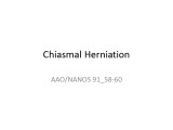 |
Chiasmal Herniation (PowerPoint) | AAO/NANOS - American Academy of Ophthalmology / North American Neuro-Ophthalmology Society | This woman was 61 years old when she underwent initial neuro-ophthalmologic evaluation. Twenty-three years earlier, she had undergone removal of a pituitary adenoma followed by radiation therapy. At that time, she had noted a preoperative visual field defect that improved somewhat after the surgery.... | Chiasmal Herniation |
| 14 |
 |
Brain Surface Anatomy | Arooj Ahmad, MD; Devin D. Mackay, MD | These images depict labeled structures of the surface anatomy of the different facies of the brain. | Neuroanatomy; Brain Surface Anatomy |
| 15 |
 |
Night Wolf | Mehdi Tavakoli, MD; Byron Lam, MD | A case presentation on radiation optic neurology. | Radiation Optic Neuropathy |
| 16 |
 |
Bony Anatomy | Ryung San Lee; James Brian Davis; Amanda Dean Henderson | These are multiple choice questions to Test Your Knowledge that go along with the Bony Anantomy topics covered in the NANOS Illustrated Curriculum. | Anatomy; Bone; Orbit; Skull |
| 17 |
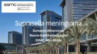 |
Suprasellar Meningioma | Sumayya Almarzouqi, MD | Description of a case of suprasellar or sellar mass causeing chiasmal compression. | Suprasellar Meningioma |
| 18 |
 |
Multiple Sclerosis | Shirley H. Wray, MD, PhD, FRCP | The patient is a 56 year old woman who presented in 1982, at the age of 48, with a one week history of painless loss of vision in the left eye. Past History: Negative for a previous attack of optic neuritis or transient neurological symptoms. Family History: Negative for CNS disease Neuro-ophthalmol... | Upbeat Nystagmus; Lid Nystagmus; Square Wave Jerks; Jerk Oscillations; Rotary Nystagmus; Saccadic Pursuit; Saccadic Dysmetria; Multiple Sclerosis; Bilateral Lid Nystagmus; Primary Position Upbeat Nystagmus; Torsional Nystagmus; Horizontal Saccadic Dysmetria |
| 19 |
 |
Optical Coherence Tomography (OCT) | Gunnar J. Goebel; Devin D. Mackay, MD | Introduction to OCT, including history, principles, interpretation, and applications. | Optical Coherence Tomography (OCT) |
| 20 |
 |
Parasellar Meningioma | Shirley H. Wray, MD, PhD, FRCP | This patient is a 58 year old woman from Peru who, in 1975, developed intermittent headaches and right retro-orbital eye pain. She was seen by several ophthalmologists in South America who were unable to make a diagnosis. In March 1977 she awoke one morning with vertical diplopia most marked on look... | Ptosis; Third Nerve Palsy; Aberrant Reinnervation of the Third Nerve; Oculomotor Nerve; Parasellar Meningioma; Cavernous Sinus Syndrome; Unilateral Oculomotor Third Nerve Palsy; Unilateral Third Nerve Palsy |
| 21 |
 |
Confrontation Visual Fields - A Concise Guide for Ophthalmology and Neurology Trainees | Stephen C. Pollock, MD | The guide describes the techniques required to competently perform confrontation visual fields. It outlines a basic screening protocol and discusses methods for further defining defects identified during the screening process. A mini-atlas of visual field defects is included as an appendix. | Confrontation Visual Fields; Visual Field Testing; Perimetry; Visual Field Loss; Visual Field Defect; Ocular Examination; Visual Sensory Evaluation; Neurologic Examination |
1 - 25 of 21