Collection of materials relating to neuro-ophthalmology as part of the Neuro-Ophthalmology Virtual Education Library.
NOVEL: https://novel.utah.edu/
TO
- NOVEL978
Filters: Collection: "ehsl_novel_novel"
| Title | Creator | Description | Subject | ||
|---|---|---|---|---|---|
| 1 |
 |
"Simon Says" Technique for Nonorganic Visual Field Constriction | Devin D. Mackay, MD | Explanation of the "Simon says" technique. | Simon Says, Visual Field Constriction |
| 2 |
 |
1% Pilocarpine Testing | Karl C. Golnik, MD | A brief description of using Pilocarpine to test the pupils in patients with anisocoria. | Pharmacological Testing; Pilocarpine |
| 3 |
 |
2013 William F Hoyt Lecture: Neuro-Ophthalmology in Review: Around the Brain with 50 Fellows | Nancy J. Newman, MD | No matter what their ultimate specialty, every ophthalmologist needs to master the basics of neuroophthalmology. To that end, we must ensure that we continue to train effective teachers of neuro-ophthalmology. This is William F. Hoyt's most important lasting legacy and charge. In this same spirit, E... | History |
| 4 |
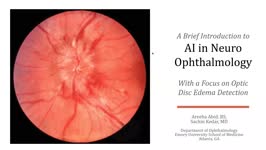 |
A Brief Introduction to AI in Neuro-ophthalmology | Areeba Abid, BS; Sachin Kedar, MD | In this video, we describe the basics of artificial intelligence and machine learning as applicable to clinical neuro-ophthalmologists. We use the example from a recent publication, where AI software was used to detect optic disc edema in fundus images. | Artificial Intelligence; Machine Learning |
| 5 |
 |
A Brief Introduction to Post-Ocular Surgery Strabismus | Srujay Pandiri, Medical Student | This is a short narrated Powerpoint that introduces concepts important to post-ocular surgery strabismus. It highlights the connection between common surgeries include cataract surgery, scleral buckle surgery, refractive surgery, among others. | Diplopia; Strabismus; Post-ocular Surgery Strabismus |
| 6 |
 |
A Case of Left Facial Pain Misdiagnosed as Migraine Headache | Hong Jiang | A 58-year-old female sought a second opinion due to a two-year duration of sporadic left facial pressure pain, along with irritation and dryness in her left eye. She has a well-documented history of episodic migraines characterized by throbbing headaches on the right side. She had been under the car... | Dry Eye; Facial Nerve Schwannoma; Facial Pain |
| 7 |
 |
A Case of Orbital Meningioma Misdiagnosed as Non-arthritic Ischemic Optic Neuropathy | Hong Jiang, MD, PhD | A 62-year-old woman with hypertension, dyslipidemia, and mild cognitive impairment presented for a second opinion of her left eye visual loss for three months. She initially noticed a black spot in the lower visual field of her left eye and sought care from her local ophthalmologist. At that time, s... | Compressive Optic Neuropathy, Orbit MRI; Orbital Meningioma |
| 8 |
 |
A Case Series of Mydriasis from an Anticholinergic Antiperspirant | Aileen Antonio, MD; Inna Bondira, DO; Cameron Holicki, DO; Christopher Glisson, DO; Tatiana Deveney, MD; Lina Nagia, DO | Causes of anisocoria span a wide range, from benign to life-threatening, making it a common indication for Neuro-Ophthalmology referrals. One such cause is related to pharmacologic mydriasis due to direct or systemic exposure. We present a case series of four patients with different presentations of... | Anisocoria; Mydriasis; Pharmacologic Anisocoria; Anticholinergic Antiperspirant |
| 9 |
 |
A Five-Minute, Intravenous Edrophonium -Window to Observe the CNS Response to Myasthenic Ophthalmoplegia | Jorge C Kattah, MD | A video showing how ocular motor adaptation may be observed. | Ocular Motor Adaptation |
| 10 |
 |
A Macro Dilemma: Demonstrations of the Anterior Chiasmal Syndrome and Wilbrand's Knee | Ariel Axelbaum, MD; Nurhan Torun, MD | Presentation reviewing two cases that demonstrate several cases of anterior chiasmal syndromes and the variability in patient presentations with sellar masses. | Anterior Chiasmal Syndromes; Masses of the Sella |
| 11 |
 |
A Suspected Case of Wildervanck Syndrome | Hari R. Anandarajah; Tejaswini K. Deshmukh; Ryan D. Walsh | A 5-year-old male with congenital right-sided hearing loss presented to the neuro-ophthalmology clinic for strabismus evaluation. He had longstanding bilateral abduction deficits with associated esotropia, for which he underwent bilateral medial rectus recessions at age three. He had persistent limi... | Cervico-oculo-acoustic Syndrome; Congenital Deafness; Duane Syndrome; Klippel-Feil Cervical Anomaly; Wildervanck Syndrome |
| 12 |
 |
Aberrant Regeneration of Third Nerve | Gregory P. Van Stavern, MD | 48 year old woman S/P rupture and repair of right sided posterior communicating artery aneurysm Video shows residual partial right third nerve palsy, with aberrant regeneration, causing a pseudo Von Graefe's sign (elevation of the right upper eyelid with attempted infraduction of the right eye) Se... | Aberrant Regeneration of Third Nerve; Third Nerve Palsy |
| 13 |
 |
Aberrant Regeneration Third Nerve | Gregory P. Van Stavern, MD | 48 year old woman S/P rupture and repair of right sided posterior communicating artery aneurysm Video shows residual partial right third nerve palsy, with aberrant regeneration, causing a pseudo Von Graefe's sign (elevation of the right upper eyelid with attempted infraduction of the right eye) | Aberrant Regeneration Third Nerve; Third Nerve Palsy |
| 14 |
 |
Acquired Hyperopia | AAO/NANOS - American Academy of Ophthalmology / North American Neuro-Ophthalmology Society | Choroidal folds may result from choroidal tumors, compression on the eye wall from thyroid ophthalmopathy, orbital pseudotumor, orbital tumor, posterior scleritis, hypotony, scleral laceration, retinal detachment, marked hyperopia, or secondary to papilledema. Intraocular pressure measurements, refr... | Acquired Hyperopia |
| 15 |
 |
Acute Idiopathic Blind Spot Enlargement (AIBSE): Case Report | Andrew R. Osborn, MD; James C. O'Brien, MD | We present a single case of Acute Idiopathic Blind Spot Enlargement (AIBSE) in a young female patient, including her clinical course and relevant imaging studies. Her case description is followed by a brief discussion surrounding the current understanding of this rare entity. Associated Images: http... | Acute Idiopathic Blind Spot Enlargement (AIBSE); Outer Retinopathy |
| 16 |
 |
Acute Idiopathic Blind Spot Enlargement (AIBSE): Images | Andrew R. Osborn, MD; James C. O'Brien, MD | We present a single case of Acute Idiopathic Blind Spot Enlargement (AIBSE) in a young female patient, including her clinical course and relevant imaging studies. Her case description is followed by a brief discussion surrounding the current understanding of this rare entity. Associated Case Report:... | Acute Idiopathic Blind Spot Enlargement (AIBSE); Outer Retinopathy |
| 17 |
 |
Acute Multifocal Pigment Epithelium Epitheliopathy (AMPEE) | Gregory P. Van Stavern, MD | Images providing example of Acute Multifocal Pigment Epithelium Epitheliopathy (AMPEE) | Acute Multifocal Pigment Epithelium Epitheliopathy (AMPEE) |
| 18 |
 |
Acute Optic Neuritis | Neil R. Miller, MD, FACS | Overview of acute optic neuritis. | Optic Neuritis |
| 19 |
 |
Acute Retinal Necrosis (ARN) | Gregory P. Van Stavern, MD | Acute Retinal Necrosis causes inflammation and subsequent retinal detachment. This powerpoint provides images depicting ARN. | Acute Retinal Necrosis (ARN) |
| 20 |
 |
Acute Zonal Occult Outer Retinopathy (AZOOR) versus Multiple Evanescent White Dot Syndrome (MEWDS) | Asim V. Farooq, MD; Michael T. Andreoli, MD; Heather E. Moss, MD | PPT case report on acute zonal occult outer retinopathy (AZOOR) versus multiple evanescent white dot syndrome (MEWDS). | AZOOR; MEWDS; Paracentral Scotoma; Goldmann Visual Field; Photoreceptor Loss |
| 21 |
 |
Afferent Visual Pathway Disorders: Typical vs Atypical Optic Neuritis | Carmen Chan, FRCP, FRCOphth, FRCSEd(Ophth), FHKAM(Ophthalmology) | Discussion of typical vs atypical optic neuritis. | Optic Neuritis |
| 22 |
 |
Age Related Macular Degeneration | Riya H. Patel; James Brian Davis; Amanda Dean Henderson | Age-related macular degeneration (AMD) is a degenerative disease of the retina that causes central vision loss, and it is the leading cause of blindness in the developed world. Age is a strong nonmodifiable risk factor for AMD. Patients may have genetic susceptibility to AMD from mutations in genes ... | AMD; AREDS2; Exudative; Geographic Atrophy; Macular Degeneration; Nonexudative |
| 23 |
 |
Age-related Macular Degeneration: The Basics | Arnav Gupta, BHSc; Rahul Sharma, MD, MPH | A presentation covering age-related macular degeneration ("ARMD" or "AMD"), an acquired, progressive, chronic, degenerative disease of the retina. | Macular Degeneration |
| 24 |
 |
Albrecht von Graefe | Kathleen B. Digre, MD; James J. Corbett, MD | Biography of Albrecht von Graefe. | Albrecht von Graefe; History |
| 25 |
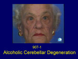 |
Alcoholic Cerebellar Degeneration | Shirley H. Wray, MD, PhD, FRCP | The patient is a 72 year old woman who presented with a 4 year history of progressive difficulty with balance, frequent falls and unsteadiness walking. She required a cane to steady herself. Past History: Significant for alcohol abuse. In 1980, she came to Boston for a second opinion and was seen in... | Square Wave Jerks; Dysmetria; Horizontal Saccadic Hypermetria; Horizontal Gaze Evoked Nystagmus; Saccadic Pursuit; Gait Ataxia; Alcoholic Cerebellar Degeneration; Gaze Evoked Horizontal Nystagmus; Horizontal Saccadic Dysmetria; Alcohol |
