Collection of materials relating to neuro-ophthalmology as part of the Neuro-Ophthalmology Virtual Education Library.
NOVEL: https://novel.utah.edu/
TO
- NOVEL193
Filters: Collection: "ehsl_novel_novel"
| Title | Creator | Description | Subject | ||
|---|---|---|---|---|---|
| 151 |
 |
The Visual Pathway: Neuroanatomy Video Lab - Brain Dissections | Suzanne S. Stensaas, PhD | A brief review of the anatomy of the eye and the photic stimulation of the receptors is followed by a gross exploration of the visual pathway from the optic nerve, chiasm, and tract to the thalamus stressing how the left part of the visual world reaches the right hemisphere. Visual fields are relate... | Visual Pathway; Brain; Dissections |
| 152 |
 |
Auditory System: Neuroanatomy Video Lab - Brain Dissections | Suzanne S. Stensaas, PhD | The anatomy of the middle ear and cochlea are shown using models and diagrams explaining the process of air-fluid transmission and finally transduction by hair cells. Gross specimens demonstrate the cochlear nerve and its brain stem relays and crossings all the way to auditory cortex. Wernicke's are... | Middle Ear; Cochlea; Auditory Cortex; Brain; Dissection |
| 153 |
 |
Basal Ganglia: Neuroanatomy Video Lab - Brain Dissections | Suzanne S. Stensaas, PhD | Structures involved in involuntary movements are shown on models, in animations, and on gross coronal and axial sections. Diagrams show the cortex-to-cortex loop and the nigrostriatal pathway. The direct and indirect pathways are shown. The direct pathway facilitates movement while the indirect path... | Basal Ganglia; Involuntary Movements; Brain; Dissection |
| 154 |
 |
Cerebellum: Neuroanatomy Video Lab - Brain Dissections | Suzanne S. Stensaas, PhD | The gross features of the cerebellum are shown. The three peduncles are demonstrated, noting their input and output to and from the cerebellum. Emphasis is given to symptoms of cerebellar disease appearing on the same side of the body. Special emphasis is given to cortical-cerebellar connections, st... | Cerebellum; Cortical-Cerebellar Connection; Brain; Dissection |
| 155 |
 |
Comparison of the Motor Systems: Neuroanatomy Video Lab - Brain Dissections | Suzanne S. Stensaas, PhD | A comparison of the three major motor systems focuses on categorizing motor problems as corticospinal tract, cerebellar, or basal ganglia. It begins with 12 minutes of gross anatomical structures and pathway review followed by clinical video clips demonstrating clinical features of disease of each o... | Corticospinal Tract; Cerebellar; Basal Ganglia; Motor Systems; Brain; Dissection |
| 156 |
 |
Control of the Eye Movements: Neuroanatomy Video Lab - Brain Dissections | Suzanne S. Stensaas, PhD | Disturbances in eye movements can provide important clues for localization of neurological damage. The role of the frontal eye fields in horizontal gaze is stressed. The need to coordinate cranial nerves on both sides of the brain stem introduces the medial longitudinal fasciculus and its role in co... | Medial Longitudinal Fasciculus; Third Cranial Nerve; Sixth Cranial Nerve; Internuclear Ophthalmoplegia; Nystagmus; Brain; Dissection |
| 157 |
 |
Three Critical Vertical Pathways: Neuroanatomy Video Lab - Brain Dissections | Suzanne S. Stensaas, PhD | There is one motor and two sensory pathways that must be mastered. Pain and temperature from the body travel together and vibration and proprioception travel in another pathway each reaching perception in the cortex. Voluntary motor control starts in the cerebral cortex and connects with a motor neu... | Spinothalamic Tract; Dorsal Column-Medical Lemniscus Pathway; Posterior Column; Vertical Pathway; Brain; Dissection |
| 158 |
 |
Vestibular System: Neuroanatomy Video Lab - Brain Dissections | Suzanne S. Stensaas, PhD | Diagrams, models and skull preparations are used to describe the vestibular apparatus. The semicircular canals, saccule and utricle are described as well as transduction by the hair cells in the ampullae and maculae. Gross material emphasizes the nerve, vestibular nuclei and connections through the ... | Vestibular; Brain; Dissection |
| 159 |
 |
White Dot Syndromes: MEWDS, AZOOR, AIBSE | Gregory P. Van Stavern, MD | Some have lumped Multiple Evanescent White Dot Syndrome (MEWDS), Acute Idiopathic Blind Spot Enlargement (AIBSE) with acute macular neuroretinopathy, and pseudo-presumed ocular histoplasmosis syndrome together with AZOOR (Acute Zonal Occult Outer Retinopathy). These conditions all present with visua... | White Dot Syndromes: MEWDS, AZOOR, AIBSE |
| 160 |
 |
The Clinical Examination of Higher Order Visual Function: Syndrome-based Approach - Visual Neglect | Victoria S. Pelak, MD; James R. Bateman, MD, MPH; Brianne Bettcher, PhD | Explanation of higher order visual function examination. See accompanying video, Double simultaneous visual field stimulation: https://collections.lib.utah.edu/ark:/87278/s6gn2h9d | Visual Function; Visual Neglect |
| 161 |
 |
Sensation from the Body: Neuroanatomy Video Lab - Brain Dissections | Suzanne S. Stensaas, PhD | Sensation consists of various modalities, which tend to travel in one of two pathways. The Anterolateral System also known as the Spinothalamic Tract carries pain and temperature. The Dorsal Column-Medical Lemniscus Pathway carries vibration, joint position, and fine 2-point discrimination. Light or... | Spinothalamic Tract; Anterolateral System; Dorsal Column-Medical Lemniscus Pathway; Body Sensation' Brain' Dissection |
| 162 |
 |
Magnetic Resonance Imaging (MRI) | Devin D. Mackay, MD | Explanation of using magnetic resonance imaging (MRI) in examinations. | Magnetic Resonance Imaging (MRI) |
| 163 |
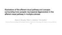 |
Illustrations of the Afferent Visual Pathway and Concepts Surrounding Trans-Synaptic Neuroaxonal Degeneration in the Visual Pathway in Multiple Sclerosis | Olwen C. Murphy; Peter A. Calabresi; Shiv Saidha | Image 1 title: Functionally-eloquent organization of the afferent visual pathway; Image 1 description: The afferent visual pathway is a sensory pathway comprised of 3 neurons. The 1st order neurons are the shortest neurons in the pathway and are entirely unmyelinated. The cell bodies of the 1st orde... | Optic Neuritis; Multiple Sclerosis; Neuroaxonal Degeneration; Trans-synaptic Degeneration; Visual Pathway; Functional Eloquence |
| 164 |
 |
Ocular Surface, Cornea, Lens | Ryung San Lee; Amanda Dean Henderson | Multiple choice questions set from the NANOS Illustrated Curriculum for Neuro-Ophthalmology. This question set is meant to accompany the video lecture on the anatomy of the ocular surface, cornea, and lens. The ocular surface is a complex topic and pertinent to the comprehensive ophthalmic care prov... | Cornea; Ocular Surface; Uvea |
| 165 |
 |
The Clinical Examination of Higher Order Visual Function: Syndrome-Based Approach | Victoria S. Pelak, MD; James R. Bateman, MD, MPH; Brianne Bettcher, PhD | Explanation of higher order visual function examination. See accompanying video, Double simultaneous visual field stimulation: https://collections.lib.utah.edu/ark:/87278/s6gn2h9d | Visual Function |
| 166 |
 |
2013 William F Hoyt Lecture: Neuro-Ophthalmology in Review: Around the Brain with 50 Fellows | Nancy J. Newman, MD | No matter what their ultimate specialty, every ophthalmologist needs to master the basics of neuroophthalmology. To that end, we must ensure that we continue to train effective teachers of neuro-ophthalmology. This is William F. Hoyt's most important lasting legacy and charge. In this same spirit, E... | History |
| 167 |
 |
Ptosis | Ethan Waisberg, MB, BCh, BAO candidate | Description of ptosis including etiology, management and treatment. | Ptosis; Blepharoptosis |
| 168 |
 |
Tolosa Hunt Syndrome | Sahil Aggarwal, MD; Jason Liss, MD | Presentation covering an overview of Tolosa Hunt Syndrome. | Tolosa Hunt Syndrome |
| 169 |
 |
Clinical Visual Electrophysiology | Gregory P. Van Stavern, MD; Byron Lam, MD | A description of the use of electrophysiology to examine the visual system. | Electrophysiology; Visual Exam |
| 170 |
 |
Skew Deviation and the Ocular Tilt Response | David Newman-Toker, MD, PhD | The objectives of this presentation are to provide an understanding of the current use of the terms "ocular tilt reaction" and "skew deviation," to create some familiarity with the anatomic and physiologic substrate of ocular tilt and skew, and to demonstrate how to distinguish between skew and isol... | Vertical Strabismus; Cyclovertical Strabismus; Skew Deviation; Vestibulo-ocular Reflex; Ocular Tilt Reaction; Vestibular Pathways; Vestibular Ocular System |
| 171 |
 |
Neuro-Ophthalmology of Multiple Sclerosis | Emily Sun, Medical Student; Amanda Henderson, MD | Multiple Sclerosis (MS) is the most common neurological disease in young people with an average age of onset between 15 and 35 years old. MS is an autoimmune inflammatory condition that causes demyelinating lesions in the CNS. The diagnosis is clinical, but MRI is typically used to support the diagn... | Multiple Sclerosis; Optic Neuritis; Uveitis; Internuclear Ophthalmoplegia; Nystagmus; Steroids; Demyelinating; Autoimmune |
| 172 |
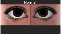 |
Normal Light Reflex and Relative Afferent Pupillary Defect (RAPD) | Marshall Huang, 4th Year Medical Student | A Relative Afferent Pupillary Defect is an examination finding in patients who have an asymmetric pupillary reaction to light when it is shined back and forth between the two eyes. It is most commonly a sign of asymmetric optic nerve disease or damage but can also present in widespread asymmetric r... | Light Reflex; RAPD |
| 173 |
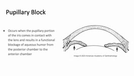 |
Pupillary Block | Sujata Dalal; James Brian Davis; Amanda Dean Henderson | Pupillary block occurs when the pupillary margin of the iris contacts the anterior surface of the lens. This creates a barrier for the outflow of aqueous humor from the posterior chamber to the anterior chamber and resultant increased pressure in the posterior chamber. This pressure can cause the ir... | Angle-Closure Glaucoma; Iris Bombe; Narrow Angle; Pupillary Block |
| 174 |
 |
Intracranial Hemorrhage: Classification and Mechanisms | Victoria S. Pelak, MD | Intracranial hemorrhages can be life-threatening events. Classification of hemorrhages depends on the anatomical location, which also determines the associated risk factors for the hemorrhage. Descriptions of each type of hemorrhage and important aspects of the pathophysiology and risk factors are p... | Intracranial Hemorrhage |
| 175 |
 |
Manuscripts: You Can Write These! | Elaine Smolock, PhD | Overview of writing techniques and parts of the manuscript, basic approach to writing results sections, what makes a good introduction, crafting a meaningful discussion, abstract and title suggestions, and how to get your editor's attention. | Writing Techniques |