Collection of materials relating to neuro-ophthalmology as part of the Neuro-Ophthalmology Virtual Education Library.
NOVEL: https://novel.utah.edu/
TO
- NOVEL194
Filters: Collection: "ehsl_novel_novel"
| Title | Creator | Description | Subject | ||
|---|---|---|---|---|---|
| 126 |
 |
Control of the Pupil: Neuroanatomy Video Lab - Brain Dissections | Suzanne S. Stensaas, PhD | Through diagrams, animations and gross specimens the constriction and dilation of the pupil by the autonomic nervous system are described. Both the parasympathetic and sympathetic control are traced and the importance of a constricted pupil, Horner's Syndrome, and temporal lobe (uncal) herniation (d... | Brain; Dissections |
| 127 |
 |
Hypothalamus: Neuroanatomy Video Lab - Brain Dissections | Suzanne S. Stensaas, PhD | Gross specimens are used to demonstrate the area of the hypothalamus and its relationship to surrounding structures. Both endocrine and autonomic functions are explored using diagrams. Mention is made of the direct hypothalamic response to circulating hormones and other substances such as sodium. Th... | Hypothalamus; Brain; Dissection |
| 128 |
 |
The Meninges: Neuroanatomy Video Lab - Brain Dissections | Suzanne S. Stensaas, PhD | The epidural, subdural and subarachnoid spaces are demonstrated and discussed with respect to trauma and disease. The relationship of the brainstem and cerebellum to the tentorium demonstrates the vulnerability of the brain stem to increased supratentorial pressure and herniation. Arachnoid granulat... | Meninges; Brain; Dissection |
| 129 |
 |
The Most Important Pathway: Motor Control: Neuroanatomy Video Lab - Brain Dissections | Suzanne S. Stensaas, PhD | The origin of the corticospinal tract in the cerebral cortex is traced through gross sections of the hemisphere and brain stem to the spinal cord. Using an animation, the terms upper and lower motor neuron are defined and clinical signs and symptom listed. | Corticospinal Tract; Cerebral Cortex; Motor Neuron; Brain; Dissection |
| 130 |
 |
NANOS Illustrated Curriculum Section D Introduction | Meagan D. Seay, DO; Victoria S. Pelak, MD | Introduction video to the resources available in Section D of the NANOS Illustrated Curriculum. | Lesions, Afferent Visual Pathway; Disorders, Afferent Visual Pathway; Pupillary Anatomy; Disorders, Pupillary Function; Lesions, Efferent Visual Pathway; Ocular Motility; Abnormal Eye Movements; Eyelids; Abnormal Facial Movements |
| 131 |
 |
The Normal Unfixed Brain: Neuroanatomy Video Lab - Brain Dissections | Suzanne S. Stensaas, PhD | The consistency and vulnerability of the brain is demonstrated along with the clear and glistening pia and arachnoid and the tough dura. The cushioning function of the CSF is stressed and the features are pointed out on the ventral surface. The uncus and temporal lobes are normal with arteries free ... | Brain; Dissections |
| 132 |
 |
Orientation: The Planes of the Brain: Neuroanatomy Video Lab - Brain Dissections | Suzanne S. Stensaas, PhD | Terms such as anterior, posterior, inferior and superior are introduced with respect to the hemispheres as well as the brain stem. Terms such as rostral and caudal or dorsal and ventral can mean different things in different areas. Sections in three planes (frontal, axial, and sagittal) are demonstr... | Frontal; Axial; Sagittal; Brain; Dissection |
| 133 |
 |
Vascular Supply of the Eye | Kathleen B. Digre, MD; James J. Corbett, MD | Slideshow describing vascular supply of the eye. | Eye Anatomy; Vascular Supply |
| 134 |
 |
A Case of Left Facial Pain Misdiagnosed as Migraine Headache | Hong Jiang | A 58-year-old female sought a second opinion due to a two-year duration of sporadic left facial pressure pain, along with irritation and dryness in her left eye. She has a well-documented history of episodic migraines characterized by throbbing headaches on the right side. She had been under the car... | Dry Eye; Facial Nerve Schwannoma; Facial Pain |
| 135 |
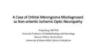 |
A Case of Orbital Meningioma Misdiagnosed as Non-arthritic Ischemic Optic Neuropathy | Hong Jiang, MD, PhD | A 62-year-old woman with hypertension, dyslipidemia, and mild cognitive impairment presented for a second opinion of her left eye visual loss for three months. She initially noticed a black spot in the lower visual field of her left eye and sought care from her local ophthalmologist. At that time, s... | Compressive Optic Neuropathy, Orbit MRI; Orbital Meningioma |
| 136 |
 |
Hydroxychloroquine Retinopathy | Zoe Rebecca Williams, MD; David Allen DiLoreto Jr., MD | This powerpoint reviews hydroxychloroquine retinopathy including the clinical findings and the updated American Academy of Ophthalmology screening guidelines (revised in 2016). It includes images depicting the typical paracentral visual field loss, spectral domain optical coherence tomography findin... | AAO Screening Guidelines; Fundus Autofluorescence; Hydroxychloroquine Retinopathy; Outer Nuclear Layer Loss; Photoreceptor Loss; Retinal Pigment Epithelium Loss; Spectral Domain Optical Coherence Tomography |
| 137 |
 |
Neuropsychological Assessment | Brianne M. Bettcher, PhD; James R. Bateman, MD, MPH; Victoria S. Pelak, MD | Introduction to neuropsychology and neuropsychological assessments. | Neuropsychological Assessment |
| 138 |
 |
Aberrant Regeneration of Third Nerve | Gregory P. Van Stavern, MD | 48 year old woman S/P rupture and repair of right sided posterior communicating artery aneurysm Video shows residual partial right third nerve palsy, with aberrant regeneration, causing a pseudo Von Graefe's sign (elevation of the right upper eyelid with attempted infraduction of the right eye) Se... | Aberrant Regeneration of Third Nerve; Third Nerve Palsy |
| 139 |
 |
Brain Stem & Reflexes: Neuroanatomy Video Lab - Brain Dissections | Suzanne S. Stensaas, PhD | The cranial nerves are reviewed again on a specimen with vessels. Next, landmarks on gross brain stem sections are shown. Stressed are the three reflexes associated with each of the three levels: pupillary, corneal and gag reflexes and their associated cranial nerves. Finally cross sections of myeli... | Brain Stem; Reflexes |
| 140 |
 |
Cortical Localization: Neuroanatomy Video Lab - Brain Dissections | Suzanne S. Stensaas, PhD | The lobes of the brain are defined together with their major functions. The visual field representation in the occipital lobe is explained with a diagram. Speech areas and the major types of aphasia are discussed in the dominant hemisphere and parietal lesions of neglect and spatial orientation are ... | Cortical Localization; Brain; Dissections |
| 141 |
 |
Introduction: Neuroanatomy Video Lab - Brain Dissections | Suzanne S. Stensaas, PhD | The regions and lobes of the brain are identified along with some of the nerves and vessels. The basic functions of the cortex of each lobe are introduced along with principal sulci and gyri. The importance of the left hemisphere for language and the temporal lobe in memory are mentioned along with ... | Brain; Dissections |
| 142 |
 |
Limbic System: Neuroanatomy Video Lab - Brain Dissections | Suzanne S. Stensaas, PhD | The decision was made to present a simplified description of a much more complex system using animations to construct a 3D image. Papez circuit is shown on gross specimens with mention of its involvement in memory. The role of the amygdala in fear and the olfactory cortex in temporal lobe epilepsy a... | Limbic System; Hippocampus; Brain; Dissection |
| 143 |
 |
Olfactory System: Neuroanatomy Video Lab - Brain Dissections | Suzanne S. Stensaas, PhD | Beginning with the location of the sensory cells within the skull the axons are traced into the cranial cavity. Demonstration of the olfactory bulb, olfactory tract and it termination in the forebrain and temporal lobe are indicated. Trauma and meningiomas can produce loss of small (anosmia). Degene... | Olfactory System; Olfactory Bulb; Anosmia; Brain; Dissection |
| 144 |
 |
The Spinal Cord & Monosynaptic Reflex: Neuroanatomy Video Lab - Brain Dissections | Suzanne S. Stensaas, PhD | The spinal cord's relationship to the foramina, discs and spinal nerves is demonstrated on a model. The dura, ganglia and rootlets are shown as well as the gray and white matter in gross sections at different levels. A model of the cord is used to demonstrate and describe the anatomy of a monosynapt... | Spinal Cord; Monosynaptic Reflex; Brain; Dissection |
| 145 |
 |
The Unfixed Spinal Cord: Neuroanatomy Video Lab - Brain Dissections | Suzanne S. Stensaas, PhD | The spinal cord's relationship to the foramina, discs and spinal nerves is demonstrated on a model. The dura, ganglia and rootlets are shown as well as the gray and white matter in gross sections at different levels. A model of the cord is used to demonstrate and describe the anatomy of a monosynapt... | Unfixed Spinal Cord; Spinal Cord; Brain; Dissection |
| 146 |
 |
The Visual Pathway: Neuroanatomy Video Lab - Brain Dissections | Suzanne S. Stensaas, PhD | A brief review of the anatomy of the eye and the photic stimulation of the receptors is followed by a gross exploration of the visual pathway from the optic nerve, chiasm, and tract to the thalamus stressing how the left part of the visual world reaches the right hemisphere. Visual fields are relate... | Visual Pathway; Brain; Dissections |
| 147 |
 |
Auditory System: Neuroanatomy Video Lab - Brain Dissections | Suzanne S. Stensaas, PhD | The anatomy of the middle ear and cochlea are shown using models and diagrams explaining the process of air-fluid transmission and finally transduction by hair cells. Gross specimens demonstrate the cochlear nerve and its brain stem relays and crossings all the way to auditory cortex. Wernicke's are... | Middle Ear; Cochlea; Auditory Cortex; Brain; Dissection |
| 148 |
 |
Basal Ganglia: Neuroanatomy Video Lab - Brain Dissections | Suzanne S. Stensaas, PhD | Structures involved in involuntary movements are shown on models, in animations, and on gross coronal and axial sections. Diagrams show the cortex-to-cortex loop and the nigrostriatal pathway. The direct and indirect pathways are shown. The direct pathway facilitates movement while the indirect path... | Basal Ganglia; Involuntary Movements; Brain; Dissection |
| 149 |
 |
Cerebellum: Neuroanatomy Video Lab - Brain Dissections | Suzanne S. Stensaas, PhD | The gross features of the cerebellum are shown. The three peduncles are demonstrated, noting their input and output to and from the cerebellum. Emphasis is given to symptoms of cerebellar disease appearing on the same side of the body. Special emphasis is given to cortical-cerebellar connections, st... | Cerebellum; Cortical-Cerebellar Connection; Brain; Dissection |
| 150 |
 |
Cerebral Circulation: Neuroanatomy Video Lab - Brain Dissections | Suzanne S. Stensaas, PhD | The major vessels of the anterior and posterior circulation are demonstrated along with the Circle of Willis on both a model and in an animation. The distribution of the three major cerebral arteries is demonstrated along with the concept of a watershed zone. A gross specimen with good vessels is al... | Cerebral Circulation; Brain; Dissections |