Collection of materials relating to neuro-ophthalmology as part of the Neuro-Ophthalmology Virtual Education Library.
NOVEL: https://novel.utah.edu/
TO
- NOVEL594
| Title | Creator | Description | Subject | ||
|---|---|---|---|---|---|
| 101 |
 |
Bilateral Acquired Brown's Syndrome | Ryan D. Walsh, MD; Collin McClelland, MD | A 27 year old female with a history of Sjogren's syndrome reported a 2 year history of a vertical binocular diplopia with looking up-and-to-the right. She has also noticed an audible "click" when positioning her eyes in this direction. As depicted in the video, when attempting to look up-and-to-the... | Brown's syndrome; Brown syndrome; hypertropia; diplopia; disorder of ocular motility; Sjogren's syndrome |
| 102 |
 |
Secchezza Degli Occhi - Dry Eye (Italian) | North American Neuro-Ophthalmology Society | People with abnormalities of the tear film are diagnosed with "dry eyes", but some patients with "dry eyes" may not feel that their eyes are "dry". Itching, burning, a scratchy sensation, a sensation that there is sand or grit in the... | Dry Eye Syndrome; Patient Brochure |
| 103 |
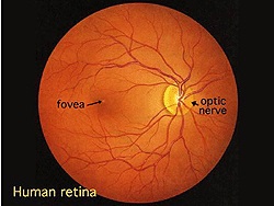 |
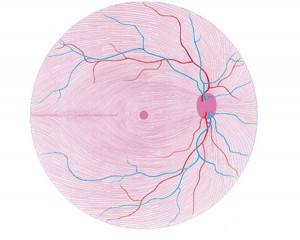
Simple Anatomy of the Retina (Webvision) | Helga Kolb, MD | Description of the anatomy of the retina with diagrams. | Retina Anatomy |
| 104 |
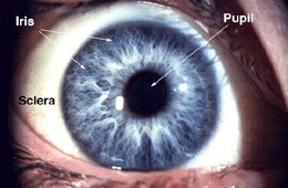 |
Gross Anatomy of the Eye (Webvision) | Helga Kolb, MD | Description of the gross anatomy of the eye, with diagrams. | Gross Anatomy Eye |
| 105 |
 |
Non-Organic Visual Loss | Omar Ozgur, MD; Rudrani Banik, MD | Power point of case presentation of 12 year old girl with recurrent monocular visual loss. Examination is normal. Differential diagnosis discussed, including non-organic visual loss. Diagnostic testing for non-organic visual loss reviewed. Slide 4: Figure 1: Table of exam findings Slide 5: Figure 2... | Non-organic Visual Loss; Monocular Visual Loss |
| 106 |
 |
Montreal Cognitive Assessment (MOCA) | Ziad Nasreddine, MD | The MoCA© is a cognitive screening test designed to assist Health Professionals in detection of mild cognitive impairment. For more information, contact: info@moca.org | Cognitive Disorders; Cognitive Assessment; MOCA Test |
| 107 |
 |
Neuromyelitis Optica | Omar Ozgur, MD; Rudrani Banik, MD | Power point of case presentation of female with bilateral, sequential atypical optic neuritis. MRI Brain normal with no demyelination; MRI Spine shows enhancement at multiple levels and NMO antibody positive, confirming diagnosis of neuromyelitis optica (NMO). History of NMO discussed, diagnostic c... | Neuromyelitis Optica; Atypical Optic Neuritis; MRI; Plasmapheresis |
| 108 |
 |
Acute Zonal Occult Outer Retinopathy (AZOOR) versus Multiple Evanescent White Dot Syndrome (MEWDS) | Asim V. Farooq, MD; Michael T. Andreoli, MD; Heather E. Moss, MD | PPT case report on acute zonal occult outer retinopathy (AZOOR) versus multiple evanescent white dot syndrome (MEWDS). | AZOOR; MEWDS; Paracentral Scotoma; Goldmann Visual Field; Photoreceptor Loss |
| 109 |
 |
Prolactinoma in Pregnancy | Timothy Sullivan, MD; Rudrani Banik, MD | Power point of case of prolactinoma which became symptomatic during pregnancy with visual field loss. Discussion of prolactinomas and their management. Patient underwent observation only. Post-partum examination revealed resolution of bitemporal field defect with reduction in size of prolactinoma ... | Prolactinoma; Pregnancy; Bitemporal Defect |
| 110 |
 |
Oculopharyngeal Muscular Dystrophy (OPMD) | Natasha Nayak, MD; Rudrani Banik, MD | Power point of case presentation of chronic, progressive ophthalmoplegia and bilateral ptosis in adult male with positive family history of similar ocular findings. Differential diagnosis with associated findings reviewed. Work up done: EMG testing consistent with myopathy. Genetic testing positiv... | Ophthalmoplegia;, Ptosis; Oculopharygneal Muscular Dystrophy; Genetic Disorder |
| 111 |
 |
Idiopathic Bilateral Neuroretinitis in a Child | Asim V. Farooq, MD; Michael T. Andreoli, MD; Molly Gilbert, MD; Heather E. Moss, MD | PPT case describing idiopathic bilateral neuroretinitis in a child. | Neuroretinitis; Pediatric; Idiopathic; Optic Atrophy |
| 112 |
 |
The History of the International Neuro-Ophthalmology Society | Klara Landau, MD, FEBO | This presentation provides an ovreview of hte hisotry of the International Neuro-ophthalmology Society (INOS), with maps and photos. | International Neuro-Ophthalmology Society: INOS |
| 113 |
 |
Palinopsia: Some Visions Never Fade | Amrita-Amanda D. Lakraj, MD; Ryan D. Walsh, MD | This is a PowerPoint presentation, which teaches the symptom of palinopsia through a video of a patient's chief complaint in which he describes the symptom almost according to a textbook. This video is followed by a brief explanation of the etiology, management, and importance of diagnosing this sym... | Palinopsia; Visual Disturbance; Ghosting |
| 114 |
 |
Pituitary Apoplexy and Hemifield Slide Phenomenon | Helen H. Yeung, MD; Rudrani Banik, MD | PowerPoint of case presentation of pituitary apoplexy. Patient presented with bilateral severe visual loss and bilateral ophthalmoplegia from partial third nerve palsies (pupil-sparing with no ptosis) from midbrain compression. After transsphenoidal surgery with decompression of mass and steroids, ... | Pituitary Apoplexy; Hemifield Slide; Bitemporal Defect; Partial Third Nerve Palsy |
| 115 |
 |
Wallenberg Syndrome and Skew Deviation | Lauren Schneider, MD; Rudrani Banik, MD | Power point of case presentation of acute Wallenberg Syndrome associated with vertical diplopia, found by 3 step and supine testing to be consistent with skew deviation. | Wallenberg Syndrome; Skew Deviation; Vertical Diplopia |
| 116 |
 |
Retinal Causes of a Neurologic-Type Visual Field Defect | Omar Ozgur, MD; Rudrani Banik, MD | Power point of case presentation of 47 year old female with history of breast cancer with new onset temporal visual field defect and photopsias. Differential diagnosis of homonymous hemianopia discussed; retinal causes of neurologic-type visual field defects reviewed including: white dot syndrome (m... | Homonymous Hemianopia; Neurologic Visual Field Defect; Temporal Visual Field Defect; White Dot Syndrome; Multiple Evanescent White Dot Syndrome (MEWDS); Cancer-Associated Retinopathy; Tamoxifen Retinopathy; Autoimmune Retinopathy |
| 117 |
 |
Pseudotumor cerebri and Chiari Malformation | Nicole Scripsema, MD; Rudrani Banik, MD | Power point of case presentation of pseudotumor cerebri with co-existing Chiari malformation. Management of severe visual loss associated with chronic papilledema discussed, as well as possible relationship between raised intracranial pressure from pseudotumor cerebri and Chiari malformation. | Pseudotumor Cerebri; Papilledema; Chiari Malformation |
| 118 |
 |
Part II: Anatomy and Physiology of the retina | Olaf Strauss, MD; Ralph Nelson; Dustin M. Graham | Section of Webvision covering the anatomy and physiology of the retina: The retinal pigment epithelium by Olaf Strauss Photoreceptors by Helga Kolb Outer Plexiform Layer by Helga Kolb Inner Plexiform Layer by Helga Kolb Morphology and Circuitry of Ganglion Cells by Helga Kolb Ganglion Cell Physiolog... | Anatomy; Physiology; Retina |
| 119 |
 |
Lemierre Syndrome - A Neuroophthalmological Approach | Vinzenz A. C. Vadasz, MD; Christina Gerth-Kahlert, MD | Case report of a twenty-two year old woman with double vision after tonsillitis, caused through multiples thrombosis by an infection with fusobacterium necrophorum known as the Lemierre-Syndrome. Fig. 1: Ocular motility at ICU (lying position) Fig. 2: white arrows show thrombosis of the right opht... | Lemierre-Syndrome; Fusobacterium Necrophorum; Septic Thrombosis |
| 120 |
 |
Cone Dystrophy | Gregory P. Van Stavern, MD | PowerPoint discussing Cone Dystrophy: Early loss of central and color vision; Color impairment often out of proportion to loss of VA; Hemeralopia ("day blindness") prominent; Light sensitivity and photophobia; Macular changes variable, and may occur late- may "Bull's Eye" pattern; Abnormal Photost... | Cone Dystrophy; Occult Macular Dystrophy; Central Cone Dystrophy |
| 121 |
 |
Pupillary Light Reflex | Wade Crow, MD | Illustration of the Pupillary Light Reflex. | Pupillary Light Reflex |
| 122 |
 |
Aberrant Regeneration of Third Nerve | Gregory P. Van Stavern, MD | 48 year old woman S/P rupture and repair of right sided posterior communicating artery aneurysm Video shows residual partial right third nerve palsy, with aberrant regeneration, causing a pseudo Von Graefe's sign (elevation of the right upper eyelid with attempted infraduction of the right eye) Se... | Aberrant Regeneration of Third Nerve; Third Nerve Palsy |
| 123 |
 |
Aberrant Regeneration Third Nerve | Gregory P. Van Stavern, MD | 48 year old woman S/P rupture and repair of right sided posterior communicating artery aneurysm Video shows residual partial right third nerve palsy, with aberrant regeneration, causing a pseudo Von Graefe's sign (elevation of the right upper eyelid with attempted infraduction of the right eye) | Aberrant Regeneration Third Nerve; Third Nerve Palsy |
| 124 |
 |
Acute Retinal Necrosis (ARN) | Gregory P. Van Stavern, MD | Acute Retinal Necrosis causes inflammation and subsequent retinal detachment. This powerpoint provides images depicting ARN. | Acute Retinal Necrosis (ARN) |
| 125 |
 |
Congenital and Secondary Syphilis | Gregory P. Van Stavern, MD | Images showing evideince of Congenital and Secondary Syphilis | Syphilis |