Collection of materials relating to neuro-ophthalmology as part of the Neuro-Ophthalmology Virtual Education Library.
NOVEL: https://novel.utah.edu/
TO
- NOVEL193
Filters: Collection: "ehsl_novel_novel"
| Title | Creator | Description | Subject | ||
|---|---|---|---|---|---|
| 26 |
 |
John Cunningham Virus | Alison Gibbons; Amanda D. Henderson, MD | This learning object is a narrated Power Point presentation describing the features of, risk factors for, and clinical presentations of the John Cunningham, or JC, virus. It includes a discussion of various immunosuppressed states, including HIV, use of natalizumab (a disease-modifying therapy that ... | John Cunningham Virus; JC Virus; Natalizumab |
| 27 |
 |
CT Venography (CTV) | Devin D. Mackay, MD | Explanation of using computed tomography venography (CTV). | CT Venography (CTV) |
| 28 |
 |
Vergence Eye Movements | Yu Hsin Chen; Amanda Dean Henderson, MD | Vergence (e.g. convergence and divergence), a class of eye movements that rotates the eyes in opposite directions (disjunctive), serves to hold image on the fovea of both eyes in order to obtain a single, clear image. This presentation overviews the neurology and examinations of vergence response, w... | Convergence; Convergence Insufficiency; Convergence Spasm; Divergence; Divergence Insufficiency |
| 29 |
 |
CT Angiography (CTA) | Devin D. Mackay, MD | Explanation of using computed tomography angiography (CTA) in examinations. | CT Angiography (CTA) |
| 30 |
 |
Doppler Ultrasonography | Devin D. Mackay, MD | Explanation of using doppler ultrasonography in examinations. | Doppler Ultrasonography |
| 31 |
 |
MR Venography (MRV) | Devin D. Mackay, MD | Explanation of using MR venography (MRV) in examinations. | MR Venography (MRV) |
| 32 |
 |
The Internal Carotid Arteries and Branches | Katherine Hutchins, MD; Devin D. Mackay, MD | Illustrations, MRA, CTA, and cerebral angiography images of the internal carotid artery and its branches. | Vascular Anatomy; Internal Carotid Artery; Anterior Cerebral Artery; Middle Cerebral Artery; Anterior Circulation |
| 33 |
 |
"Simon Says" Technique for Nonorganic Visual Field Constriction | Devin D. Mackay, MD | Explanation of the "Simon says" technique. | Simon Says, Visual Field Constriction |
| 34 |
 |
Functional MRI | Devin D. Mackay, MD | Explanation of using functional MRI in examinations. | Functional MRI |
| 35 |
 |
The Clinical Examination of Higher Order Visual Function: Syndrome-based Approach - Visual Constructional Apraxia | Victoria S. Pelak, MD; James R. Bateman, MD, MPH; Brianne Bettcher, PhD | Explanation of higher order visual function examination. | Visual Function; Visual Constructional Apraxia |
| 36 |
 |
Diffusion Tensor Imaging (DTI) | Devin D. Mackay, MD | Explanation of using diffusion tensor imaging (DTI) in examinations. | Diffusion Tensor Imaging (DTI) |
| 37 |
 |
Digital Subtraction Angiography | Devin D. Mackay, MD | Explanation of using digital subtraction angiography in examinations. | Digital Subtraction Angiography |
| 38 |
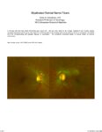 |
Myelinated Retinal Nerve Fibers | Scott N. Grossman, MD | A 33 year old man has noted chronically poor vision OS - left eye color noted to be 'orange' instead of red. fundus photos revealed myelinated retinal nerve fiber layer OU (OS>OD) with corresponding linear paracentral scotoma on Humphrey visual field 24-2 OS corresponding with greatest degree of my... | Myelinated Retinal Nerve Fibers |
| 39 |
|
The Clinical Examination of Higher Order Visual Function: Syndrome-based Approach - Visual Central Achromatopsia | Victoria S. Pelak, MD; James R. Bateman, MD, MPH; Brianne Bettcher, PhD | Explanation of higher order visual function examination. | Visual Function; Visual Central Achromatopsia |
| 40 |
 |
Computed Tomography (CT) | Devin D. Mackay, MD | Explantation of computed tomography (CT) examinations. | Computed Tomography (CT) |
| 41 |
 |
MR Angiography (MRA) | Devin D. Mackay, MD | Explanation of using MR angiography in examinations. | MR Angiography (MRA) |
| 42 |
 |
MRI Orbital Protocol | Devin D. Mackay, MD | Description of the MRI orbital protocol. | MRI Orbital Protocol |
| 43 |
 |
Positron Emission Tomography (PET) | Devin D. Mackay, MD | Explanation of using positron emission tomography (PET) in examinations. | Positron Emission Tomography (PET) |
| 44 |
 |
The Anatomic Course of Cranial Nerve VI | Divya Chauhan, MD | Overview of the intracranial course of the abducens nerve. | Cranial Nerve VI; Abducens Nerve; Anatomy |
| 45 |
 |
Chorioretinal Coloboma | Kirstyn Taylor; Drew Scoles, MD | Coloboma is a term used to describe defects seen in various ocular structures due to incomplete embryologic development. Fundus coloboma specifically is due to failure of the embryonal fissure to close, which typically occurs by 5-7 weeks gestation. Posterior colobomas, involving the retina, choroid... | Chorioretinal; Coloboma; Optic Nerve |
| 46 |
|
The Clinical Examination of Higher Order Visual Function: Syndrome-based Approach - Visual Prosopagnosia | Victoria S. Pelak, MD; James R. Bateman, MD, MPH; Brianne Bettcher, PhD | Explanation of higher order visual function examination. | Visual Function; Visual Prosopagnosia |
| 47 |
 |
The Anatomic Course of Cranial Nerve IV | Divya Chauhan, MD | Overview of the intracranial course of the trochlear nerve. | Cranial Nerve IV; Trochlear Nerve; Anatomy |
| 48 |
 |
CSF Composition | Divya Chauhan, MD | Overview of the composition of cerebrospinal fluid. | Cerebrospinal Fluid; CSF |
| 49 |
 |
Cerebellar Anatomy on MRI | Joshua East, MD; Nicholas A. Koontz, MD; Devin D. Mackay, MD | Overview of structural anatomy of the cerebellum and surround structures on MRI images of the brain. | Cerebellar Anatomy; MRI |
| 50 |
 |
Introduction to Funduscopic Examination | Valérie Biousse, MD | Introduction to the funduscopic examination section of the NExT curriculum. | Exams; Funduscopy |