Collection of materials relating to neuro-ophthalmology as part of the Neuro-Ophthalmology Virtual Education Library.
NOVEL: https://novel.utah.edu/
TO
1 - 25 of 14
| Title | Creator | Description | Subject | ||
|---|---|---|---|---|---|
| 1 |
 |
A Suspected Case of Wildervanck Syndrome | Hari R. Anandarajah; Tejaswini K. Deshmukh; Ryan D. Walsh | A 5-year-old male with congenital right-sided hearing loss presented to the neuro-ophthalmology clinic for strabismus evaluation. He had longstanding bilateral abduction deficits with associated esotropia, for which he underwent bilateral medial rectus recessions at age three. He had persistent limi... | Cervico-oculo-acoustic Syndrome; Congenital Deafness; Duane Syndrome; Klippel-Feil Cervical Anomaly; Wildervanck Syndrome |
| 2 |
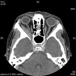 |
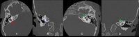
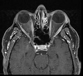
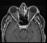
Sixth nerve palsy or not? (Image) | Vivian Paraskevi Douglas; Konstantinos Douglas; Nurhan Torun | Here in we present a case of a 72-yearold Caucasian female with remote history of breast cancer who presented with diplopia in the right gaze over the past several months. There had been no improvement after a 5-day course of steroids by an outside ophthalmologist. On neuro-ophthalmic examination, t... | Abduction deficit; Diplopia; Orbital imaging |
| 3 |
 |
Chorioretinal Coloboma | Kirstyn Taylor; Drew Scoles, MD | Coloboma is a term used to describe defects seen in various ocular structures due to incomplete embryologic development. Fundus coloboma specifically is due to failure of the embryonal fissure to close, which typically occurs by 5-7 weeks gestation. Posterior colobomas, involving the retina, choroid... | Chorioretinal; Coloboma; Optic Nerve |
| 4 |
 |
Curtain Sign (Enhanced Ptosis) - Associated Image 2 | Bashaer Aldhahwani, MD; Hong Jiang, MD, PhD | This is a 78-year-old male patient who presented with diplopia, right eyelid ptosis, and ophthalmoplegia. He had severe ptosis OD and pseudo-proptosis (lid retraction) OS at baseline, but when the right eyelid was manually elevated, there was marked enhanced ptosis of the left eyelid (Video). He was... | Myasthenia GravIs; Clinical Signs |
| 5 |
 |
Curtain Sign (Enhanced Ptosis) - Associated Image 1 | Bashaer Aldhahwani, MD; Hong Jiang, MD, PhD | This is a 78-year-old male patient who presented with diplopia, right eyelid ptosis, and ophthalmoplegia. He had severe ptosis OD and pseudo-proptosis (lid retraction) OS at baseline, but when the right eyelid was manually elevated, there was marked enhanced ptosis of the left eyelid (Video). He was... | Myasthenia GravIs; Clinical Signs |
| 6 |
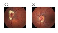 |
Myelinated Retinal Nerve Fiber Layer (MRNFL) | Sparsh Jain, BS; Ryan D. Walsh, MD | Fundus photos demonstrating bilateral (right > left) peripapillary myelinated retinal nerve fiber layer (MRNFL) in a 14-year old boy. Note the typical appearance of MRNFL of a white patch with feathered margins involving the inner retina. In this case, the MRNFL is more prominent in the right eye, a... | Myelinated Retinal Nerve Fiber Layer; MRNFL; Congenital Anomalies |
| 7 |
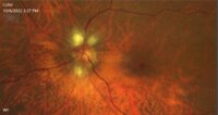 |
Myelinated Retinal Nerve Fiber Layer | Bashaer Aldhahwani, MD; Hong Jiang, MD, PhD | A 78 YOF with no visual symptoms has an incidental finding of yellow-white well-demarcated patches with ragged borders at the peripapillary area of her left eye (see the fundus photo). | Myelinated Retinal Nerve Fiber Layer |
| 8 |
 |
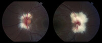
Myelinated Nerve Fibers | Carmen Chan,RN, PhD, FAAN | Fundus photos from a patient with extensive myelinated nerve fibers. The patient had normal visual functions. | Myelinated Nerve Fibers |
| 9 |
 |
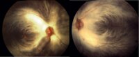
Myelinated Nerve Fibers | John J. Chen, MD, PhD | Fundus photographs of a 19-year old female with prominent peripapillary myelinated nerve fibers in both eyes that was incidentally found on routine eye examination. | Myelinated Nerve Fibers |
| 10 |
|
Peripapillary Myelinated Nerve Fibers | John J. Chen, MD, PhD | Fundus photographs of a 19-year old female with prominent peripapillary myelinated nerve fibers in both eyes that was incidentally found on routine eye examination. | Myelinated Nerve Fibers |
| 11 |
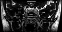 |
Cavernous Sinus | Andrew R. Carey, MD | Cavernous sinus imaging with labels. | Cavernous Sinus |
| 12 |
 |
2013 William F Hoyt Lecture: Neuro-Ophthalmology in Review: Around the Brain with 50 Fellows | Nancy J. Newman, MD | No matter what their ultimate specialty, every ophthalmologist needs to master the basics of neuroophthalmology. To that end, we must ensure that we continue to train effective teachers of neuro-ophthalmology. This is William F. Hoyt's most important lasting legacy and charge. In this same spirit, E... | History |
| 13 |
 |
Pseudotumor Cerebri | Deborah I. Friedman, MD | This one hour presentation on Pseudotumor cerebri is the first in a series of Neuro-Ophthalmology All Star Grand Rounds. The videolecture is accompanied by written material and is intended as a teaching tool for medical residents. Studies in the 1980s calculated the annual incidence of pseudotumor c... | Pseudotumor Cerebri; Idiopathic Intracranial Hypertension |
| 14 |
 |
Animated Lessons on the Neurology of Eye Movements and Pupillary Disorders | Dario Beltran, MD; Douglas Woo, MD; Elliot Frohman, MD, PhD; Steven L. Galetta, MD; Lewis E. Calver, MS; Kim Hoggatt Krumwiede, MA | This interactive training guide correlates clinical eye exams with lesion localization using illustrations, animations, and MRI's to enhance the learning of various common neuro-ophthalmologic lesions that are found in patients with Multiple Sclerosis (MS), stroke, tumor, or infection. Neurologists,... | Pupil Abnormalities; Ocular Movement Abnormalities; Diplopia |
1 - 25 of 14