Collection of materials relating to neuro-ophthalmology as part of the Neuro-Ophthalmology Virtual Education Library.
NOVEL: https://novel.utah.edu/
TO
- NOVEL136
| Title | Creator | Description | Subject | ||
|---|---|---|---|---|---|
| 1 |
 |
Optical Coherence Tomography (OCT) | Gunnar J. Goebel; Devin D. Mackay, MD | Introduction to OCT, including history, principles, interpretation, and applications. | Optical Coherence Tomography (OCT) |
| 2 |
 |
Susac's Syndrome | David O. Sohutskay; Kevin E. Lai, MD; Linda S. Williams, MD; Devin D. Mackay, MD | Overview of a case of Susac Syndrome. | Susac Syndrome |
| 3 |
 |
Vitamin B12 Deficiency and Neuro-Ophthalmic Manifestations | Jourdan Carroll; Devin D. Mackay, MD | This presentation covers vitamin B12 deficiency, including etiology, signs and symptoms, neurologic and ophthalmic findings, a case presentation and treatment. | Vitamin B12 Deficiency and Neuro-Ophthalmic Manifestations |
| 4 |
 |
Recurrent Painful Ophthalmoplegic Neuropathy | Jay Chopra, BS; Devin D. Mackay, MD | Overview of recurrent painful ophthalmoplegic neuropathy with an illustrative case example and discussion of clinical presentation, possible mechanisms, and treatment. | Recurrent Painful Ophthalmoplegic Neuropathy; RPON; Ophthalmoplegic Migraine; Ophthalmoparesis; Painful Cranial Nerve Palsy |
| 5 |
 |
Management of Non-Organic Vision Loss | Aumer Shughoury, BA; Devin D. Mackay, MD | A description of the management of non-organic visual loss. | Non-Organic Vision Loss; NOVL |
| 6 |
 |
Thiamine (Vitamin B1) Deficiency (Presentation) | Nirupama Devanathan; Devin D. Mackay, MD | Overview of thiamine deficiency and its neuro-ophthalmic manifestations with an illustrative case example and discussion of clinical presentation, relevant biochemistry, testing, risk factors, and treatment. Corresponding Video: https://collections.lib.utah.edu/details?id=2297569 | Thiamine; Upbeat Nystagmus; Nutritional Optic neuropathy; Wernicke Encephalopathy |
| 7 |
 |
Genetic Causes of Isolated Optic Neuropathy | Kathleen Ho; Devin Mackay | Overview of isolated genetic optic neuropathies and their neuro-ophthalmic manifestations with an illustrative case example and discussion of clinical presentation, relevant biochemistry, and testing. | Genetics; Hereditary; MFN2; NDUFV1; OPA1; OPA3; Optic Atrophy; Optic Neuropathy; POLG; SPG7; WFS1 |
| 8 |
 |
Shaken Baby Syndrome | Kathleen B. Digre, MD; James J. Corbett, MD | Neuro-ophthalmologic implications of shaken baby syndrome. | Shaken Baby Syndrome; Child Abuse; Abuse |
| 9 |
 |
Brain Surface Anatomy | Arooj Ahmad, MD; Devin D. Mackay, MD | These images depict labeled structures of the surface anatomy of the different facies of the brain. | Neuroanatomy; Brain Surface Anatomy |
| 10 |
 |
The Vertebrobasilar System | Katherine Hutchins, MD; Devin D. Mackay, MD | Illustrations, MRA, and CTA images of the vertebrobasilar system and branches. | Vascular Anatomy; Basilar Artery; Vertebral Artery; AICA; PICA; Superior Cerebellar Artery; Posterior Cerebral Artery; Posterior Circulation |
| 11 |
 |
Indirect Ophthalmoscope | Devin D. Mackay, MD; Valérie Biousse, MD | Explanation of using the indirect ophthalmoscope in examinations. | Indirect Ophthalmoscope |
| 12 |
|
Slit Lamp Binocular | Devin D. Mackay, MD; Valérie Biousse, MD | Description of the slit lamp binocular examination. | Slit Lamp Binocular |
| 13 |
|
Fundus Photography | Devin D. Mackay, MD; Valérie Biousse, MD, | Explanation of using fundus photography in examinations. | Fundus Photography |
| 14 |
|
Ocular Fundus Examination | Devin D. Mackay, MD; Valérie Biousse, MD | Explanation of using the direct ophthalmoscope in examinations. | Direct Ophthalmoscope |
| 15 |
 |
CT Venography (CTV) | Devin D. Mackay, MD | Explanation of using computed tomography venography (CTV). | CT Venography (CTV) |
| 16 |
 |
CT Angiography (CTA) | Devin D. Mackay, MD | Explanation of using computed tomography angiography (CTA) in examinations. | CT Angiography (CTA) |
| 17 |
 |
Doppler Ultrasonography | Devin D. Mackay, MD | Explanation of using doppler ultrasonography in examinations. | Doppler Ultrasonography |
| 18 |
 |
MR Venography (MRV) | Devin D. Mackay, MD | Explanation of using MR venography (MRV) in examinations. | MR Venography (MRV) |
| 19 |
 |
The Internal Carotid Arteries and Branches | Katherine Hutchins, MD; Devin D. Mackay, MD | Illustrations, MRA, CTA, and cerebral angiography images of the internal carotid artery and its branches. | Vascular Anatomy; Internal Carotid Artery; Anterior Cerebral Artery; Middle Cerebral Artery; Anterior Circulation |
| 20 |
 |
The Clinical Examination of Higher Order Visual Function: Syndrome-based Approach - Visual Constructional Apraxia | Victoria S. Pelak, MD; James R. Bateman, MD, MPH; Brianne Bettcher, PhD | Explanation of higher order visual function examination. | Visual Function; Visual Constructional Apraxia |
| 21 |
 |
"Simon Says" Technique for Nonorganic Visual Field Constriction | Devin D. Mackay, MD | Explanation of the "Simon says" technique. | Simon Says, Visual Field Constriction |
| 22 |
 |
Diffusion Tensor Imaging (DTI) | Devin D. Mackay, MD | Explanation of using diffusion tensor imaging (DTI) in examinations. | Diffusion Tensor Imaging (DTI) |
| 23 |
 |
Digital Subtraction Angiography | Devin D. Mackay, MD | Explanation of using digital subtraction angiography in examinations. | Digital Subtraction Angiography |
| 24 |
 |
Functional MRI | Devin D. Mackay, MD | Explanation of using functional MRI in examinations. | Functional MRI |
| 25 |
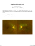 |
Myelinated Retinal Nerve Fibers | Scott N. Grossman, MD | A 33 year old man has noted chronically poor vision OS - left eye color noted to be 'orange' instead of red. fundus photos revealed myelinated retinal nerve fiber layer OU (OS>OD) with corresponding linear paracentral scotoma on Humphrey visual field 24-2 OS corresponding with greatest degree of my... | Myelinated Retinal Nerve Fibers |