Collection of materials relating to neuro-ophthalmology as part of the Neuro-Ophthalmology Virtual Education Library.
NOVEL: https://novel.utah.edu/
TO
- NOVEL966
Filters: Collection: "ehsl_novel_novel"
| Title | Creator | Description | Subject | ||
|---|---|---|---|---|---|
| 1 |
 |
Branch Retinal Artery Occlusion with Multiple Retinal Emboli | Kathleen B. Digre, MD; James J. Corbett, MD | Slideshow describing condition. | Retinal Emboli; Emboli |
| 2 |
 |
Branch Retinal Vein Occlusion (BRVO) | Kathleen B. Digre, MD; James J. Corbett, MD | Slideshow describing condition. | Occlusion |
| 3 |
 |
Branch Retinal Artery Occlusion | Kathleen B. Digre, MD; James J. Corbett, MD | Slideshow describing condition. | Occlusion |
| 4 |
 |
Branch Retinal Emboli | Kathleen B. Digre, MD; James J. Corbett, MD | Slideshow describing condition. | Emboli |
| 5 |
 |
Calcific Emboli | Kathleen B. Digre, MD; James J. Corbett, MD | Slideshow describing condition. | Emboli |
| 6 |
 |
Central/Branch Retinal Artery Occlusion (PowerPoint) | AAO/NANOS - American Academy of Ophthalmology / North American Neuro-Ophthalmology Society | Occlusion of a branch or central retinal artery may result in acute visual loss. The ophthalmoscopic findings are retinal whitening due to ischemic retina in the distribution of the occluded artery. Sparing or selective involvement of cilioretinal artery branches may occur. Patients with a central r... | Central/Branch Retinal Artery Occlusion |
| 7 |
 |
Central Retinal Vein Occlusion | Kathleen B. Digre, MD | Slideshow describing condition. | Occlusion |
| 8 |
 |
Chest CT Thymoma (Guest Lecture) | Robert A. Novelline, MD | The patient is a 46 year old woman who presented in July 1977 with horizontal double vision lasting two weeks. Three weeks later the left upper eyelid started to droop and by the end of the day the eye was closed. She had no ptosis of the right eye and no generalized fatigue. She consulted an intern... | Unilateral Ptosis; Unilateral Lid Retraction; Myasthenic Lid Twitch; External Ophthalmoplegia; Ocular Myasthenia Gravis; Tensilon Test; Thymolipoma; Generalized Myasthenia Gravis; Unilateral Myasthenia Gravis; Myasthenic Ptosis; Lid Retraction; Lid Twitch |
| 9 |
 |
Chiasmal Herniation (PowerPoint) | AAO/NANOS - American Academy of Ophthalmology / North American Neuro-Ophthalmology Society | This woman was 61 years old when she underwent initial neuro-ophthalmologic evaluation. Twenty-three years earlier, she had undergone removal of a pituitary adenoma followed by radiation therapy. At that time, she had noted a preoperative visual field defect that improved somewhat after the surgery.... | Chiasmal Herniation |
| 10 |
 |
Clinical Characteristics of Ocular Lateropulsion | Jorge C Kattah, MD | A discussion of the normal mechanism that maintain the eyes in normal horizontal position. | Ocular Lateropulsion; Unilateral Gaze Palsy; Radiographic h-CGD |
| 11 |
 |
Cogan's Lid Twitch Sign | Raed Behbehani, MD | Cogan's lid twitch sign is a twitch sign of he upper lid upon looking straight from a sustained downgaze position. It is associated with Ocular Myasthenia Gavis. | Myasthenia; Ptosis; Lid Twitch |
| 12 |
 |
Cone Dystrophy | Gregory P. Van Stavern, MD | PowerPoint discussing Cone Dystrophy: Early loss of central and color vision; Color impairment often out of proportion to loss of VA; Hemeralopia ("day blindness") prominent; Light sensitivity and photophobia; Macular changes variable, and may occur late- may "Bull's Eye" pattern; Abnormal Photost... | Cone Dystrophy; Occult Macular Dystrophy; Central Cone Dystrophy |
| 13 |
 |
Common Patterns of Visual Field Defects | Sean Gratton, MD; Sarah Lam, 6th year BA/MD | Lecture covering common visual field defects, including those of the retina, optic nerve, chiasm, and retrochiasmal. | Visual Field Defects |
| 14 |
 |
Complications of Strabismus Surgery and Botox | W. Walker Motley, MD | A narrated video slideshow outlining complications associated with strabismus surgery. | Strabismus; Surgery; Surgical Complications; Botox |
| 15 |
 |
Cotton Wool Spots: The Basics | Arnav Gupta, BHSc; Rahul Sharma, MD, MPH | A presentation describing cotton wool spots, an abnormal finding on funduscopic exam of the retina of the eye. | Cotton Wool Spots; Retina |
| 16 |
 |
Cranial Nerves: Neuroanatomy Video Lab - Brain Dissections | Suzanne S. Stensaas, PhD | The approach is to learn to associate the cranial nerves with their brainstem level and blood supply. Emphasis is given to the midbrain (3, 4), pons (5, 6, 7, 8), medulla (9, 10, 11, 12) and their most important functions. | Cranial Nerves; Brain; Dissection |
| 17 |
 |
Craniopharyngioma and Optic Atrophy | Kathleen B. Digre, MD; James J. Corbett, MD | Slideshow describing condition. | Craniopharayngioma; Otpic Atrophy |
| 18 |
 |
CRAO with Ciliary Artery Sparing | Kathleen B. Digre, MD; James J. Corbett, MD | Slideshow describing condition. | CRAO |
| 19 |
 |
Diffusion Tensor Imaging (DTI) | Devin D. Mackay, MD | Explanation of using diffusion tensor imaging (DTI) in examinations. | Diffusion Tensor Imaging (DTI) |
| 20 |
 |
Diffusion Weighted Imaging (DWI) | John Pula, MD | Diffusion weighted imaging sequences are often included as part of a routine brain MRI protocol. Imaging provides examples of DWI. | Diffusion Weighted Imaging; DWI |
| 21 |
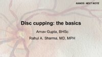 |
Disc Cupping: The Basics | Arnav Gupta, BHSc; Rahul Sharma, MD, MPH | A presentation describing optic disc cupping, due to damage of optic nerve fibres. | Disc Cupping; Optic Disc |
| 22 |
 |
Dry Eye Syndrome (Danish) | NANOS | People with abnormalities of the tear film are diagnosed with "dry eyes", but some patients with "dry eyes" may not feel that their eyes are "dry". Itching, burning, a scratchy sensation, a sensation that there is sand or grit in the eye, or intermittent blurring of the vision can all be symptoms of... | Dry Eye Syndrome; Patient Brochure |
| 23 |
 |
Central Cone Dystrophy Occult Macular Dystrophy | Gregory Van Stavern, MD | Slideshow describing condition of Central Cone Dystrophy Occult Macular Dystrophy | Central Cone Distrophy; Macular Dystrophy; Occult Macular Dystrophy |
| 24 |
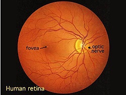 |
Simple Anatomy of the Retina (Webvision) | Helga Kolb, MD | Description of the anatomy of the retina with diagrams. | Retina Anatomy |
| 25 |
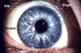 |
Gross Anatomy of the Eye (Webvision) | Helga Kolb, MD | Description of the gross anatomy of the eye, with diagrams. | Gross Anatomy Eye |