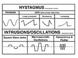
1 - 25 of 60
Number of results to display per page
TitleSetnameType
1 A Comparison of Nystagmus and Saccadic Intrusions/Oscillationsehsl_novel_goldImage
2 An Approach to the Patient with (Recent Onset) Spontaneous Episodic Vestibular Syndromeehsl_novel_goldImage
3 An Approach to the Patient with Acute Onset Prolonged Vertigoehsl_novel_goldImage
4 Atypical PC BPPV Variant Figuresehsl_novel_goldImage
5 CANVAS (Cerebellar Ataxia, Neuropathy, and Vestibular Areflexia Syndrome) Video Head Impulse Test (vHIT) Figureehsl_novel_goldImage
6 Central Anatomy of the Fourth Nerveehsl_novel_goldImage
7 Central Anatomy of the Third Nerveehsl_novel_goldImage
8 Coronal Section of the Brainstem Showing Ocular Motor Nuclei and Anatomy of the Vestibular Nucleus (with SCC Inputs)ehsl_novel_goldImage
9 Eyelid Anatomyehsl_novel_goldImage
10 Figure 17: Bony Structures Relevant to the Orbitehsl_novel_goldImage
11 Figure 1: Oculosympathetic Pathway for Pupillary Dilationehsl_novel_goldImage
12 Figure 24: Typical Visual Field Defects Associated with Discrete Lesions Along the Visual Pathwaysehsl_novel_goldImage
13 Figure 27: Vascular Supply of the Optic Nerve Head, Choroid and Retinaehsl_novel_goldImage
14 Figure 2: Parasympathetic Pathway for Pupillary Constrictionehsl_novel_goldImage
15 Figure 43: How the Brain Makes Sense of What It Sees - The Dorsal and Ventral Visual Pathways, and a 3 Tiered Approach to Visionehsl_novel_goldImage
16 Figure 46: The Course of the 6th (VI) Nerveehsl_novel_goldImage
17 Figure 50: Anatomy and Physiology of the Saccadic Pathwaysehsl_novel_goldImage
18 Figure 51: Lateral Medullary Lesion Causing Saccadic Dysmetriaehsl_novel_goldImage
19 Figure 51: Lateral Medullary Lesion Causing Saccadic Dysmetria (Supplement)ehsl_novel_goldImage
20 Figure 51: Lateral Medullary Lesion Causing Saccadic Dysmetria (Supplement)ehsl_novel_goldImage
21 Figure 53: Vascular Distribution and Anatomy Relevant to the Lateral Medullary (Wallenberg) Syndromeehsl_novel_goldImage
22 Figure 53: Vascular Distribution and Anatomy Relevant to the Lateral Medullary (Wallenberg) Syndrome (Supplement)ehsl_novel_goldImage
23 Figure 53: Vascular Distribution and Anatomy Relevant to the Lateral Medullary (Wallenberg) Syndrome (Supplement)ehsl_novel_goldImage
24 Figure 61: Vascular Distribution and Anatomy (Including 6th, 7th, 8th Nerves, MLF) of the Ponsehsl_novel_goldImage
25 Figure 61: Vascular Distribution and Anatomy (Including 6th, 7th, 8th Nerves, MLF) of the Pons (Supplement)ehsl_novel_goldImage
1 - 25 of 60