A collection of videos relating to the diagnosis and treatment of eye movement disorders. This collection includes many demonstrations of examination techniques.
Dan Gold, D.O., Associate Professor of Neurology, Ophthalmology, Neurosurgery, Otolaryngology - Head & Neck Surgery, Emergency Medicine, and Medicine, The Johns Hopkins School of Medicine.
A collection of videos relating to the diagnosis and treatment of eye movement disorders.
NOVEL: https://novel.utah.edu/
TO
1 - 25 of 8
| Title | Description | Type | ||
|---|---|---|---|---|
| 1 |
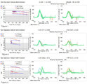 |
Ocular Motor & Vestibular Features of the MLF Syndrome (Figures 1, 2, and 3) | This 61-year-old woman with HTN and DM presented for evaluation of acute onset diagonal diplopia. Adduction OS was about 60% of normal while medialization OS improved with convergence. In right gaze, dissociated abducting nystagmus was present OD, and there was a clear adduction lag when asking he... | Image |
| 2 |
 |
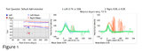
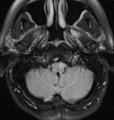
Periodic Alternating Nystagmus Due to Nodulus Stroke (Figure 1) | This is a 70-year-old woman who experienced the acute onset of vertigo and imbalance. MRI demonstrated a diffusion-weighted imaging hyperintensity involving the nodulus (with corresponding ADC hypointensity) consistent with an acute stroke. On examination several weeks after the stroke, periodic alt... | Image |
| 3 |
 |
Vestibular Neuritis with + Head Impulse Test and Unidirectional Nystagmus (Figure 1) | Vestibular neuritis is the most common cause of the acute vestibular syndrome, which is characterized by continuous vertigo and spontaneous nystagmus lasting days. It may be mimicked by central causes, including stroke, but in the hands of subspecialists, the HINTS+ (Head Impulse, Nystagmus, Test of... | Image |
| 4 |
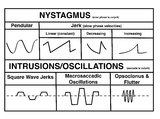 |
A Comparison of Nystagmus and Saccadic Intrusions/Oscillations | Nystagmus can be classified into pendular and jerk waveforms, where both are generated by a slow, pathologic phase. Corrective phase (the position reset mechanism) differs. In pendular nystagmus, the eyes move back and forth with about the same velocity and amplitude, similar to that of a pendulum... | Image |
| 5 |
 |
Subtle Torsional Pendular Nystagmus in Oculopalatal Tremor (OPT) (Figure 1) | This is a 50-year-old woman who presented with imbalance, and MRI demonstrated a right cerebellar cavernous malformation. She underwent surgery to resect the malformation, and post-operatively experienced right hemiparesis and ataxia. Six months after the surgery, balance worsened and vision became ... | Image |
| 6 |
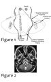 |
Triangle of Guillain-Mollaret | Seen here is a schematic representation of the Gullain-Mollaret triangle (Figure 1), also referred to as the dentato-olivary pathway, reflecting the 3 points of this imaginary triangle - 1) dentate nucleus, 2) red nucleus, and 3) inferior olivary nucleus. The olive sends decussating climbing fibers ... | Image |
| 7 |
 |
Neuro-Ophthalmic Features and Pseudo-MG Lid Signs in Miller Fisher Syndrome (Figure 1) | This is a 51-year-old woman who presented with imbalance, acute onset dizziness and diplopia that developed over three days following two weeks of upper respiratory infection and bacterial conjunctivitis. When she was initially seen as an outpatient, nystagmus was noted to the right and left, and a ... | Image |
| 8 |
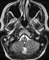 |
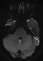
Oculopalatal Tremor with Prominent Nystagmus, Bilateral Horizontal Gaze Palsy, and Bilateral Facial Palsies (Figure 1) | Figure 1, MRI T2 sequence demonstrating hyperintensities involving bilateral inferior olives of the medulla. This is a 50-year-old woman who experienced the acute onset of right sixth and seventh nerve palsies and left hemiparesis. Two cavernomas within the right pons (one in the region of the facia... | Image |
1 - 25 of 8