A collection of videos relating to the diagnosis and treatment of eye movement disorders. This collection includes many demonstrations of examination techniques.
Dan Gold, D.O., Associate Professor of Neurology, Ophthalmology, Neurosurgery, Otolaryngology - Head & Neck Surgery, Emergency Medicine, and Medicine, The Johns Hopkins School of Medicine.
A collection of videos relating to the diagnosis and treatment of eye movement disorders.
NOVEL: https://novel.utah.edu/
TO
| Title | Description | Type | ||
|---|---|---|---|---|
| 1 |
 |
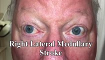
Horizontal Gaze Palsy, Facial Nerve Palsy, and Nystagmus Due to Dorsal Pontine Ischemia | 𝗢𝗿𝗶𝗴𝗶𝗻𝗮𝗹 𝗗𝗲𝘀𝗰𝗿𝗶𝗽𝘁𝗶𝗼𝗻: Presented here are two patients with horizontal gaze and facial palsies due to stroke. The first patient is a 60-year-old man who presented with double vision and hemiparesis due to a right dorsal pontine ischemic stroke.... | Image/MovingImage |
| 2 |
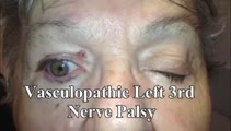 |
3rd Nerve Palsy With Preserved 4th Nerve Function | 80-yo-woman with a left vasculopathic 3rd nerve palsy (minimal pupil involvement of about 1 mm relative mydriasis OS - other etiologies ruled out and resolved as expected over months). Although the inferior rectus is paretic, intact superior oblique muscle function can be demonstrated by asking the ... | Image/MovingImage |
| 3 |
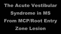 |
The Acute Vestibular Syndrome in MS Due to Middle Cerebellar Peduncle/Root Entry Zone Lesion | This is a 13 year-old girl with relatively abrupt onset vertigo and oscillopsia. On exam, there was primarily right-beating nystagmus in primary gaze with a slight upward (upbeat) component, giving the nystagmus an oblique appearance. The upward component and lack of a clear torsional component acut... | Image/MovingImage |
| 4 |
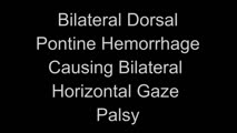 |
Bilateral Horizontal Gaze Palsy and Oculopalatal Tremor Due to Pontine Hemorrhage | This 70-yo-woman experienced headache and diplopia and was found to have a hemorrhage centrally within the dorsal pons. Months after the onset, the patient was seen in clinic and had no horizontal eye movements (pursuit, saccades, VOR) in either eye, suggestive of bilateral nuclear 6th nerve palsies... | Image/MovingImage |
| 5 |
 |
Complete Peripheral Vestibulopathy & Ipsilateral Facial Palsy | 𝗢𝗿𝗶𝗴𝗶𝗻𝗮𝗹 𝗗𝗲𝘀𝗰𝗿𝗶𝗽𝘁𝗶𝗼𝗻: 60-yo-man who suffered the fairly abrupt onset (over hours) of right lower motor neuron facial nerve palsy (7th cranial nerve), vertigo and deafness in the right ear (8th cranial nerve). Vesicles were noted on otoscopy, a... | Image/MovingImage |
| 6 |
 |
Latent Nystagmus and DVD in Infantile Esotropia | This is a 20-year-old woman with infantile esotropia (s/p strabismus surgery as a child) who demonstrated latent nystagmus and presumed dissociated vertical deviation (DVD) OS, which are commonly seen with infantile esotropia (also inferior oblique overaction and monocular nasotemporal asymmetry to ... | Image/MovingImage |
| 7 |
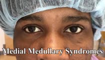 |
Medial Medullary Syndromes | This is a video of two patients who suffered small strokes involving the right medial medulla, and who presented with acute vertigo and oscillopsia. The first patient in the video had pure upbeat nystagmus, while the second patient had upbeat-torsional (towards the right ear) nystagmus in addition t... | Image/MovingImage |
| 8 |
 |
Posterior Canal BPPV with Fixation and with Fixation Removed | This is a 60-yo-woman with positional vertigo. In the right Dix-Hallpike position with fixation removed, there was clear upbeat-torsional nystagmus (towards the lowermost right ear) which led to the diagnosis of right posterior canal BPPV. In right Dix-Hallpike with fixation there was mainly torsion... | Image/MovingImage |
| 9 |
 |
Unidirectional Nystagmus in Lateral Medullary Syndrome | This is a 70-yo-man who presented with acute vertigo. Examination demonstrated very mild spontaneous torsional nystagmus (towards the right ear) in primary (not seen well in this video), with robust downbeat-torsional (towards right ear) nystagmus in right gaze and (less robust) almost pure torsiona... | Image/MovingImage |
| 10 |
 |
Unidirectional Vestibular Nystagmus | 60-yo-man with recurrent vertigo attacks - this video was taken during one of his typical attacks, and shows left-beating nystagmus that stayed left-beating in all directions of gaze, more in left gaze (in accordance with Alexander's Law), and less in right gaze. This pattern is more commonly seen w... | Image/MovingImage |
| 11 |
 |
Wallenberg Syndrome in MS | 𝗢𝗿𝗶𝗴𝗶𝗻𝗮𝗹 𝗗𝗲𝘀𝗰𝗿𝗶𝗽𝘁𝗶𝗼𝗻: 30-yo-woman with MS presenting with acute vertigo and vertical diplopia. Examination demonstrated several aspects of the Wallenberg syndrome (her acute demyelinating lesion was in the left lateral medulla): ipsilesional (... | Image/MovingImage |
| 12 |
 |
Anterior Canal - BPPV: Deep Head Hanging | 𝗢𝗿𝗶𝗴𝗶𝗻𝗮𝗹 𝗗𝗲𝘀𝗰𝗿𝗶𝗽𝘁𝗶𝗼𝗻: Regardless or whether it is thought that the patient has right or left anterior canal (AC) involvement, the deep head hanging maneuver is performed in the same way. • First the patient is placed in the long-sitting posi... | Image/MovingImage |
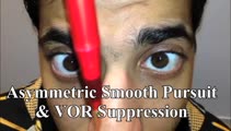
| 13 |
 |
Periodic Alternating Nystagmus Due to Spinocerebellar Ataxia Type 6 | 𝗢𝗿𝗶𝗴𝗶𝗻𝗮𝗹 𝗗𝗲𝘀𝗰𝗿𝗶𝗽𝘁𝗶𝗼𝗻: This 50-yo-man complained of imbalance for several years and more recently oscillopsia. On examination, there was saccadic pursuit and VOR suppression in addition to gaze-evoked nystagmus with rebound, raising suspicion f... | Image/MovingImage |
| 14 |
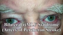 |
riMLF Syndrome from Artery of Percheron Stroke | 𝗢𝗿𝗶𝗴𝗶𝗻𝗮𝗹 𝗗𝗲𝘀𝗰𝗿𝗶𝗽𝘁𝗶𝗼𝗻: This is a 65-yo-man who suffered the abrupt onset of loss of consciousness followed by difficulty looking down. MRI showed bilateral rostral midbrain strokes in the distribution of the artery of Percheron. He could not in... | Image/MovingImage |
| 15 |
 |
Central 4th Nerve Palsy with Contralateral Horner's Syndrome | This is a 60-yo-woman who presented with a complaint of diplopia. Examination demonstrated a left hypertropia that worsened in right and down gaze as well as in left head tilt, and a left 4th nerve palsy was diagnosed. There was also evidence of a mild motility deficit in down/medial gaze OS, consis... | Image/MovingImage |
| 16 |
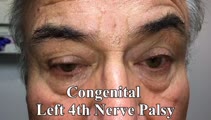 |
Inferior Oblique Overaction in a Congenital 4th Nerve Palsy | 60-yo-man complaining of intermittent oblique diplopia. There was a left hypertropia that worsened in down gaze, right gaze and in left head tilt. There was a large vertical fusional amplitude in addition to a longstanding rightward head tilt, and on examination there was left inferior oblique overa... | Image/MovingImage |
| 17 |
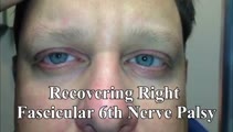 |
Slow Abducting Saccade in 6th Nerve Palsy | 40-yo-man with a right fascicular 6th nerve palsy due to stroke. There was improvement and only a minimal residual right abduction paresis OD by this visit, but still a relatively slow right abducting saccade seen in the video, especially apparent in the slow motion segment. Video shows slow abduct... | Image/MovingImage |
| 18 |
 |
Saccadic Smooth Pursuit and Vestibulo-ocular Reflex Suppression (VORS) | 𝗢𝗿𝗶𝗴𝗶𝗻𝗮𝗹 𝗗𝗲𝘀𝗰𝗿𝗶𝗽𝘁𝗶𝗼𝗻: This is a 20-yo-man who suffered a left MCA stroke years prior. Upon evaluation of his eye movements, saccades and all classes of eye movements were normal, although his smooth pursuit and VORS were choppy to the left (ip... | Image/MovingImage |
| 19 |
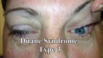 |
Duane's Syndrome Type III | This is a 40-yo-woman seen in neurology clinic for a complaint unrelated to her eyes. On exam, there was impaired adduction and abduction OS. In adduction, there was narrowing of the palpebral fissure OS, a result of her globe retraction due to co-contraction of the medial and lateral rectus muscles... | Image/MovingImage |
| 20 |
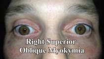 |
Superior Oblique Myokymia (SOM) | 𝗢𝗿𝗶𝗴𝗶𝗻𝗮𝗹 𝗗𝗲𝘀𝗰𝗿𝗶𝗽𝘁𝗶𝗼𝗻: This is a patient with transient monocular oscillopsia OD and vertical diplopia noted to have many episodes of SOM in the office. There was not only myokymia OD, but also a 4 prism diopter left hypertropia during episodes... | Image/MovingImage |
| 21 |
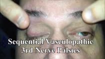 |
Sequential Vasculopathic 3rd Nerve Palsies with Preserved 4th Nerve Function | 65-yo-man with uncontrolled diabetes who developed sequential vasculopathic 3rd nerve palsies. In attempted downgaze, there's clear incyclotorsion OU suggestive of preserved 4th nerve function on both sides. There was complete recovery over months. Video shows bilateral 3rd nerve palsies with intact... | Image/MovingImage |
| 22 |
 |
Trigeminal, Facial (with Aberrant Regeneration), and Vestibulocochlear Nerve Palsies Following Tumor Resection | This is a 30-yo-woman who underwent resection of a right trigeminal schwannoma. Post-operatively, she was vertiginous with a clearly + head impulse test to the right (and spontaneous left-beating nystagmus), had lost hearing in the right ear, had no facial sensation on the right, and had a right low... | Image/MovingImage |
| 23 |
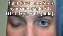 |
Cavernous Sinus Mass Causing Right 3rd and 4th Nerve Palsies | 𝗢𝗿𝗶𝗴𝗶𝗻𝗮𝗹 𝗗𝗲𝘀𝗰𝗿𝗶𝗽𝘁𝗶𝗼𝗻: 25-yo-man who complained of diplopia and was initially found to have right 4th and 6th nerve palsies in the setting of a right cavernous sinus mass (subsequently diagnosed as Ewing's sarcoma). When seen in follow-up (this... | Image/MovingImage |
| 24 |
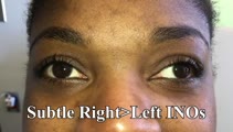 |
Wall-eyed Bilateral INO in Caudal Midbrain Lesion | This is a 30-yo-woman with the relatively acute onset of diplopia. There was a large angle exotropia, very subtle lag of the adducting saccades OD>OS, suggestive of bilateral INOs. This was best seen with rapid horizontal saccades, and a lesion involving bilateral MLFs in the caudal midbrain was dem... | Image/MovingImage |
| 25 |
 |
Pseudo-INOs in Myasthenia Gravis | This is a 55-yo-woman with an intermittent exotropia who had normal adduction OU, but clear lag of adducting saccades OD>OS with rapid horizontal saccades. This was much more apparent after repeat testing (ie, it was fatigable), and she wound up having ocular MG. | Image/MovingImage |