A collection of videos relating to the diagnosis and treatment of eye movement disorders. This collection includes many demonstrations of examination techniques.
Dan Gold, D.O., Associate Professor of Neurology, Ophthalmology, Neurosurgery, Otolaryngology - Head & Neck Surgery, Emergency Medicine, and Medicine, The Johns Hopkins School of Medicine.
A collection of videos relating to the diagnosis and treatment of eye movement disorders.
NOVEL: https://novel.utah.edu/
TO
Filters: Collection: "ehsl_novel_gold"
| Title | Description | Type | ||
|---|---|---|---|---|
| 1 |
 |
Assessing for Hyperventilation-induced Nystagmus | 𝗢𝗿𝗶𝗴𝗶𝗻𝗮𝗹 𝗗𝗲𝘀𝗰𝗿𝗶𝗽𝘁𝗶𝗼𝗻: Hyperventilation induced nystagmus is tested by asking the patient to take quick deep breaths (~1/s) for 40-60 seconds. This decreases ICP and increases CSF pH. This can be helpful in diagnosing irritative conditions of ... | Image/MovingImage |
| 2 |
 |
Dynamic Visual Acuity | 𝗢𝗿𝗶𝗴𝗶𝗻𝗮𝗹 𝗗𝗲𝘀𝗰𝗿𝗶𝗽𝘁𝗶𝗼𝗻: After assessing static binocular visual acuity, dynamic visual acuity (DVA) is determined by repeating the test during horizontal and vertical head shaking at 2-3 Hz. Dynamic visual acuity is most important to test when ... | Image/MovingImage |
| 3 |
 |
Evaluation of Convergence | 𝗢𝗿𝗶𝗴𝗶𝗻𝗮𝗹 𝗗𝗲𝘀𝗰𝗿𝗶𝗽𝘁𝗶𝗼𝗻: The assessment of convergence includes measuring alignment at near versus distance (see video, https://collections.lib.utah.edu/details?id=187677), near point of convergence and convergence amplitude. Near point of conve... | Image/MovingImage |
| 4 |
 |
Optokinetic Nystagmus | 𝗢𝗿𝗶𝗴𝗶𝗻𝗮𝗹 𝗗𝗲𝘀𝗰𝗿𝗶𝗽𝘁𝗶𝗼𝗻: During the bedside evaluation of optokinetic nystagmus (OKN), the patient is instructed to look at each red (or white) square as it moves past. Because this is not a full-field visual stimuli, using an optokinetic flag m... | Image/MovingImage |
| 5 |
 |
Pressure Testing for Superior Canal Dehiscence Syndrome | 𝗢𝗿𝗶𝗴𝗶𝗻𝗮𝗹 𝗗𝗲𝘀𝗰𝗿𝗶𝗽𝘁𝗶𝗼𝗻: Superior semicircular canal dehiscence syndrome (SCDS) is caused by a third mobile window in the inner ear. This allows for transmission of sound or pressure to the superior canal. Tragal compression and/or glottic and ... | Image/MovingImage |
| 6 |
 |
Skew Deviation and Spontaneous Nystagmus Due to Posterior Fossa Lesions | This is a 50-year-old woman who reported the abrupt onset of imbalance, right upper extremity incoordination and binocular vertical diplopia several months prior to her presentation to our clinic. On examination, she had a left hypertropia that was fairly comitant (measuring 5 prism diopters) assoc... | Image/MovingImage |
| 7 |
 |
Slow Saccades Due to Unilateral Paramedian Pontine Reticular Formation (PPRF) Injury with Preserved Movements Using the Vestibulo-Ocular Reflex | 𝗢𝗿𝗶𝗴𝗶𝗻𝗮𝗹 𝗗𝗲𝘀𝗰𝗿𝗶𝗽𝘁𝗶𝗼𝗻: This is a 60-year-old man who presented for imbalance and oscillopsia 10 months after surgery and 8 months after radiation for Merkel cell carcinoma of the neck. He developed imbalance after surgery and diplopia and osci... | Image/MovingImage |
| 8 |
 |
Test Your Knowledge - Bilateral 4th Nerve Palsies | Watch the video until instructed to stop. Which of the following features is likely to be present given her exam findings? A. Gaze-evoked nystagmus and impaired smooth pursuit B. History of traumatic brain injury C. History of blepharoplasty or brow lift surgery and prominence of superior sulcus on ... | Image/MovingImage |
| 9 |
|
Test Your Knowledge - Optokinetic Nystagmus with a Parietal Lesion | 𝗢𝗿𝗶𝗴𝗶𝗻𝗮𝗹 𝗗𝗲𝘀𝗰𝗿𝗶𝗽𝘁𝗶𝗼𝗻: Given the finding seen in the first part of the video, which of the following associated features are most likely? (more than one answer may be correct) A. Left homonymous visual field defect B. Right homonymous visual fi... | Image/MovingImage |
| 10 |
 |
Testing for Adduction Lag in Partial INO Using an Optokinetic Stimulus | In this patient we demonstrate the use of an optokinetic stimulus to elicit an internuclear ophthalmoplegia (INO). Occasionally adduction appears to be normal with an INO, and an adduction lag with horizontal saccades should be sought as a confirmatory sign. Optokinetic tape is an easy way to assess... | Image/MovingImage |
| 11 |
 |
Video Head Impulse Testing | 𝗢𝗿𝗶𝗴𝗶𝗻𝗮𝗹 𝗗𝗲𝘀𝗰𝗿𝗶𝗽𝘁𝗶𝗼𝗻: The video head impulse test (vHIT) is a clinical assessment technique used to assess the function of the semicircular canals-the angular acceleration detectors-which initiate the vestibulo-ocular reflex (VOR). The HIT and... | Text |
| 12 |
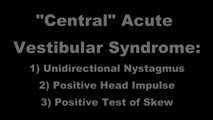 |
Acute Vestibular Syndrome with Ocular Tilt Reaction Due to Bacterial Labyrinthitis | This is a patient who initially presented with the acute vestibular syndrome (AVS, e.g., acute prolonged vertigo, spontaneous nystagmus) and right sided hearing loss, and was diagnosed with bacterial labyrinthritis. Her HINTS (Head Impulse, Nystagmus, Test of Skew) testing indicated a central etiolo... | Image/MovingImage |
| 13 |
 |
Saccades | 𝗢𝗿𝗶𝗴𝗶𝗻𝗮𝗹 𝗗𝗲𝘀𝗰𝗿𝗶𝗽𝘁𝗶𝗼𝗻: The examiner should note: conjugacy (a lag of the adducting eye may be seen with an INO); accuracy (posterior fossa lesions commonly produce dysmetria (overshooting or undershooting); velocity (if slow, may suggest a lesi... | Image/MovingImage |
| 14 |
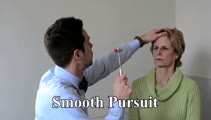 |
Smooth Pursuit | 𝗢𝗿𝗶𝗴𝗶𝗻𝗮𝗹 𝗗𝗲𝘀𝗰𝗿𝗶𝗽𝘁𝗶𝗼𝗻: A pursuit deficit in one direction suggests an ipsilesional localization, but beware of a superimposed spontaneous nystagmus; a pursuit deficit in all directions is commonly seen with cerebellar lesions. 𝗡𝗲𝘂𝗿�... | Image/MovingImage |
| 15 |
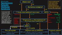 |
An Approach to the Patient with (Recent Onset) Spontaneous Episodic Vestibular Syndrome | 𝗢𝗿𝗶𝗴𝗶𝗻𝗮𝗹 𝗗𝗲𝘀𝗰𝗿𝗶𝗽𝘁𝗶𝗼𝗻: A vascular etiology should always be on the differential diagnosis of the recent onset of the spontaneous (unprovoked) episodic vestibular syndrome (EVS), especially in the older population and when vascular risk factors ... | Image |
| 16 |
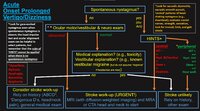 |
An Approach to the Patient with Acute Onset Prolonged Vertigo | 𝗢𝗿𝗶𝗴𝗶𝗻𝗮𝗹 𝗗𝗲𝘀𝗰𝗿𝗶𝗽𝘁𝗶𝗼𝗻: A vascular etiology should always be on the differential diagnosis of the acute onset prolonged vertigo, especially in the older population and when vascular risk factors are present. However, young patients can suffer fr... | Image |
| 17 |
|
Ocular Motor Signs in Brainstem Demyelinating Disease - Spontaneous Upbeat, Vertical Gaze-Evoked Nystagmus, Slow Saccades, Bilateral Vestibular Loss, INOs | 𝗢𝗿𝗶𝗴𝗶𝗻𝗮𝗹 𝗗𝗲𝘀𝗰𝗿𝗶𝗽𝘁𝗶𝗼𝗻: This is a 25-year-old woman who presented with painful vision loss bilaterally two years prior to this video recording, which was diagnosed as optic neuritis. Months later, she experienced oscillopsia and binocular horizo... | Image/MovingImage |
| 18 |
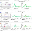 |
Ocular Motor & Vestibular Features of the MLF Syndrome (Figures 1, 2, and 3) | This 61-year-old woman with HTN and DM presented for evaluation of acute onset diagonal diplopia. Adduction OS was about 60% of normal while medialization OS improved with convergence. In right gaze, dissociated abducting nystagmus was present OD, and there was a clear adduction lag when asking he... | Image |
| 19 |
 |
Horizontal Gaze Palsy, Facial Nerve Palsy, and Nystagmus Due to Dorsal Pontine Ischemia | 𝗢𝗿𝗶𝗴𝗶𝗻𝗮𝗹 𝗗𝗲𝘀𝗰𝗿𝗶𝗽𝘁𝗶𝗼𝗻: Presented here are two patients with horizontal gaze and facial palsies due to stroke. The first patient is a 60-year-old man who presented with double vision and hemiparesis due to a right dorsal pontine ischemic stroke.... | Image/MovingImage |
| 20 |
|
Test Your Knowledge - Vertical-Torsional Nystagmus | Question #1: Watch the first portion of the video until you are told to stop. Is this vestibular nystagmus more likely to be peripheral or central? A. Peripheral B. Central Answer for #1: A. Incorrect. While the patient has upbeat-torsional (top poles beating toward the right ear) nystagmus which is... | Image/MovingImage |
| 21 |
 |
Ocular Motor Signs in Early Progressive Supranuclear Palsy | 𝗢𝗿𝗶𝗴𝗶𝗻𝗮𝗹 𝗗𝗲𝘀𝗰𝗿𝗶𝗽𝘁𝗶𝗼𝗻: This is a 64-year old man who experienced imbalance and falls (usually backwards) for the last 6 months. He experienced difficulty navigating stairs and had become a messy eater (thought to be in large part due to his ver... | Image/MovingImage |
| 22 |
 |
Gaze-Evoked, Rebound, and Centripetal Nystagmus in Cerebellar Degeneration | A 68-year-old female reported a 2-year history of progressive gait imbalance, falls, dizziness and vertical oscillopsia. She described that dizziness and oscillopsia were worst when looking down. There was no family history of ataxia. Composite gaze with fixation was recorded with video-oculography ... | Image/MovingImage |
| 23 |
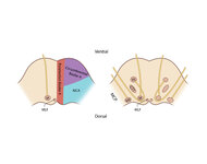 |
Figure 61: Vascular Distribution and Anatomy (Including 6th, 7th, 8th Nerves, MLF) of the Pons | In this axial section of the pons, the proximity of the 7th (VII) and 8th (VIII) fascicles can be appreciated, and a lateral inferior pontine syndrome (anterior inferior cerebellar artery, AICA territory), which could involve both of these fascicles, could cause acute prolonged vertigo accompanied b... | Image |
| 24 |
 |
Cerebellar Ataxia, Neuropathy, & Vestibular Areflexia Syndrome (CANVAS): Impaired Visually-Enhanced VOR and Abnormal Head Impulse Testing | A 67 year old woman presented with 1 year of progressive numbness, gait instability, and oscillopsia when walking or with head movements. Examination showed excessive square-wave jerks, bilateral horizontal gaze-evoked nystagmus, impairment of the visually-enhanced vestibular ocular reflex (vVOR - s... | Image/MovingImage |
| 25 |
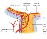 |
Figure 27: Vascular Supply of the Optic Nerve Head, Choroid and Retina | The ophthalmic artery is a branch of the internal carotid artery, which in turn, supplies the posterior ciliary (to choroid and outer retina) and central retinal (to inner retina) arteries. The central retinal artery (CRA) enters the optic nerve about 1 cm posterior to the globe, and an embolus may ... | Image |