AAO-NANOS Neuro-Ophthalmology Clinical Collection: Derived from the AAO-NANOS Clinical Neuro-Ophthalmology collection produced on CD. The images are of selected cases from the NANOS teaching slide exchange, and the CD was produced under the direction of Larry Frohman, MD and Andrew Lee, MD.
The American Academy of Ophthalmology (AAO); The North American Neuro-Ophthalmology Association (NANOS).
NOVEL: https://novel.utah.edu/
TO
| Title | Creator | Description | ||
|---|---|---|---|---|
| 1 |
 |
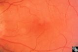
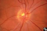
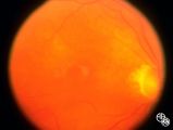
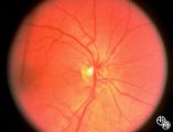
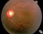
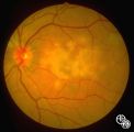
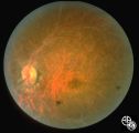
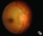
Isolated Congenital Optic Disc Anomalies | Anthony C. Arnold, MD | This patient has known pseudoxanthoma elasticum (an uncommon elastic tissue disorder characterized by plaque-like skin folds [plucked chicken skin] and degeneration of collagen fibers involving multiple systems, including the GI tract and heart), angioid streaks, and optic disc drusen. |
| 2 |
 |
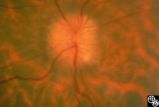
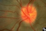
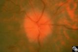
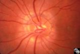
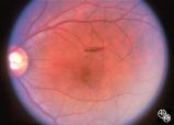
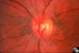
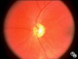
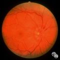
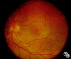
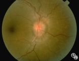
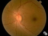
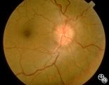
Isolated Optic Neuritis/Neuropathy | Anthony C. Arnold, MD | This 48-year-old man presented with a 1-month history of headache. Both discs had the appearance seen in this image, with prominent peripapillary nerve fiber layer myelination; the disc itself is hyperemic, with dilated, telangiectatic surface vasculature, suggesting true disc edema as well. |
| 3 |
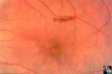
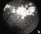
 |
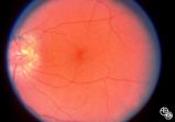
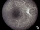
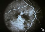
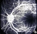
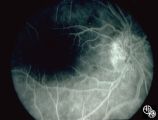
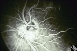
Optic Disc Drusen, Fluorescein Angiogram | Anthony C. Arnold, MD | Images 92_64 and 92_67 demonstrate the characteristics of optic disc drusen on flourescein angiography. This image shows the early arteriovenous phase, with irregular dye uptake and focal hypoflourescence superotemporally. Pair with 92_63 and 92_67 |
| 4 |
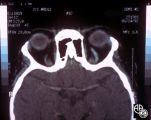
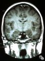
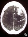
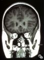
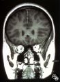
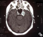
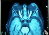
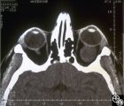
 |
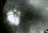
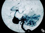
Optic Nerve Drusen, Late Fluorescein Angiogram | Anthony C. Arnold, MD | Images 92_64 and 92_67 demonstrate the characteristics of optic disc drusen on flourescein angiography. This image displays the nodular staining of the drusen without leakage. Pair with 92_63 and 92_64. |
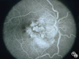
| 5 |
 |
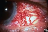
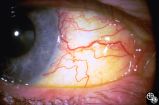
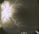
Neuro-Ophthalmic Vascular Disease | Anthony C. Arnold, MD | This 76-year-old woman has a 7-month history of redness and pressure sensation in both eyes that is worse in the morning. She has noted intermittent horizontal diplopia during this time. Angiography demonstrated a right dural cavernous sinus fistula, which was successfully occluded with direct injec... |
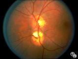
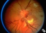
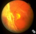
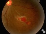
| 6 |
 |
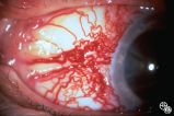
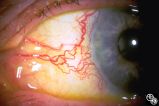
Neuro-Ophthalmic Vascular Disease | Anthony C. Arnold, MD | This 76-year-old woman has a 7-month history of redness and pressure sensation in both eyes that is worse in the morning. She has noted intermittent horizontal diplopia during this time. Angiography demonstrated a right dural cavernous sinus fistula, which was successfully occluded with direct injec... |
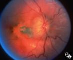
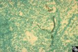
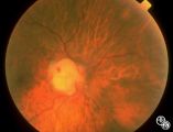
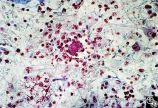
| 7 |
 |
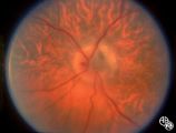
Neuro-Ophthalmic Vascular Disease | Anthony C. Arnold, MD | This 76-year-old woman has a 7-month history of redness and pressure sensation in both eyes that is worse in the morning. She has noted intermittent horizontal diplopia during this time. Angiography demonstrated a right dural cavernous sinus fistula, which was successfully occluded with direct injec... |
| 8 |
 |
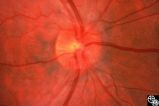
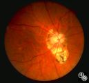
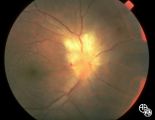
Congenitally Tilted Optic Disc | Anthony C. Arnold, MD | Colobomas or defects of the optic nerve may exhibit spontaneous pulsations. Disease/Diagnosis: Coloboma. |
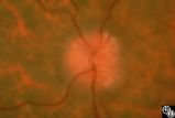
| 9 |
 |
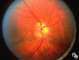
Isolated Optic Neuritis/Neuropathy | Anthony C. Arnold, MD | This 42-year-old male with pseudotumor cerebri and chronic papilledema demonstrated refractile bodies, which can be seen with chronic optic disc edema. This image exhibits decreased disc edema and resolution of the refractile bodies OD after therapy. Pair with 96_01, 96_02, 96_03, 96_05, and 96_06. |
| 10 |
 |
Isolated Optic Neuritis/Neuropathy | Anthony C. Arnold, MD | This 42-year-old male with pseudotumor cerebri and chronic papilledema demonstrated refractile bodies, which can been seen with chronic optic disc edema. This image demonstrates later recurrence of the refractile bodies with worsening papilledema OD. Pair with 96_01, 96_02, 96_03, 96_04, and 96_06. |
| 11 |
 |
Isolated Optic Neuritis/Neuropathy | Anthony C. Arnold, MD | This 42-year-old male with pseudotumor cerebri and chronic papilledema demonstrated refractile bodies, which can be seen with chronic optic disc edema. This image demonstrates later recurrence of the refractile bodies with worsening papilledema OD. Pair with 96_01, 96_02, 96_03, 96_04, and 96_05. |
| 12 |
 |
Isolated Optic Neuritis/Neuropathy | Anthony C. Arnold, MD | This 42-year-old male with pseudotumor cerebri and chronic papilledema demonstrated refractile bodies, which can be seen with chronic optic disc edema. This image shows the chronic papilledema at presentation, with associated refractile hyaline bodies at the disc periphery in both eyes. Pair with 96... |
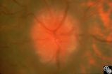
| 13 |
 |
Isolated Optic Neuritis/Neuropathy | Anthony C. Arnold, MD | This image demonstrates Paton's lines in a 34-year-old patient with pseudotumor cerebri and chronic papilledema. |
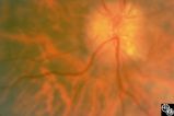
| 14 |
 |
Neuro-Ophthalmic Vascular Disease | Anthony C. Arnold, MD | This 76-year-old woman has a 7-month history of redness and pressure sensation in both eyes that is worse in the morning. She has noted intermittent horizontal diplopia during this time. Angiography demonstrated a right dural cavernous sinus fistula, which was successfully occluded with direct injec... |
| 15 |
 |
Neuro-Ophthalmic Vascular Disease | Anthony C. Arnold, MD | This 76-year-old woman has a 7-month history of redness and pressure sensation in both eyes that is worse in the morning. She has noted intermittent horizontal diplopia during this time. Angiography demonstrated a right dural cavernous sinus fistula, which was successfully occluded with direct injec... |
| 16 |
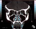 |
Neuro-Ophthalmic Vascular Disease | Anthony C. Arnold, MD | This coronal CT scan shows the enlarged superior ophthalmic vein in the left orbit. This 76-year-old woman has a 7-month history of redness and pressure sensation in both eyes that is worse in the morning. She has noted intermittent horizontal diplopia during this time. Angiography demonstrated a ri... |
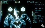
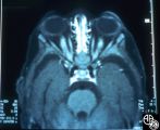
| 17 |
 |
Isolated Optic Neuritis/Neuropathy | Anthony C. Arnold, MD | This 42-year-old male with pseudotumor cerebri and chronic papilledema demonstrated refractile bodies, which can been seen with chronic optic disc edema. This image exhibits decreased disc edema and resolution of the refractile bodies OD after therapy. Pair with 96_01, 96_02, 96_04, 96_05, and 96_06... |
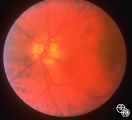
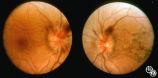
| 18 |
 |
Bridge Coloboma | Anthony C. Arnold, MD | The patient is a 35-year-old asymptomatic woman with a bridge coloboma, also known as double optic disc. Disease/Diagnosis: Coloboma. |
| 19 |
 |
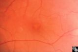
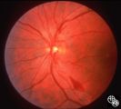
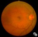
Isolated Congenital Optic Disc Anomalies | Anthony C. Arnold, MD | This image shows drusen that are especially prominent superotemporally. Pair with 92_64 and 92_67. |
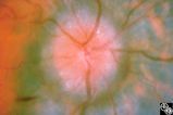
| 20 |
 |
Isolated Optic Neuritis/Neuropathy | Anthony C. Arnold, MD | This 42-year-old male with pseudotumor cerebri and chronic papilledema demonstrated refractile bodies, which can be seen with chronic optic disc edema. This image shows the chronic papilledema at presentation, with associated refractile hyaline bodies at the disc periphery in both eyes. Pair with 96... |
| 21 |
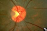 |
Isolated Congenital Optic Disc Anomalies | Anthony C. Arnold, MD | A 10-year-old girl had central visual loss due to this optic pit. Disease/Diagnosis: Optic Pit. |
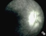
| 22 |
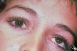
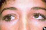
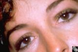
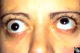
 |
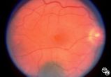
Isolated Congenital Optic Disc Anomalies | Anthony C. Arnold, MD | This is a photograph of peripheral drusen. The paired image 92_69 demonstrates the typical autofluorescence. |
| 23 |
 |
Optic Disc Drusen Autofluorescence | Anthony C. Arnold, MD | This case of optic disc drusen demonstrates the typical autofluorescence. Pair with 92_68. Imaging: Autoflourescence? |
| 24 |
 |
Ocular Manifestations of Congenital/Inherited Diseases | Anthony C. Arnold, MD | Note the retinal/optic nerve head astrocytoma in a 12-year-old boy with tuberous sclerosis. Disease/Diagnosis: Tuberous Sclerosis. |
| 25 |
 |
Optic Neuropathies | Daniel M. Jacobson MD | This 28-year-old otherwise-healthy woman was referred to for treatment of what was thought to be optic neuritis OD. Three weeks earlier she had noted a dark inferior scotoma OD that progressed to involve fixation over the next 10-12 days. She experienced photopsias OD at the onset. She had no viral ... |
| 26 |
 |
Optic Neuropathies | Daniel M. Jacobson MD | This 28-year-old otherwise-healthy woman was referred to for treatment of what was thought to be optic neuritis OD. Three weeks earlier she had noted a dark inferior scotoma OD that progressed to involve fixation over the next 10-12 days. She experienced photopsias OD at the onset. She had no viral ... |
| 27 |
 |
Optic Neuropathies | Daniel M. Jacobson MD | This 34-year-old otherwise-healthy woman was referred for a neuro-ophthalmologic consultation for unexplained loss of vision OD following head trauma. Two months earlier, she was involved in a motor vehicle accident that resulted in a closed head injury complicated by a spontaneously resolving small... |
| 28 |
 |
Optic Neuropathies | Daniel M. Jacobson MD | This 34-year-old otherwise-healthy woman was referred for a neuro-ophthalmologic consultation for unexplained loss of vision OD following head trauma. Two months earlier, she was involved in a motor vehicle accident that resulted in a closed head injury complicated by a spontaneously resolving small... |
| 29 |
 |
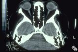
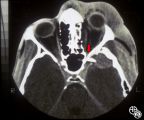
Optic Neuropathies | Daniel M. Jacobson MD | This 34-year-old otherwise-healthy woman was referred for a neuro-ophthalmologic consultation for unexplained loss of vision OD following head trauma. Two months earlier, she was involved in a motor vehicle accident that resulted in a closed head injury complicated by a spontaneously resolving small... |
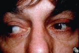
| 30 |
 |
Optic Neuropathies | Daniel M. Jacobson MD | This 34-year-old otherwise-healthy woman was referred for a neuro-ophthalmologic consultation for unexplained loss of vision OD following head trauma. Two months earlier, she was involved in a motor vehicle accident that resulted in a closed head injury complicated by a spontaneously resolving small... |
| 31 |
 |
Ocular Manifestations of Systemic Disorders | Daniel M. Jacobson MD | This 59-year-old man had a 4-week history of malaise, myalgias, night sweats, weight loss, fatigue, and generalized headaches, and a few days before admission he developed a red eye OS secondary to exposure keratitis associated with a left-sided seventh nerve palsy. Subsequent evaluation disclosed a... |
| 32 |
 |
Isolated Optic Neuritis/Neuropathy | Daniel M. Jacobson MD | This 35-year-old otherwise-healthy woman developed typical optic neuritis OD with excellent recovery. She had no clinical evidence of multiple sclerosis at that time. She presented in August of 1991, at which time perivenous sheathing was seen in the retinal periphery OU. A limited workup was negati... |
| 33 |
 |
Isolated Optic Neuritis/Neuropathy | Daniel M. Jacobson MD | This 35-year-old otherwise-healthy woman developed typical optic neuritis OD with excellent recovery. She had no clinical evidence of multiple sclerosis at that time. She presented in August of 1991, at which time perivenous sheathing was seen in the retinal periphery OU. A limited workup was negati... |
| 34 |
 |
Isolated Optic Neuritis/Neuropathy | Daniel M. Jacobson MD | This 35-year-old otherwise-healthy woman developed typical optic neuritis OD with excellent recovery. She had no clinical evidence of multiple sclerosis at that time. She presented in August of 1991, at which time perivenous sheathing was seen in the retinal periphery OU. A limited workup was negati... |
| 35 |
 |
Optic Neuropathies | Daniel M. Jacobson MD | This 28-year-old otherwise-healthy woman was referred to for treatment of what was thought to be optic neuritis OD. Three weeks earlier she had noted a dark inferior scotoma OD that progressed to involve fixation over the next 10-12 days. She experienced photopsias OD at the onset. She had no viral ... |
| 36 |
 |
Optic Neuropathies | Daniel M. Jacobson MD | This 28-year-old otherwise-healthy woman was referred to for treatment of what was thought to be optic neuritis OD. Three weeks earlier she had noted a dark inferior scotoma OD that progressed to involve fixation over the next 10-12 days. She experienced photopsias OD at the onset. She had no viral ... |
| 37 |
 |
Optic Neuropathies | Daniel M. Jacobson MD | This 28-year-old otherwise-healthy woman was referred to for treatment of what was thought to be optic neuritis OD. Three weeks earlier she had noted a dark inferior scotoma OD that progressed to involve fixation over the next 10-12 days. She experienced photopsias OD at the onset. She had no viral ... |
| 38 |
 |
Optic Neuropathies | Daniel M. Jacobson MD | This 28-year-old otherwise-healthy woman was referred to for treatment of what was thought to be optic neuritis OD. Three weeks earlier she had noted a dark inferior scotoma OD that progressed to involve fixation over the next 10-12 days. She experienced photopsias OD at the onset. She had no viral ... |
| 39 |
 |
Optic Neuropathies | Daniel M. Jacobson MD | A previously healthy 28-year-old woman (except for a history of optic neuritis OS 9 years previously with full recovery) was referred for management of optic neuritis OD. Three weeks earlier she had noted a dark inferior scotoma OD that progressed to involve fixation over the next 10-12 days. She ex... |
| 40 |
 |
Optic Neuropathies | Daniel M. Jacobson MD | This 28-year-old otherwise-healthy woman was referred to for treatment of what was thought to be optic neuritis OD. Three weeks earlier she had noted a dark inferior scotoma OD that progressed to involve fixation over the next 10-12 days. She experienced photopsias OD at the onset. She had no viral ... |
| 41 |
 |
Isolated Optic Neuritis/Neuropathy | Daniel M. Jacobson MD | This 35-year-old otherwise-healthy woman developed typical optic neuritis OD with excellent recovery. She had no clinical evidence of multiple sclerosis at that time. She presented in August of 1991, at which time perivenous sheathing was seen in the retinal periphery OU. A limited workup was negati... |
| 42 |
 |
Ocular Manifestations of Congenital/Inherited Diseases | Daniel M. Jacobson MD | This 38-year-old man had unexplained poor vision in his left eye all his life and was told that he had some type of congenital vascular anomaly. He had no neurocutaneous problems. The photographs demonstrate a complex arteriovenous malformation with dilated loops of veins extending out from the nerv... |
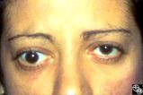
| 43 |
 |
Motility Disturbances | Don Bienfang, MD | This patient displays a posttraumatic left fourth nerve palsy sustained after having struck her head on the dashboard. |
| 44 |
 |
Motility Disturbances | Don Bienfang, MD | This patient displays a posttraumatic left fourth nerve palsy sustained after having struck her head on the dashboard. |
| 45 |
 |
Motility Disturbances | Don Bienfang, MD | This patient displays a posttraumatic left fourth nerve palsy sustained after having struck her head on the dashboard. |
| 46 |
 |
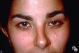
Motility Disturbances | Don Bienfang, MD | This patient displays a posttraumatic left fourth nerve palsy sustained after having struck her head on the dashboard. |
| 47 |
 |
Neuro-Ophthalmic Consequences of Therapy | Don Bienfang, MD | An elderly woman was first seen in 1991, after being on Plaquenil (Hydroxychloroquine Sulfate) for 5 to 6 years, with a new symptomatic disturbance in her reading. Because her blue/yellow color perception was disturbed, and because she had symptoms, the medication was stopped. However the fundi and ... |
| 48 |
 |
Neuro-Ophthalmic Consequences of Therapy | Don Bienfang, MD | An elderly woman was first seen in 1991, after being on Plaquenil (Hydroxychloroquine Sulfate) for 5 to 6 years, with a new symptomatic disturbance in her reading. Because her blue/yellow color perception was disturbed, and because she had symptoms, the medication was stopped. However the fundi and ... |
| 49 |
 |
Neuro-Ophthalmic Consequences of Therapy | Don Bienfang, MD | An elderly woman was first seen in 1991, after being on Plaquenil (Hydroxychloroquine Sulfate) for 5 to 6 years, with a new symptomatic disturbance in her reading. Because her blue/yellow color perception was disturbed, and because she had symptoms, the medication was stopped. However the fundi and ... |
| 50 |
 |
Neuro-Ophthalmic Consequences of Therapy | Don Bienfang, MD | An elderly woman was first seen in 1991, after being on Plaquenil (Hydroxychloroquine Sulfate) for 5 to 6 years, with a new symptomatic disturbance in her reading. Because her blue/yellow color perception was disturbed, and because she had symptoms, the medication was stopped. However the fundi and ... |
| 51 |
 |
Neuro-Ophthalmic Consequences of Therapy | Don Bienfang, MD | An elderly woman was first seen in 1991, after being on Plaquenil (Hydroxychloroquine Sulfate) for 5 to 6 years, with a new symptomatic disturbance in her reading. Because her blue/yellow color perception was disturbed, and because she had symptoms, the medication was stopped. However the fundi and ... |
| 52 |
 |
Neuro-Ophthalmic Consequences of Therapy | Don Bienfang, MD | An elderly woman was first seen in 1991, after being on Plaquenil (Hydroxychloroquine Sulfate) for 5 to 6 years, with a new symptomatic disturbance in her reading. Because her blue/yellow color perception was disturbed, and because she had symptoms, the medication was stopped. However the fundi and ... |
| 53 |
 |
Neuro-Ophthalmic Consequences of Therapy | Don Bienfang, MD | An elderly woman was first seen in 1991, after being on Plaquenil (Hydroxychloroquine Sulfate) for 5 to 6 years, with a new symptomatic disturbance in her reading. Because her blue/yellow color perception was disturbed, and because she had symptoms, the medication was stopped. However the fundi and ... |
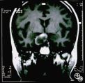
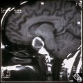
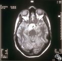
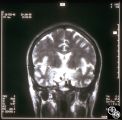
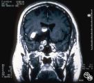
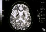
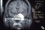
| 54 |
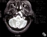
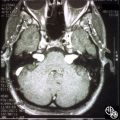
 |
Neuro-Ophthalmic Imaging-MRI | Don Bienfang, MD | A 59-year-old untreated hypertensive man had a sudden onset of vomiting, gait ataxia, dysarthria, and left-sided weakness. The eyes were deviated downward and to the left. (Note position of gaze in 94_11). Extraocular motility was full. There was rotary nystagmus with the fast phase to the left; the... |
| 55 |
 |
Trochlear Motility Disturbances | Don Bienfang, MD | This patient displays a posttraumatic left fourth nerve palsy sustained after having struck her head on the dashboard. |
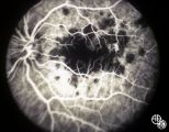
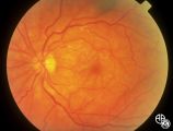
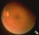
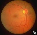
| 56 |
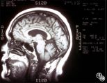
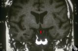
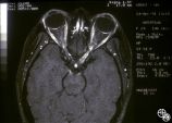
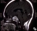
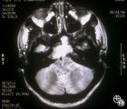
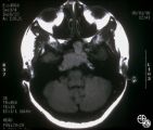
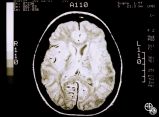
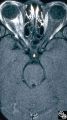
 |
Motility Disturbances | Eric L. Berman, MD | This is a 60-year-old albino woman with chronic progressive external ophthalmoplegia and strabismus fixus. Her extreme bilateral esotropia caused her acuity to be 20/400 OU, no view of fundus could be obtained except for the far periphery. CPEO is the most common manifestation of mitochondrial myopa... |
| 57 |
 |
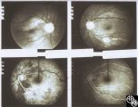
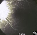
Isolated Hereditary Optic Neuritis/Neuropathy | Gregory S. Kosmorsky, MD | Leber's hereditary optic neuropathy is a mitochondrial hereditary optic neuropathy that usually affects young males but may occur at any age and in males or females. The clinical features are usually acute bilateral simultaneous or sequential visual loss with a central acuity and central visual fiel... |
| 58 |
 |
Isolated Hereditary Optic Neuritis/Neuropathy | Gregory S. Kosmorsky, MD | Leber's hereditary optic neuropathy is a mitochondrial hereditary optic neuropathy that usually affects young males but may occur at any age and in males or females. The clinical features are usually acute bilateral simultaneous or sequential visual loss with a central acuity and central visual fiel... |
| 59 |
 |
Isolated Hereditary Optic Neuritis/Neuropathy | Gregory S. Kosmorsky, MD | Leber's hereditary optic neuropathy is a mitochondrial hereditary optic neuropathy that usually affects young males but may occur at any age and in males or females. The clinical features are usually acute bilateral simultaneous or sequential visual loss with a central acuity and central visual fiel... |
| 60 |
 |
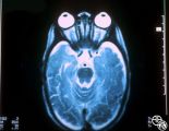
Ocular Manifestations of Congenital/Inherited Diseases | Jacqueline A. Leavitt, MD | This 22-year-old woman has neurofibromatosis, type 2. Acuity, color plates, pupillary responses, slit-lamp examination, IOP, fields, and funduscopy are all normal. There is a 3 mm proptosis OS. The patient has recently undergone gamma knife for the acoustic tumor, and she has residual facial nerve p... |
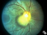
| 61 |
 |
Neuro-Ophthalmic Manifestations of Brain Tumors | Jacqueline A. Leavitt, MD | Chordomas of the clivus may result in diplopia due to a sixth nerve palsy. The sixth nerve runs up the clivus and may be the presenting manifestation of the lesion. Pair with Images 97_33, 97_34, and 97_35. |
| 62 |
 |
Ocular Manifestations of Congenital/Inherited Diseases | Jacqueline A. Leavitt, MD | This 22-year-old woman has neurofibromatosis, type 2. Acuity, color plates, pupillary responses, slit-lamp examination, IOP, fields, and funduscopy are all normal. There is a 3 mm proptosis OS. The patient has recently undergone gamma knife for the acoustic tumor, and she has residual facial nerve p... |
| 63 |
 |
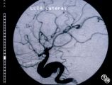
Neuro-Ophthalmic Vascular Disease | Jacqueline A. Leavitt, MD | Arteriovenous malformations are dysplastic vascular channels. They may result in neuro-ophthalmologic findings from direct compressive effects, secondary hemorrhage, or secondary seizure disorder. CT and/or MRI may demonstrate the lesion, but cerebral arteriography is usually required to better defi... |
| 64 |
 |
Ocular Manifestations of Congenital/Inherited Diseases | Jacqueline A. Leavitt, MD | This 22-year-old woman has neurofibromatosis, type 2. Acuity, color plates, pupillary responses, slit-lamp examination, IOP, fields, and funduscopy are all normal. There is a 3 mm proptosis OS. The patient has recently undergone gamma knife for the acoustic tumor, and she has residual facial nerve p... |
| 65 |
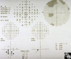
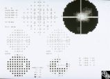
 |
Ocular Manifestations of Congenital/Inherited Diseases | Jacqueline A. Leavitt, MD | This 22-year-old woman has neurofibromatosis, type 2. Acuity, color plates, pupillary responses, slit-lamp examination, IOP, fields, and funduscopy are all normal. There is a 3 mm proptosis OS. The patient has recently undergone gamma knife for the acoustic tumor, and she has residual facial nerve p... |
| 66 |
 |
Ocular Manifestations of Congenital/Inherited Diseases | Jacqueline A. Leavitt, MD | This 22-year-old woman has neurofibromatosis, type 2. Acuity, color plates, pupillary responses, slit-lamp examination, IOP, fields, and funduscopy are all normal. There is a 3 mm proptosis OS. The patient has recently undergone gamma knife for the acoustic tumor, and she has residual facial nerve p... |
| 67 |
 |
Neuro-Ophthalmic Manifestations of Brain Tumors | Jacqueline A. Leavitt, MD | Chordomas of the clivus may result in diplopia due to a sixth nerve palsy. The sixth nerve runs up the clivus and may be the presenting manifestation of the lesion. Pair with Images 97_34, 97_35, and 97_36. |
| 68 |
 |
Neuro-Ophthalmic Manifestations of Brain Tumors | Jacqueline A. Leavitt, MD | Chordomas of the clivus may result in diplopia due to a sixth nerve palsy. The sixth nerve runs up the clivus and may be the presenting manifestation of the lesion. Pair with Images 97_33, 97_34, and 97_36. |
| 69 |
 |
Neuro-Ophthalmic Manifestations of Brain Tumors | Jacqueline A. Leavitt, MD | Chordomas of the clivus may result in diplopia due to a sixth nerve palsy. The sixth nerve runs up the clivus and may be the presenting manifestation of the lesion. Imaging: MRI, T-1 axial with contrast. Pair with Images 97_33, 97_34, and 97_36. Anatomy: Clivus, Sixth nerve. Pathology: Sixth nerve p... |
| 70 |
 |
Pupillary Syndrome | Jeffrey G. Odel, MD | Intermittent dilation of the pupils may occur as a benign phenomenon in healthy young adults. In the absence of other third nerve signs, (eg, ptosis, diplopia, ophthalmoplegia), an isolated transient dilation of the pupil in an otherwise healthy adult is unlikely to represent a third nerve palsy. Tr... |
| 71 |
 |
Pupillary Syndrome | Jeffrey G. Odel, MD | Intermittent dilation of the pupils may occur as a benign phenomenon in healthy young adults. In the absence of other third nerve signs, (eg, ptosis, diplopia, ophthalmoplegia), an isolated transient dilation of the pupil in an otherwise healthy adult is unlikely to represent a third nerve palsy. Tr... |
| 72 |
 |
Isolated Congenital Optic Disc Anomalies | Joel M. Weinstein, MD | This 8-year-old boy presented with a 2-week history of decreased vision in the right eye. He had undergone a normal MRI and CSF examination, including intracranial pressure, before neuro-ophthalmologic assessment. The fundus photographs and fluorescein angiograms show subretinal neovascularization a... |
| 73 |
 |
Neuro-Ophthalmic Consequences of Therapy | Joel M. Weinstein, MD | The patient is a 38-year-old woman with oligodendroglioma of the left cerebral hemisphere. The patient received intracarotid BCNU and developed BCNU retinopathy, optic neuropathy, and partial ophthalmoplegia. Image 92_21is from 1 month later and demonstrates hemorrhagic swelling of the disc, retinal... |
| 74 |
 |
Neuro-Ophthalmic Consequences of Therapy | Joel M. Weinstein, MD | The patient is a 38-year-old woman with oligodendroglioma of the left cerebral hemisphere. The patient received intracarotid BCNU and developed BCNU retinopathy, optic neuropathy, and partial ophthalmoplegia. Image 92_22 Shows late changes, with more severe retinal ischemia, vascular attenuation, an... |
| 75 |
 |
Pseudopapilledema With Subretinal Neovascularization on Fluorescein Angiogram | Joel M. Weinstein, MD | This 8-year-old boy presented with a 2-week history of decreased vision in the right eye. He had undergone a normal MRI and CSF examination, including intracranial pressure, before neuro-ophthalmologic assessment. The fundus photographs and fluorescein angiograms show subretinal neovascularization a... |
| 76 |
 |
Pseudopapilledema With Retinal Neovascularization, Fluorescein Angiogram | Joel M. Weinstein, MD | This 8-year-old boy presented with a 2-week history of decreased vision in the right eye. He had undergone a normal MRI and CSF examination, including intracranial pressure, before neuro-ophthalmologic assessment. The fundus photographs and fluorescein angiograms show subretinal neovascularization a... |
| 77 |
 |
Isolated Congenital Optic Disc Anomalies | Joel M. Weinstein, MD | This 8-year-old boy presented with a 2-week history of decreased vision in the right eye. He had undergone a normal MRI and CSF examination, including intracranial pressure, before neuro-ophthalmologic assessment. The fundus photographs and fluorescein angiograms show subretinal neovascularization a... |
| 78 |
 |
Systemic Disorders With Optic Nerve and Retinal Findings | Joel M. Weinstein, MD | This 46-year-old woman noted progressive bilateral visual loss over a 10-month period. She had a malignant melanoma removed from her right thigh 2 years ago and excisional biopsy of an inguinal node metastasis 8 months ago. She also complained of poor night vision and rare intermittent ""sparkles"" ... |
| 79 |
 |
Systemic Disorders With Optic Nerve and Retinal Findings | Joel M. Weinstein, MD | This 46-year-old woman noted progressive bilateral visual loss over a 10-month period. She had a malignant melanoma removed from her right thigh 2 years ago and excisional biopsy of an inguinal node metastasis 8 months ago. She also complained of poor night vision and rare intermittent ""sparkles"" ... |
| 80 |
 |
Systemic Disorders With Optic Nerve and Retinal Findings | Joel M. Weinstein, MD | This 46-year-old woman noted progressive bilateral visual loss over a 10-month period. She had a malignant melanoma removed from her right thigh 2 years ago and excisional biopsy of an inguinal node metastasis 8 months ago. She also complained of poor night vision and rare intermittent ""sparkles"" ... |
| 81 |
 |
Systemic Disorders With Optic Nerve and Retinal Findings | Joel M. Weinstein, MD | This 46-year-old woman noted progressive bilateral visual loss over a 10-month period. She had a malignant melanoma removed from her right thigh 2 years ago and excisional biopsy of an inguinal node metastasis 8 months ago. She also complained of poor night vision and rare intermittent ""sparkles"" ... |
| 82 |
 |
Systemic Disorders With Optic Nerve and Retinal Findings | John A. Charley, MD | A hypercoagulable state may result from deficiency of various factors involved in the clotting pathway, such as factor V. This may result in venous thrombosis and a branch or central retinal vein occlusion. Consideration for testing for an underlying coagulopathy, including the factor V deficiency, ... |
| 83 |
 |
Systemic Disorders With Optic Nerve and Retinal Findings | John A. Charley, MD | A hypercoagulable state may result from deficiency of various factors involved in the clotting pathway, such as factor V. This may result in venous thrombosis and a branch or central retinal vein occlusion. Consideration for testing for an underlying coagulopathy, including the factor V deficiency, ... |
| 84 |
 |
Isolated Optic Neuritis/Neuropathy | John A. Charley, MD | Several Primary Mutations result in Leber's hereditary optic neuropathy, including mitochondrial deletions at positions 11778, 3460, and 14484. Although the 11778 is the most common mutation, the 11484 has the best prognosis for spontaneous recovery. This case exhibits the 3460 mutation. |
| 85 |
 |
Isolated Optic Neuritis/Neuropathy | John A. Charley, MD | Several Primary Mutations result in Leber's hereditary optic neuropathy, including mitochondrial deletions at positions 11778, 3460, and 14484. Although the 11778 is the most common mutation, the 11484 has the best prognosis for spontaneous recovery. This case exhibits the 3460 mutation. |
| 86 |
 |
Isolated Optic Neuritis/Neuropathy | John A. Charley, MD | Several Primary Mutations result in Leber's hereditary optic neuropathy, including mitochondrial deletions at positions 11778, 3460, and 14484. Although the 11778 is the most common mutation, the 11484 has the best prognosis for spontaneous recovery. This case exhibits the 3460 mutation. |
| 87 |
 |
Isolated Optic Neuritis/Neuropathy | John A. Charley, MD | Several Primary Mutations result in Leber's hereditary optic neuropathy, including mitochondrial deletions at positions 11778, 3460, and 14484. Although the 11778 is the most common mutation, the 11484 has the best prognosis for spontaneous recovery. This case exhibits the 3460 mutation. |
| 88 |
 |
Isolated Optic Neuritis/Neuropathy | John A. Charley, MD | Several Primary Mutations result in Leber's hereditary optic neuropathy, including mitochondrial deletions at positions 11778, 3460, and 14484. Although the 11778 is the most common mutation, the 11484 has the best prognosis for spontaneous recovery. This case exhibits the 3460 mutation. |
| 89 |
 |
Chiasmal Syndromes | Larry P. Frohman, MD | This woman was 61 years old when she underwent initial neuro-ophthalmologic evaluation. Twenty-three years earlier, she had undergone removal of a pituitary adenoma followed by radiation therapy. At that time, she had noted a preoperative visual field defect that improved somewhat after the surgery.... |
| 90 |
 |
Chiasmal Syndromes | Larry P. Frohman, MD | This woman was 61 years old when she underwent initial neuro-ophthalmologic evaluation. Twenty-three years earlier, she had undergone removal of a pituitary adenoma followed by radiation therapy. At that time, she had noted a preoperative visual field defect that improved somewhat after the surgery.... |
| 91 |
 |
Chiasmal Syndromes | Larry P. Frohman, MD | This woman was 61 years old when she underwent initial neuro-ophthalmologic evaluation. Twenty-three years earlier, she had undergone removal of a pituitary adenoma followed by radiation therapy. At that time, she had noted a preoperative visual field defect that improved somewhat after the surgery.... |
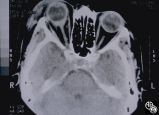
| 92 |
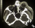 |
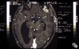
Neuro-Ophthalmic Imaging-CT Scan | Larry P. Frohman, MD | This patient was assaulted with a blunt object and suffered acute blindness due to traumatic optic neuropathy. Note how the lateral orbital wall has been fractured and displaced posteromedially into the region of the anterior optic canal. |
| 93 |
 |
Motility Disturbances | Larry P. Frohman, MD | A 27-year-old healthy man presented for neuro-ophthalmologic evaluation for recurrent vertical binocular diplopia. In 1982, he had a minor blow to the head, and 12 days later he sustained vertical binocular diplopia, worse on downgaze, without ever having diurnal variation. His sense of taste was al... |
| 94 |
 |
Motility Disturbances | Larry P. Frohman, MD | A 27-year-old healthy man presented for neuro-ophthalmologic evaluation for recurrent vertical binocular diplopia. In 1982, he had a minor blow to the head, and 12 days later he sustained vertical binocular diplopia, worse on downgaze, without ever having diurnal variation. His sense of taste was al... |
| 95 |
 |
Motility Disturbances | Larry P. Frohman, MD | A 27-year-old healthy man presented for neuro-ophthalmologic evaluation for recurrent vertical binocular diplopia. In 1982, he had a minor blow to the head, and 12 days later he sustained vertical binocular diplopia, worse on downgaze, without ever having diurnal variation. His sense of taste was al... |
| 96 |
 |
Motility Disturbances | Larry P. Frohman, MD | A 27-year-old healthy man presented for neuro-ophthalmologic evaluation for recurrent vertical binocular diplopia. In 1982, he had a minor blow to the head, and 12 days later he sustained vertical binocular diplopia, worse on downgaze, without ever having diurnal variation. His sense of taste was al... |
| 97 |
 |
Systemic Disorders With Optic Nerve and Retinal Findings | Larry P. Frohman, MD | Cogan's syndrome is a vasculitic-autoimmune process with critical ocular features. In its original classical form it consists of nonluetic interstitial keratitis, with audiovestibular dysfunction, often with pain. More recently, it has been pointed out that there are nonclassical forms as well, part... |
| 98 |
 |
Motility Disturbances | Larry P. Frohman, MD | A 27-year-old healthy man presented for neuro-ophthalmologic evaluation for recurrent vertical binocular diplopia. In 1982, he had a minor blow to the head, and 12 days later he sustained vertical binocular diplopia, worse on downgaze, without ever having diurnal variation. His sense of taste was al... |
| 99 |
 |
Motility Disturbances | Larry P. Frohman, MD | A 27-year-old healthy man presented for neuro-ophthalmologic evaluation for recurrent vertical binocular diplopia. In 1982, he had a minor blow to the head, and 12 days later he sustained vertical binocular diplopia, worse on downgaze, without ever having diurnal variation. His sense of taste was al... |
| 100 |
 |
Motility Disturbances | Larry P. Frohman, MD | This 5-year-old child presented with a 70 PD exotropia OS and a right face turn. She had a CT scan of the head at age 4 months that was normal , and she was felt to have an isolated left medial rectus paresis. Her acuity was 20/20 OU. She could fuse with a large face turn, and was orthomorphic is ex... |
| 101 |
 |
Motility Disturbances | Larry P. Frohman, MD | This 5-year-old child presented with a 70 PD exotropia OS and a right face turn. She had a CT scan of the head at age 4 months that was normal , and she was felt to have an isolated left medial rectus paresis. Her acuity was 20/20 OU. She could fuse with a large face turn, and was orthomorphic is ex... |
| 102 |
 |
Motility Disturbances | Larry P. Frohman, MD | This 5-year-old child presented with a 70 PD exotropia OS and a right face turn. She had a CT scan of the head at age 4 months that was normal , and she was felt to have an isolated left medial rectus paresis. Her acuity was 20/20 OU. She could fuse with a large face turn, and was orthomorphic is ex... |
| 103 |
 |
Motility Disturbances | Larry P. Frohman, MD | This 5-year-old child presented with a 70 PD exotropia OS and a right face turn. She had a CT scan of the head at age 4 months that was normal , and she was felt to have an isolated left medial rectus paresis. Her acuity was 20/20 OU. She could fuse with a large face turn, and was orthomorphic is ex... |
| 104 |
 |
Systemic Disorders With Optic Nerve and Retinal Findings | Larry P. Frohman, MD | A 29-year-old African American woman presented with headaches, bilateral transient visual obscurations, blurred vision, numbness, and weakness of the lower extremities with myalgia and joint pains. She had an unplanned 12-pound weight loss over 2 months. A neurologist and internist diagnosed her wit... |
| 105 |
 |
Systemic Disorders With Optic Nerve and Retinal Findings | Larry P. Frohman, MD | A 25-year-old African-American woman presented with a 20/200 optic neuropathology and no other illness. Because this disc appearance of a granuloma led us to suspect occult sarcoidosis, she underwent systemic evaluation. She was ultimately shown to have systemic sarcoidosis, including pulmonary invo... |
| 106 |
 |
Ocular Manifestations of Systemic Disorders | Larry P. Frohman, MD | A 17-year-old girl had undergone multiple superficial biopsies of the orbit for what was felt to be refractory orbital pseudotumor. Initial evaluation revealed the saddle-nose deformity, which the patient confirmed was acquired. More extensive biopsy was consistent with lymphomatoid granulomatosis. ... |
| 107 |
 |
Optic Tract Syndrome Due to Carotid Artery Dolichoectasia | Larry P. Frohman, MD | This 43-year-old man was referred for evaluation of 6 months of visual loss OU. In retrospect, he had noticed increasing difficulty with his tennis game dating back over 3 years, as balls would pass him unexpectedly when hit to his backhand (left) side. The patient did not think this was progressive... |
| 108 |
 |
Optic Tract Syndrome Due to Carotid Artery Dolichoectasia | Larry P. Frohman, MD | This 43-year-old man was referred for evaluation of 6 months of visual loss OU. In retrospect, he had noticed increasing difficulty with his tennis game dating back over 3 years, as balls would pass him unexpectedly when hit to his backhand (left) side. The patient did not think this was progressive... |
| 109 |
 |
Isolated Optic Neuritis/Neuropathy | Larry P. Frohman, MD | The patient is a 62-year-old female who presented in August 1996 with visual loss OD that she first noted as loss of her superior field in May 1996. She felt that it had been static since, and perhaps was even a little better in the week before she was seen. There was no pain, even with ocular rotat... |
| 110 |
 |
Isolated Optic Neuritis/Neuropathy | Larry P. Frohman, MD | The patient is a 62-year-old female who presented in August 1996 with visual loss OD that she first noted as loss of her superior field in May 1996. She felt that it had been static since, and perhaps was even a little better in the week before she was seen. There was no pain, even with ocular rotat... |
| 111 |
 |
Isolated Optic Neuritis/Neuropathy | Larry P. Frohman, MD | The patient is a 62-year-old female who presented in August 1996 with visual loss OD that she first noted as loss of her superior field in May 1996. She felt that it had been static since, and perhaps was even a little better in the week before she was seen. There was no pain, even with ocular rotat... |
| 112 |
 |
Isolated Optic Neuritis/Neuropathy | Larry P. Frohman, MD | The patient is a 62-year-old female who presented in August 1996 with visual loss OD that she first noted as loss of her superior field in May 1996. She felt that it had been static since, and perhaps was even a little better in the week before she was seen. There was no pain, even with ocular rotat... |
| 113 |
 |
Isolated Optic Neuritis/Neuropathy | Larry P. Frohman, MD | The patient is a 62-year-old female who presented in August 1996 with visual loss OD that she first noted as loss of her superior field in May 1996. She felt that it had been static since, and perhaps was even a little better in the week before she was seen. There was no pain, even with ocular rotat... |
| 114 |
 |
Isolated Optic Neuritis/Neuropathy | Larry P. Frohman, MD | The patient is a 62-year-old female who presented in August 1996 with visual loss OD that she first noted as loss of her superior field in May 1996. She felt that it had been static since, and perhaps was even a little better in the week before she was seen. There was no pain, even with ocular rotat... |
| 115 |
 |
Orbital Tumors | Larry P. Frohman, MD | This 30-year-old man had a retrobulbar intraconal mass OS. The CT scans showed a heterogeneous lobulated enhancing mass, 2.2 x 1.9 x 1.8 cm. The case beautifully exhibits chorodial folds. The ultrasound showed internal reflectivity. The patient refused surgery. Pair with Images 97_60, 97_62, 97_63, ... |
| 116 |
 |
Orbital Tumors | Larry P. Frohman, MD | This 30-year-old man had a retrobulbar intraconal mass OS. The CT scans showed a heterogeneous lobulated enhancing mass, 2.2 x 1.9 x 1.8 cm. The case beautifully exhibits chorodial folds. The ultrasound showed internal reflectivity. The patient refused surgery. Pair with Images 97_60, 97_61, 97_63, ... |
| 117 |
 |
Orbital Tumors | Larry P. Frohman, MD | This 30-year-old man had a retrobulbar intraconal mass OS. The CT scans showed a heterogeneous lobulated enhancing mass, 2.2 x 1.9 x 1.8 cm. The case beautifully exhibits chorodial folds. The ultrasound showed internal reflectivity. The patient refused surgery. Pair with Images 97_61, 97_62, 97_63, ... |
| 118 |
 |
Isolated Congenital Optic Disc Anomalies | Larry P. Frohman, MD | This 63-year-old man with amblyopia OD was seen for a question of ischemic optic neuropathy with a pale, swollen disc OD. The correct diagnosis is an exophytic capillary angioma of the optic nerve head. Disease/Diagnosis: Capillary Angioma. |
| 119 |
 |
Optic Neuropathies | Larry P. Frohman, MD | This healthy 29-year-old man with dense amblyopia OS presented with a foreign-body sensation OS and further visual loss in his amblyopic eye. He was noted to have bilateral disc edema and lesions in the left eye consistent with unilateral acute multifocal placoid pigment epitheliopathy (AMPPE). He r... |
| 120 |
 |
Optic Neuropathies | Larry P. Frohman, MD | This healthy 29-year-old man with dense amblyopia OS presented with a foreign-body sensation OS and further visual loss in his amblyopic eye. He was noted to have bilateral disc edema and lesions in the left eye consistent with unilateral acute multifocal placoid pigment epitheliopathy (AMPPE). He r... |
| 121 |
 |
Optic Neuropathies | Larry P. Frohman, MD | This healthy 29-year-old man with dense amblyopia OS presented with a foreign-body sensation OS and further visual loss in his amblyopic eye. He was noted to have bilateral disc edema and lesions in the left eye consistent with unilateral acute multifocal placoid pigment epitheliopathy (AMPPE). He r... |
| 122 |
 |
Optic Neuropathies | Larry P. Frohman, MD | This healthy 29-year-old man with dense amblyopia OS presented with a foreign-body sensation OS and further visual loss in his amblyopic eye. He was noted to have bilateral disc edema and lesions in the left eye consistent with unilateral acute multifocal placoid pigment epitheliopathy (AMPPE). He r... |
| 123 |
 |
Systemic Disorders With Optic Nerve and Retinal Findings | Larry P. Frohman, MD | At age 41, in 1984, this woman, who grew up in the Ohio River Valley, had 3 days of ocular pain OD, and her vision declined to 20/80 OD she has had no visual changes since, nor has she had any other neurologic symptoms. The ""presumed"" diagnosis is optic neuropathy in presumed ocular histoplasmosis... |
| 124 |
 |
Systemic Disorders With Optic Nerve and Retinal Findings | Larry P. Frohman, MD | This 57-year-old man had a neuro-ophthalmology consult, requested the night before his 2-cm pituitary tumor was to be resected. His examination revealed his acuities to be 20/70 OU, with a visual field not consistent with chiasmal compression. The fundus appearance, with peripheral salt and pepperin... |
| 125 |
 |
Neuro-Ophthalmic Vascular Disease | Larry P. Frohman, MD | This 27-year-old woman had no past ocular history and presented with 3 weeks of redness OS that has been treated by the referring doctor as allergic conjunctivitis. She was referred for evaluation when she developed binocular diplopia. Her past medical history included phlebitis and one miscarriage ... |
| 126 |
 |
Neuro-Ophthalmic Vascular Disease | Larry P. Frohman, MD | This 27-year-old woman had no past ocular history and presented with 3 weeks of redness OS that has been treated by the referring doctor as allergic conjunctivitis. She was referred for evaluation when she developed binocular diplopia. Her past medical history included phlebitis and one miscarriage ... |
| 127 |
 |
Neuro-Ophthalmic Consequences of Therapy | Larry P. Frohman, MD | This woman presented at age 52, 3 years after radiation therapy for a salivary gland carcinoma extending into the right maxillary sinus. She had received 6000 rads in 30 fractions over 45 days. She presented with 3 weeks of visual loss, with acuity of 20/30, normal color plates, normal fields, and n... |
| 128 |
 |
Neuro-Ophthalmic Consequences of Therapy | Larry P. Frohman, MD | This woman presented at age 52, 3 years after radiation therapy for a salivary gland carcinoma extending into the right maxillary sinus. She had received 6000 rads in 30 fractions over 45 days. She presented with 3 weeks of visual loss, with acuity of 20/30, normal color plates, normal fields, and n... |
| 129 |
 |
Optic Tract Syndrome Due to Carotid Artery Dolichoectasia | Larry P. Frohman, MD | This 43-year-old man was referred for evaluation of 6 months of visual loss OU. In retrospect, he had noticed increasing difficulty with his tennis game dating back over 3 years, as balls would pass him unexpectedly when hit to his backhand (left) side. The patient did not think this was progressive... |
| 130 |
 |
Chiasmal Syndromes | Larry P. Frohman, MD | A 50-year-old right-handed woman with no significant past medical history underwent liposuction on her abdomen, hips, and thighs under general anesthesia. Her height and weight were 167 cm and 63 kg, respectively. Subcutaneous fat was injected with 2.5 L of 0.05% xylocaine and 1:10,000,000 epinephri... |
| 131 |
 |
Chiasmal Syndromes | Larry P. Frohman, MD | A 50-year-old right-handed woman with no significant past medical history underwent liposuction on her abdomen, hips, and thighs under general anesthesia. Her height and weight were 167 cm and 63 kg, respectively. Subcutaneous fat was injected with 2.5 L of 0.05% xylocaine and 1:10,000,000 epinephri... |
| 132 |
 |
Chiasmal Syndromes | Larry P. Frohman, MD | A 50-year-old right-handed woman with no significant past medical history underwent liposuction on her abdomen, hips, and thighs under general anesthesia. Her height and weight were 167 cm and 63 kg, respectively. Subcutaneous fat was injected with 2.5 L of 0.05% xylocaine and 1:10,000,000 epinephri... |
| 133 |
 |
Optic Neuropathies | Larry P. Frohman, MD | This healthy 29-year-old man with dense amblyopia OS presented with a foreign-body sensation OS and further visual loss in his amblyopic eye. He was noted to have bilateral disc edema and lesions in the left eye consistent with unilateral acute multifocal placoid pigment epitheliopathy (AMPPE). He r... |
| 134 |
 |
Optic Neuropathies | Larry P. Frohman, MD | This healthy 29-year-old man with dense amblyopia OS presented with a foreign-body sensation OS and further visual loss in his amblyopic eye. He was noted to have bilateral disc edema and lesions in the left eye consistent with unilateral acute multifocal placoid pigment epitheliopathy (AMPPE). He r... |
| 135 |
 |
Optic Neuropathies | Larry P. Frohman, MD | The patient is a 66-year-old man with a history of ethanol abuse. He presented with 3 months of right-sided headache and a few days of progressive visual loss OD to hand motions only. When seen by the orbital service, he had nearly complete ophthalmoplegia and ptosis. Sinus biopsy showed fungus, whi... |
| 136 |
 |
Optic Neuropathies | Larry P. Frohman, MD | The patient is a 66-year-old man with a history of ethanol abuse. He presented with 3 months of right-sided headache and a few days of progressive visual loss OD to hand motions only. When seen by the orbital service, he had nearly complete ophthalmoplegia and ptosis. Sinus biopsy showed fungus, whi... |
| 137 |
 |
Optic Neuropathies | Larry P. Frohman, MD | The patient is a 66-year-old man with a history of ethanol abuse. He presented with 3 months of right-sided headache and a few days of progressive visual loss OD to hand motions only. When seen by the orbital service, he had nearly complete ophthalmoplegia and ptosis. Sinus biopsy showed fungus, whi... |
| 138 |
 |
Systemic Disorders With Optic Nerve and Retinal Findings | Larry P. Frohman, MD | This 48-year-old female was seen in May 1996 with a history of 2 months of diplopia from a right abducens palsy. This was due to the recurrence of myeloma that had initially been diagnosed and treated with radiation and chemotherapy 9 years before and required further therapy, including bone marrow ... |
| 139 |
 |
Systemic Disorders With Optic Nerve and Retinal Findings | Larry P. Frohman, MD | This 48-year-old female was seen in May 1996 with a history of 2 months of diplopia from a right abducens palsy. This was due to the recurrence of myeloma that had initially been diagnosed and treated with radiation and chemotherapy 9 years before and required further therapy, including bone marrow ... |
| 140 |
 |
Systemic Disorders With Optic Nerve and Retinal Findings | Larry P. Frohman, MD | This 48-year-old female was seen in May 1996 with a history of 2 months of diplopia from a right abducens palsy. This was due to the recurrence of myeloma that had initially been diagnosed and treated with radiation and chemotherapy 9 years before and required further therapy, including bone marrow ... |
| 141 |
 |
Isolated Optic Neuritis/Neuropathy | Larry P. Frohman, MD | The patient is a 62-year-old female who presented in August 1996 with visual loss OD that she first noted as loss of her superior field in May 1996. She felt that it had been static since, and perhaps was even a little better in the week before she was seen. There was no pain, even with ocular rotat... |
| 142 |
 |
Systemic Disorders With Optic Nerve and Retinal Findings | Larry P. Frohman, MD | This 74-year-old asthmatic male had acute visual loss OS while watching the Super Bowl in 1994. He was seen the next day by a retina specialist, who noted that his optic disc was normal and referred the patient to a neuro-ophthalmologist, who evaluated him about 40 hours after his visual loss. He wa... |
| 143 |
 |
Neuro-Ophthalmic Vascular Disease | Larry P. Frohman, MD | This 23-year-old woman has had insulin-dependent diabetes mellitus since age 3. She was diagnosed with Sydenham's chorea in early childhood and had grand mal seizures from age 13 to 15. She has been hypertensive since age 18. Her vision was 20/25 OD and 20/40 OS, with dyschromatopsia OS, and a 1.8 l... |
| 144 |
 |
Systemic Disorders With Optic Nerve and Retinal Findings | Larry P. Frohman, MD | This 74-year-old asthmatic male had acute visual loss OS while watching the Super Bowl in 1994. He was seen the next day by a retina specialist, who noted that his optic disc was normal and referred the patient to a neuro-ophthalmologist, who evaluated him about 40 hours after his visual loss. He wa... |
| 145 |
 |
Systemic Disorders With Optic Nerve and Retinal Findings | Larry P. Frohman, MD | This 74-year-old asthmatic male had acute visual loss OS while watching the Super Bowl in 1994. He was seen the next day by a retina specialist, who noted that his optic disc was normal and referred the patient to a neuro-ophthalmologist, who evaluated him about 40 hours after his visual loss. He wa... |
| 146 |
 |
Optic Neuropathies | Larry P. Frohman, MD | The patient is a 66-year-old man with a history of ethanol abuse. He presented with 3 months of right-sided headache and a few days of progressive visual loss OD to hand motions only. When seen by the orbital service, he had nearly complete ophthalmoplegia and ptosis. Sinus biopsy showed fungus, whi... |
| 147 |
 |
Ocular Manifestations of Systemic Disorders | Larry P. Frohman, MD | The same young woman from Case 77 (Image 92_57) presented 8 years after the orbital biopsy with this retinal lesion, presumably from her underlying disorder. The eye has 20/20 vision and no other disturbances. This is a fundus photo. |
| 148 |
 |
Neuro-Ophthalmic Imaging-CT Scan | Larry P. Frohman, MD | This 39-year-old woman's initial sign was painless, progressive, symmetric ptosis OU, without diurnal variation, that manifested when she was age 17 living in the Dominican Republic. At that time, she had no diplopia or systemic signs. She had no family history of ocular or muscle disease, and no ot... |
| 149 |
 |
Ocular Manifestations of Congenital/Inherited Diseases | Larry P. Frohman, MD | This 14-year-old boy presented with sudden visual loss of the right eye that occurred 3 weeks before and due to a central retinal vein occlusion. His ocular history was quite complicated. He had had a resection of a lymphangioma of the left upper lid at age 7 months and underwent left orbitotomy for... |
| 150 |
 |
Isolated Optic Neuritis/Neuropathy | Larry P. Frohman, MD | The patient is a 62-year-old female who presented in August 1996 with visual loss OD that she first noted as loss of her superior field in May 1996. She felt that it had been static since, and perhaps was even a little better in the week before she was seen. There was no pain, even with ocular rotat... |
| 151 |
 |
Neuro-Ophthalmic Vascular Disease | Larry P. Frohman, MD | This 32-year-old woman was referred with a history of 4 days of loss of vision OD. She had a history of manic depressive illness and IV drug abuse; she had been HIV tested 4 weeks before and was negative. She said she last injected cocaine 5 days before being seen, the night before she awoke with th... |
| 152 |
 |
Neuro-Ophthalmic Vascular Disease | Larry P. Frohman, MD | This 32-year-old woman was referred with a history of 4 days of loss of vision OD. She had a history of manic depressive illness and IV drug abuse; she had been HIV tested 4 weeks before and was negative. She said she last injected cocaine 5 days before being seen, the night before she awoke with th... |
| 153 |
 |
Neuro-Ophthalmic Vascular Disease | Larry P. Frohman, MD | This 32-year-old woman was referred with a history of 4 days of loss of vision OD. She had a history of manic depressive illness and IV drug abuse; she had been HIV tested 4 weeks before and was negative. She said she last injected cocaine 5 days before being seen, the night before she awoke with th... |
| 154 |
 |
Motility Disturbances | Larry P. Frohman, MD | This young woman had bilateral sixth nerve paresis from a motor vehicle accident. The images show the results of a successful Jensen procedure. |
| 155 |
 |
Motility Disturbances | Larry P. Frohman, MD | This young woman had bilateral sixth nerve paresis from a motor vehicle accident. The images show the results of a successful Jensen procedure. |
| 156 |
 |
Motility Disturbances | Larry P. Frohman, MD | This patient sustained a traumatic avulsion of the left medial rectus. Image 94_75 shows the successful postoperative result. |
| 157 |
 |
Motility Disturbances | Larry P. Frohman, MD | This man had a posttraumatic right sixth nerve paresis. Image 94_66 demonstrates the adduction deficit that the Botox induced. |
| 158 |
 |
Motility Disturbances | Larry P. Frohman, MD | This man had a posttraumatic right sixth nerve paresis. He is shown in primary gaze before Botox (botulinum toxin; image 94_64) |
| 159 |
 |
Motility Disturbances | Larry P. Frohman, MD | This man had a posttraumatic right sixth nerve paresis. He is shown in primary gaze after Botox (image 94_65). |
| 160 |
 |
Motility Disturbances | Larry P. Frohman, MD | This patient sustained a traumatic avulsion of the left medial rectus. |
| 161 |
 |
Motility Disturbances | Larry P. Frohman, MD | This patient sustained a traumatic avulsion of the left medial rectus. |
| 162 |
 |
Neuro-Ophthalmic Vascular Disease | Larry P. Frohman, MD | This 32-year-old woman was referred with a history of 4 days of loss of vision OD. She had a history of manic depressive illness and IV drug abuse; she had been HIV tested 4 weeks before and was negative. She said she last injected cocaine 5 days before being seen, the night before she awoke with th... |
| 163 |
 |
Neuro-Ophthalmic Vascular Disease | Larry P. Frohman, MD | This 32-year-old woman was referred with a history of 4 days of loss of vision OD. She had a history of manic depressive illness and IV drug abuse; she had been HIV tested 4 weeks before and was negative. She said she last injected cocaine 5 days before being seen, the night before she awoke with th... |
| 164 |
 |
Chiasmal Syndromes | Larry P. Frohman, MD | A 50-year-old right-handed woman with no significant past medical history underwent liposuction on her abdomen, hips, and thighs under general anesthesia. Her height and weight were 167 cm and 63 kg, respectively. Subcutaneous fat was injected with 2.5 L of 0.05% xylocaine and 1:10,000,000 epinephri... |
| 165 |
 |
Systemic Disorders With Optic Nerve and Retinal Findings | Larry P. Frohman, MD | This 74-year-old asthmatic male had acute visual loss OS while watching the Super Bowl in 1994. He was seen the next day by a retina specialist, who noted that his optic disc was normal and referred the patient to a neuro-ophthalmologist, who evaluated him about 40 hours after his visual loss. He wa... |
| 166 |
 |
Optic Tract Syndrome Due to Carotid Artery Dolichoectasia | Larry P. Frohman, MD | This 43-year-old man was referred for evaluation of 6 months of visual loss OU. In retrospect, he had noticed increasing difficulty with his tennis game dating back over 3 years, as balls would pass him unexpectedly when hit to his backhand (left) side. The patient did not think this was progressive... |
| 167 |
 |
Neuro-Ophthalmic Vascular Disease | Larry P. Frohman, MD | This 23-year-old woman has had insulin-dependent diabetes mellitus since age 3. She was diagnosed with Sydenham's chorea in early childhood and had grand mal seizures from age 13 to 15. She has been hypertensive since age 18. Her vision was 20/25 OD and 20/40 OS, with dyschromatopsia OS, and a 1.8 l... |
| 168 |
 |
Neuro-Ophthalmic Vascular Disease | Larry P. Frohman, MD | This 27-year-old woman had no past ocular history and presented with 3 weeks of redness OS that has been treated by the referring doctor as allergic conjunctivitis. She was referred for evaluation when she developed binocular diplopia. Her past medical history included phlebitis and one miscarriage ... |
| 169 |
 |
Ocular Manifestations of Congenital/Inherited Diseases | Larry P. Frohman, MD | This 14-year-old boy presented with sudden visual loss of the right eye that occurred 3 weeks before and due to a central retinal vein occlusion. His ocular history was quite complicated. He had had a resection of a lymphangioma of the left upper lid at age 7 months and underwent left orbitotomy for... |
| 170 |
 |
Ocular Manifestations of Congenital/Inherited Diseases | Larry P. Frohman, MD | This 14-year-old boy presented with sudden visual loss of the right eye that occurred 3 weeks before and due to a central retinal vein occlusion. His ocular history was quite complicated. He had had a resection of a lymphangioma of the left upper lid at age 7 months and underwent left orbitotomy for... |
| 171 |
 |
Systemic Disorders With Optic Nerve and Retinal Findings | Larry P. Frohman, MD | A patient with small-cell lung carcinoma that was metastatic to the optic nerves, ciliary body, and brain. This is a fundus photo. |
| 172 |
 |
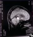
Systemic Disorders With Optic Nerve and Retinal Findings | Larry P. Frohman, MD | This 1-year-old child with familial erythrophagocytic lymphohistiocytosis was readmitted with a fever and was noted to have bilateral blindness. The spinal tap showed a protein of 148, with 178 WBC with 98% ""lymphocytes."" This MRI image demonstrates the optic nerve infiltration. He was treated wit... |
| 173 |
 |
Optic Tract Syndrome Due to Carotid Artery Dolichoectasia | Larry P. Frohman, MD | This 43-year-old man was referred for evaluation of 6 months of visual loss OU. In retrospect, he had noticed increasing difficulty with his tennis game dating back over 3 years, as balls would pass him unexpectedly when hit to his backhand (left) side. The patient did not think this was progressive... |
| 174 |
 |
Neuro-Ophthalmic Consequences of Therapy | Larry P. Frohman, MD | This woman presented at age 52, 3 years after radiation therapy for a salivary gland carcinoma extending into the right maxillary sinus. She had received 6000 rads in 30 fractions over 45 days. She presented with 3 weeks of visual loss, with acuity of 20/30, normal color plates, normal fields, and n... |
| 175 |
 |
Neuro-Ophthalmic Vascular Disease | Larry P. Frohman, MD | This 23-year-old woman has had insulin-dependent diabetes mellitus since age 3. She was diagnosed with Sydenham's chorea in early childhood and had grand mal seizures from age 13 to 15. She has been hypertensive since age 18. Her vision was 20/25 OD and 20/40 OS, with dyschromatopsia OS, and a 1.8 l... |
| 176 |
 |
Neuro-Ophthalmic Vascular Disease | Larry P. Frohman, MD | This 23-year-old woman has had insulin-dependent diabetes mellitus since age 3. She was diagnosed with Sydenham's chorea in early childhood and had grand mal seizures from age 13 to 15. She has been hypertensive since age 18. Her vision was 20/25 OD and 20/40 OS, with dyschromatopsia OS, and a 1.8 l... |
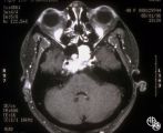
| 177 |
 |
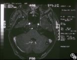
Neuro-Ophthalmic Imaging-CT Scan | Larry P. Frohman, MD | This 70-year-old woman sustained traumatic optic neuropathy in a motor vehicle accident. Note the funnel-shaped hemorrhage within the optic nerve sheath just posterior to the globe. |
| 178 |
 |
Systemic Disorders With Optic Nerve and Retinal Findings | Larry P. Frohman, MD | This 48-year-old female was seen in May 1996 with a history of 2 months of diplopia from a right abducens palsy. This was due to the recurrence of myeloma that had initially been diagnosed and treated with radiation and chemotherapy 9 years before and required further therapy, including bone marrow ... |
| 179 |
 |
Systemic Disorders With Optic Nerve and Retinal Findings | Larry P. Frohman, MD | This 1-year-old child with familial erythrophagocytic lymphohistiocytosis was readmitted with a fever and was noted to have bilateral blindness. The spinal tap showed a protein of 148, with 178 WBC with 98% ""lymphocytes."" This MRI image demonstrates the optic nerve infiltration. He was treated wit... |
| 180 |
 |
Chiasmal Syndromes | Larry P. Frohman, MD | This 36-year-old woman presented in 1988 with 3 weeks of vertical binocular diplopia. She was a known amblyope OD. Her examination was notable for a right hyperdeviation of 1 PD present in right gaze and a subtle left noncongruous homonymous field defect. She was lost to follow-up, but 5 years later... |
| 181 |
 |
Ocular Manifestations of Congenital/Inherited Diseases | Larry P. Frohman, MD | This 14-year-old boy presented with sudden visual loss of the right eye that occurred 3 weeks before and due to a central retinal vein occlusion. His ocular history was quite complicated. He had had a resection of a lymphangioma of the left upper lid at age 7 months and underwent left orbitotomy for... |
| 182 |
 |
Ocular Manifestations of Congenital/Inherited Diseases | Larry P. Frohman, MD | This 14-year-old boy presented with sudden visual loss of the right eye that occurred 3 weeks before and due to a central retinal vein occlusion. His ocular history was quite complicated. He had had a resection of a lymphangioma of the left upper lid at age 7 months and underwent left orbitotomy for... |
| 183 |
 |
Systemic Disorders With Optic Nerve and Retinal Findings | Larry P. Frohman, MD | This 1-year-old child with familial erythrophagocytic lymphohistiocytosis was readmitted with a fever and was noted to have bilateral blindness. The spinal tap showed a protein of 148, with 178 WBC with 98% ""lymphocytes."" This MRI image demonstrates the optic nerve infiltration. He was treated wit... |
| 184 |
 |
Orbital Tumors | Larry P. Frohman, MD | This 30-year-old man had a retrobulbar intraconal mass OS. The CT scans showed a heterogeneous lobulated enhancing mass, 2.2 x 1.9 x 1.8 cm. The case beautifully exhibits chorodial folds. The ultrasound showed internal reflectivity. The patient refused surgery. Pair with Images 97_60, 97_61, 97_62, ... |
| 185 |
 |
Orbital Tumors | Larry P. Frohman, MD | This 30-year-old man had a retrobulbar intraconal mass OS. The CT scans showed a heterogeneous lobulated enhancing mass, 2.2 x 1.9 x 1.8 cm. The case beautifully exhibits chorodial folds. The ultrasound showed internal reflectivity. The patient refused surgery. Pair with Images 97_60, 97_61, 97_62, ... |
| 186 |
 |
Isolated Congenital Optic Disc Anomalies | Larry P. Frohman, MD | This 63-year-old man with amblyopia OD was seen for a question of ischemic optic neuropathy with a pale, swollen disc OD. The correct diagnosis is an exophytic capillary angioma of the optic nerve head. Disease/Diagnosis: Capillary Angioma. |
| 187 |
 |
Neuro-Ophthalmic Imaging-CT Scan | Larry P. Frohman, MD | This patient was assaulted with a blunt object and suffered acute blindness due to traumatic optic neuropathy. Note how the lateral orbital wall has been fractured and displaced posteromedially into the region of the anterior optic canal. |
| 188 |
 |
Optic Tract Syndrome Due to Carotid Artery Dolichoectasia | Larry P. Frohman, MD | This 43-year-old man was referred for evaluation of 6 months of visual loss OU. In retrospect, he had noticed increasing difficulty with his tennis game dating back over 3 years, as balls would pass him unexpectedly when hit to his backhand (left) side. The patient did not think this was progressive... |
| 189 |
 |
Optic Tract Syndrome Due to Carotid Artery Dolichoectasia | Larry P. Frohman, MD | This 43-year-old man was referred for evaluation of 6 months of visual loss OU. In retrospect, he had noticed increasing difficulty with his tennis game dating back over 3 years, as balls would pass him unexpectedly when hit to his backhand (left) side. The patient did not think this was progressive... |
| 190 |
 |
Optic Tract Syndrome Due to Carotid Artery Dolichoectasia | Larry P. Frohman, MD | This 43-year-old man was referred for evaluation of 6 months of visual loss OU. In retrospect, he had noticed increasing difficulty with his tennis game dating back over 3 years, as balls would pass him unexpectedly when hit to his backhand (left) side. The patient did not think this was progressive... |
| 191 |
 |
Systemic Disorders With Optic Nerve and Retinal Findings | Larry P. Frohman, MD | This 8-year-old girl had a history of acute lymphoblastic leukemia with neurologic involvement, with remission induced with radiation and chemotherapy. Her relapse showed as an isolated optic neuropathy with this fundus appearance. The other eye was normal. The MRI and spinal tap at this point were ... |
| 192 |
 |
Systemic Disorders With Optic Nerve and Retinal Findings | Larry P. Frohman, MD | A 42-year old woman presented with a history of severe brow pain and 4 days of progressive visual loss OD. There was no increased pain on ocular rotation. Aside from heavy menses, she denied any significant past medical history. Her examination revealed acuity NLP OD, 20/25 OS; color vision 9/10 OS;... |
| 193 |
 |
Systemic Disorders With Optic Nerve and Retinal Findings | Larry P. Frohman, MD | A 42-year old woman presented with a history of severe brow pain and 4 days of progressive visual loss OD. There was no increased pain on ocular rotation. Aside from heavy menses, she denied any significant past medical history. Her examination revealed acuity NLP OD, 20/25 OS; color vision 9/10 OS;... |
| 194 |
 |
Systemic Disorders With Optic Nerve and Retinal Findings | Larry P. Frohman, MD | A 42-year old woman presented with a history of severe brow pain and 4 days of progressive visual loss OD. There was no increased pain on ocular rotation. Aside from heavy menses, she denied any significant past medical history. Her examination revealed acuity NLP OD, 20/25 OS; color vision 9/10 OS;... |
| 195 |
 |
Systemic Disorders With Optic Nerve and Retinal Findings | Larry P. Frohman, MD | A 42-year old woman presented with a history of severe brow pain and 4 days of progressive visual loss OD. There was no increased pain on ocular rotation. Aside from heavy menses, she denied any significant past medical history. Her examination revealed acuity NLP OD, 20/25 OS; color vision 9/10 OS;... |
| 196 |
 |
Systemic Disorders With Optic Nerve and Retinal Findings | Larry P. Frohman, MD | A 42-year old woman presented with a history of severe brow pain and 4 days of progressive visual loss OD. There was no increased pain on ocular rotation. Aside from heavy menses, she denied any significant past medical history. Disease/Diagnosis: Syphilitic optic neuritis/perineuritis, fundus, OS -... |
| 197 |
 |
Systemic Disorders With Optic Nerve and Retinal Findings | Larry P. Frohman, MD | This is a 32-year-old HIV-positive man with anterior uveitis, vitritis, and bilateral papillitis from syphilis. With intravenous penicillin treatment, the optic discs and vision returned to normal. |
| 198 |
 |
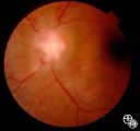
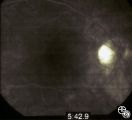
Chiasmal Syndromes | Larry P. Frohman, MD | This 39-year-old HIV-positive man presented in 1982 with 1 month of bilateral vision loss. His prior evaluation had included 2 CT scans, which suggested a fullness to the chiasm, and a spinal tap that showed 6 monocytes, 14 red cells, a protein of 81 mg/dl, and a glucose of 56 mg/dl, with a negative... |
| 199 |
 |
Systemic Disorders With Optic Nerve and Retinal Findings | Larry P. Frohman, MD | This 25-year-old man presented to the eye service with a history of 3 days of decreased vision OD. His past medical history was unremarkable. His examination showed acuities of 20/25 OU, with intact color plates, a 0.3 log unit of RAPD OD, and an inferior arcuate scotoma. The photos (Images 95_42, 9... |
| 200 |
 |
Systemic Disorders With Optic Nerve and Retinal Findings | Larry P. Frohman, MD | A 29-year-old African American woman presented with headaches, bilateral transient visual obscurations, blurred vision, numbness, and weakness of the lower extremities with myalgia and joint pains. She had an unplanned 12-pound weight loss over 2 months. A neurologist and internist diagnosed her wit... |