AAO-NANOS Neuro-Ophthalmology Clinical Collection: Derived from the AAO-NANOS Clinical Neuro-Ophthalmology collection produced on CD. The images are of selected cases from the NANOS teaching slide exchange, and the CD was produced under the direction of Larry Frohman, MD and Andrew Lee, MD.
The American Academy of Ophthalmology (AAO); The North American Neuro-Ophthalmology Association (NANOS).
NOVEL: https://novel.utah.edu/
TO
Filters: Collection: "ehsl_novel_aao_nanos"
1 - 25 of 3
| Title | Description | Subject | ||
|---|---|---|---|---|
| 1 |
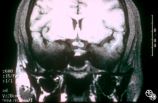 |
Neuro-Ophthalmic Imaging-MRI | This 23-year-old right-handed man had a history of idiopathic recurrent optic neuritis. The patient presented with acuity of 20/400 OD and 20/100 OS, with a central scotoma OD and a complete temporal defect OS. MRI with fat suppression and gadolinium revealed enhancement of the intracranial nerve an... | Chiasmal Optic Neuritis |
| 2 |
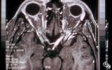 |
Neuro-Ophthalmic Imaging-MRI | This 23-year-old right-handed man had a history of idiopathic recurrent optic neuritis. The patient presented with acuity of 20/400 OD and 20/100 OS, with a central scotoma OD and a complete temporal defect OS. MRI with fat suppression and gadolinium revealed enhancement of the intracranial nerve an... | Chiasmal Optic Neuritis |
| 3 |
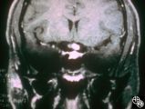 |
Neuro-Ophthalmic Imaging-MRI | This 23-year-old right-handed man had a history of idiopathic recurrent optic neuritis. The patient presented with acuity of 20/400 OD and 20/100 OS, with a central scotoma OD and a complete temporal defect OS. MRI with fat suppression and gadolinium revealed enhancement of the intracranial nerve an... | Chiasmal Optic Neuritis |
1 - 25 of 3