AAO-NANOS Neuro-Ophthalmology Clinical Collection: Derived from the AAO-NANOS Clinical Neuro-Ophthalmology collection produced on CD. The images are of selected cases from the NANOS teaching slide exchange, and the CD was produced under the direction of Larry Frohman, MD and Andrew Lee, MD.
The American Academy of Ophthalmology (AAO); The North American Neuro-Ophthalmology Association (NANOS).
NOVEL: https://novel.utah.edu/
TO
Filters: Collection: "ehsl_novel_aao_nanos"
1 - 25 of 22
| Title | Creator | Description | ||
|---|---|---|---|---|
| 1 |
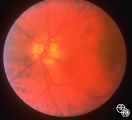
 |
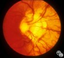
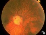
Isolated Congenital Optic Disc Anomalies | Thomas R. Wolf, MD | Benign tumors of blood vessels (hemangiomas) may occur on the optic nerve and may mimic optic disc edema. Disease/Diagnosis: Optic Nerve Hemangioma. |
| 2 |
 |
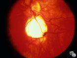
Isolated Congenital Optic Disc Anomalies | Thomas R. Wolf, MD | Patients with midline closure defects may exhibit abnormalities in the optic nerve, choroid, retinal pigment epithelium or retina. Anterior closure defects may result in colobomas of the structures of the anterior segment, such as the iris. Disease/Diagnosis: Coloboma. |
| 3 |
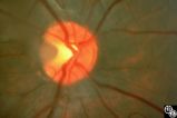 |
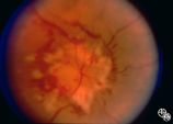
Isolated Congenital Optic Disc Anomalies | Anthony C. Arnold, MD | A 10-year-old girl had central visual loss due to this optic pit. Disease/Diagnosis: Optic Pit. |
| 4 |
 |
Retinal Coloboma Underneath a Relatively Normal Optic Nerve | Thomas R. Wolf, MD | Optic nerve colobomas appear as enlarged, white optic discs that are deeply excavated, often with some sapring of the superior rim. They result from an abnormal fusion of the proximal embryonic fissure. Optic nerve colobomas occur unilaterally or bilaterally with a similar frequency and can result i... |
| 5 |
 |
Systemic Disorders With Optic Nerve and Retinal Findings | Rosa A. Tang, MD | Neoplasms may result in an optic neuropathy by direct metastatic involvement. In this patient, a lung adenocarcinoma was metastatic to the optic nerve.This is a fundus photo. |
| 6 |
 |
Systemic Disorders With Optic Nerve and Retinal Findings | Larry P. Frohman, MD | A patient with small-cell lung carcinoma that was metastatic to the optic nerves, ciliary body, and brain. This is a fundus photo. |
| 7 |
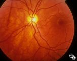 |
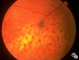
Isolated Congenital Optic Disc Anomalies | Thomas R. Wolf, MD | Patients with hypoplasia of the optic nerve may have normal or subnormal visual acuity or visual field. The condition may be unilateral or bilateral. Optic nerve hypoplasia is usually idiopathic, but maternal diabetes, or maternal use of anti-epileptic drugs or alcohol are predisposing factors. Opti... |
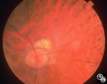
| 8 |
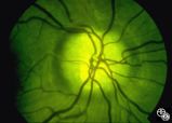 |
Isolated Congenital Optic Disc Anomalies | Thomas R. Wolf, MD | This optic disc displays multiple drusen. Note the pseudopapilledema here. One can differentiate this from true papilledema in that there is no obscuration of the vessel by the peripapillary nerve fiber layer as they cross the disc margin. This photograph was taken with barrier filters in place, but... |
| 9 |
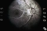
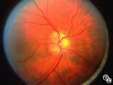 |
Isolated Congenital Optic Disc Anomalies | Anthony C. Arnold, MD | This image shows drusen that are especially prominent superotemporally. Pair with 92_64 and 92_67. |
| 10 |
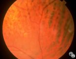
 |
Isolated Congenital Optic Disc Anomalies | Anthony C. Arnold, MD | This is a photograph of peripheral drusen. The paired image 92_69 demonstrates the typical autofluorescence. |
| 11 |
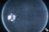 |
Optic Disc Drusen Autofluorescence | Anthony C. Arnold, MD | This case of optic disc drusen demonstrates the typical autofluorescence. Pair with 92_68. Imaging: Autoflourescence? |
| 12 |
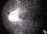
 |
Systemic Disorders With Optic Nerve and Retinal Findings | Robert L. Lesser, MD | Intraocular lymphoma may present with an unexplained vitritis, optic disc infiltration, or choroidal infiltration. One unusual manifestation of large-cell lymphoma is this leopard-spot appearance. Pair with 94_32, 94_33, and 94_35. This is a fundus photo. |
| 13 |
 |
Systemic Disorders With Optic Nerve and Retinal Findings | Robert L. Lesser, MD | Intraocular lymphoma may present with an unexplained vitritis, optic disc infiltration, or choroidal infiltration. One unusual manifestation of large-cell lymphoma is this leopard-spot appearance. Pair with 94_32, 94_34, and 94_35. This is a fundus photo. |
| 14 |
 |
Systemic Disorders With Optic Nerve and Retinal Findings | Robert L. Lesser, MD | Intraocular lymphoma may present with an unexplained vitritis, optic disc infiltration, or choroidal infiltration. One unusual manifestation of large-cell lymphoma is this leopard-spot appearance. Pair with 94_33, 94_34, and 94_35. This is a fundus photo. |
| 15 |
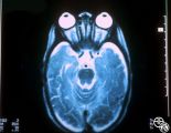 |
Systemic Disorders With Optic Nerve and Retinal Findings | Larry P. Frohman, MD | This 1-year-old child with familial erythrophagocytic lymphohistiocytosis was readmitted with a fever and was noted to have bilateral blindness. The spinal tap showed a protein of 148, with 178 WBC with 98% ""lymphocytes."" This MRI image demonstrates the optic nerve infiltration. He was treated wit... |
| 16 |
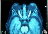 |
Systemic Disorders With Optic Nerve and Retinal Findings | Larry P. Frohman, MD | This 1-year-old child with familial erythrophagocytic lymphohistiocytosis was readmitted with a fever and was noted to have bilateral blindness. The spinal tap showed a protein of 148, with 178 WBC with 98% ""lymphocytes."" This MRI image demonstrates the optic nerve infiltration. He was treated wit... |
| 17 |
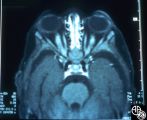 |
Systemic Disorders With Optic Nerve and Retinal Findings | Larry P. Frohman, MD | This 1-year-old child with familial erythrophagocytic lymphohistiocytosis was readmitted with a fever and was noted to have bilateral blindness. The spinal tap showed a protein of 148, with 178 WBC with 98% ""lymphocytes."" This MRI image demonstrates the optic nerve infiltration. He was treated wit... |
| 18 |
 |
Isolated Congenital Optic Disc Anomalies | Rosa A. Tang, MD | This patient has optic disc drusen and evidence of a superimposed optic neuropathy, including loss of visual field, an ipsilateral afferent pupillary defect, and optic atrophy. Although optic disc drusen typically causes visual field loss without visual acuity loss superimposed, ischemic optic neuro... |
| 19 |
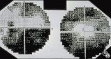 |
Optic Disc Drusen Visual Fields | Thomas R. Wolf, MD | This is the visual field of patient with optic nerve drusen. Whereas they typically do not cause central field loss, optic disc drusen may cause nerve fiber bundle layer defects and, thus, peripheral field defects, including altitudinal defects (seen inferiorly in the left eye) or arcuate defects (s... |
| 20 |
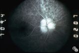
 |
Optic Disc Drusen, Fluorescein Angiogram | Anthony C. Arnold, MD | Images 92_64 and 92_67 demonstrate the characteristics of optic disc drusen on flourescein angiography. This image shows the early arteriovenous phase, with irregular dye uptake and focal hypoflourescence superotemporally. Pair with 92_63 and 92_67 |
| 21 |
 |
Optic Nerve Drusen, Late Fluorescein Angiogram | Anthony C. Arnold, MD | Images 92_64 and 92_67 demonstrate the characteristics of optic disc drusen on flourescein angiography. This image displays the nodular staining of the drusen without leakage. Pair with 92_63 and 92_64. |
| 22 |
 |
Systemic Disorders With Optic Nerve and Retinal Findings | Robert L. Lesser, MD | Intraocular lymphoma may present with an unexplained vitritis, optic disc infiltration, or choroidal infiltration. One unusual manifestation of large-cell lymphoma is this leopard-spot appearance. Pair with 94_32, 94_33, and 94_34. This is a fluorescein angiogram. |
1 - 25 of 22