AAO-NANOS Neuro-Ophthalmology Clinical Collection: Derived from the AAO-NANOS Clinical Neuro-Ophthalmology collection produced on CD. The images are of selected cases from the NANOS teaching slide exchange, and the CD was produced under the direction of Larry Frohman, MD and Andrew Lee, MD.
The American Academy of Ophthalmology (AAO); The North American Neuro-Ophthalmology Association (NANOS).
NOVEL: https://novel.utah.edu/
TO
Filters: Collection: "ehsl_novel_aao_nanos"
| Title | Creator | Description | ||
|---|---|---|---|---|
| 1 |
 | Motility Disturbances | Larry P. Frohman, MD | This patient sustained a traumatic avulsion of the left medial rectus. |
| 2 |
 | Motility Disturbances | Larry P. Frohman, MD | This patient sustained a traumatic avulsion of the left medial rectus. Image 94_75 shows the successful postoperative result. |
| 3 |
 | Motility Disturbances | Larry P. Frohman, MD | This man had a posttraumatic right sixth nerve paresis. Image 94_66 demonstrates the adduction deficit that the Botox induced. |
| 4 |
 | Motility Disturbances | Larry P. Frohman, MD | This man had a posttraumatic right sixth nerve paresis. He is shown in primary gaze after Botox (image 94_65). |
| 5 |
 | Motility Disturbances | Larry P. Frohman, MD | This patient sustained a traumatic avulsion of the left medial rectus. |
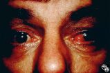
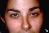
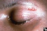
| 6 |
 | Motility Disturbances | Larry P. Frohman, MD | This young woman had bilateral sixth nerve paresis from a motor vehicle accident. The images show the results of a successful Jensen procedure. |
| 7 |
 | Motility Disturbances | Larry P. Frohman, MD | This young woman had bilateral sixth nerve paresis from a motor vehicle accident. The images show the results of a successful Jensen procedure. |
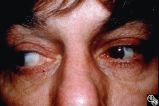
| 8 |
 | Motility Disturbances | Larry P. Frohman, MD | This man had a posttraumatic right sixth nerve paresis. He is shown in primary gaze before Botox (botulinum toxin; image 94_64) |
| 9 |
 | Neuro-Ophthalmic Imaging-CT Scan | Larry P. Frohman, MD | This 70-year-old woman sustained traumatic optic neuropathy in a motor vehicle accident. Note the funnel-shaped hemorrhage within the optic nerve sheath just posterior to the globe. |
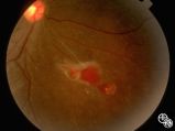
| 10 |
 | Systemic Disorders With Optic Nerve and Retinal Findings | Larry P. Frohman, MD | A patient with small-cell lung carcinoma that was metastatic to the optic nerves, ciliary body, and brain. This is a fundus photo. |
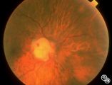
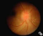
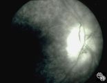
| 11 |
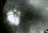 | Isolated Congenital Optic Disc Anomalies | Larry P. Frohman, MD | This 63-year-old man with amblyopia OD was seen for a question of ischemic optic neuropathy with a pale, swollen disc OD. The correct diagnosis is an exophytic capillary angioma of the optic nerve head. Disease/Diagnosis: Capillary Angioma. |
| 12 |
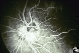 | Isolated Congenital Optic Disc Anomalies | Larry P. Frohman, MD | This 63-year-old man with amblyopia OD was seen for a question of ischemic optic neuropathy with a pale, swollen disc OD. The correct diagnosis is an exophytic capillary angioma of the optic nerve head. Disease/Diagnosis: Capillary Angioma. |
| 13 |
 | Neuro-Ophthalmic Imaging-CT Scan | Larry P. Frohman, MD | This patient was assaulted with a blunt object and suffered acute blindness due to traumatic optic neuropathy. Note how the lateral orbital wall has been fractured and displaced posteromedially into the region of the anterior optic canal. |
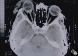
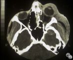
| 14 |
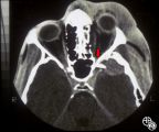 | Neuro-Ophthalmic Imaging-CT Scan | Larry P. Frohman, MD | This patient was assaulted with a blunt object and suffered acute blindness due to traumatic optic neuropathy. Note how the lateral orbital wall has been fractured and displaced posteromedially into the region of the anterior optic canal. |
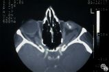
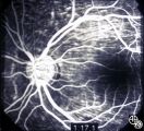
| 15 |
 | Ocular Manifestations of Congenital/Inherited Diseases | Larry P. Frohman, MD | On optic nerve CT scan, this patient with neurofibromatosis, type 1, shows the classic railroad-track sign of optic nerve meningioma and the kink sign of optic nerve glioma. Disease/Diagnosis: Neurofibromatosis, Type 1. |
| 16 |
 | Systemic Disorders With Optic Nerve and Retinal Findings | Larry P. Frohman, MD | This is a 32-year-old HIV-positive man with anterior uveitis, vitritis, and bilateral papillitis from syphilis. With intravenous penicillin treatment, the optic discs and vision returned to normal. |
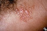
| 17 |
 | Systemic Disorders With Optic Nerve and Retinal Findings | Larry P. Frohman, MD | Skin rashes occur in about 30 percent of patients with sarcoid. When seen, the rashes offer an accessible site for obtaining histologic material for confirmation of the clinical diagnosis. Pair with 91_68. |
| 18 |
 | Systemic Disorders With Optic Nerve and Retinal Findings | Larry P. Frohman, MD | Skin rashes occur in about 30 percent of patients with sarcoid. When seen, the rashes offer an accessible site for obtaining histologic material for confirmation of the clinical diagnosis. Pair with 91_69. |
| 19 |
 | Systemic Disorders With Optic Nerve and Retinal Findings | Larry P. Frohman, MD | This is a 32-year-old HIV-positive man with anterior uveitis, vitritis, and bilateral papillitis from syphilis. With intravenous penicillin treatment, the optic discs and vision returned to normal. |
| 20 |
 | Neuro-Ophthalmic Consequences of Therapy | Larry P. Frohman, MD | This woman presented at age 52, 3 years after radiation therapy for a salivary gland carcinoma extending into the right maxillary sinus. She had received 6000 rads in 30 fractions over 45 days. She presented with 3 weeks of visual loss, with acuity of 20/30, normal color plates, normal fields, and n... |
| 21 |
 | Neuro-Ophthalmic Consequences of Therapy | Larry P. Frohman, MD | This woman presented at age 52, 3 years after radiation therapy for a salivary gland carcinoma extending into the right maxillary sinus. She had received 6000 rads in 30 fractions over 45 days. She presented with 3 weeks of visual loss, with acuity of 20/30, normal color plates, normal fields, and n... |
| 22 |
 | Neuro-Ophthalmic Consequences of Therapy | Larry P. Frohman, MD | This woman presented at age 52, 3 years after radiation therapy for a salivary gland carcinoma extending into the right maxillary sinus. She had received 6000 rads in 30 fractions over 45 days. She presented with 3 weeks of visual loss, with acuity of 20/30, normal color plates, normal fields, and n... |
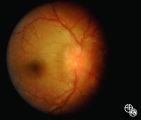
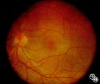
| 23 |
 | Ocular Manifestations of Systemic Disorders | Larry P. Frohman, MD | The same young woman from Case 77 (Image 92_57) presented 8 years after the orbital biopsy with this retinal lesion, presumably from her underlying disorder. The eye has 20/20 vision and no other disturbances. This is a fundus photo. |
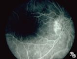
| 24 |
 | Orbital Tumors | Larry P. Frohman, MD | This 30-year-old man had a retrobulbar intraconal mass OS. The CT scans showed a heterogeneous lobulated enhancing mass, 2.2 x 1.9 x 1.8 cm. The case beautifully exhibits chorodial folds. The ultrasound showed internal reflectivity. The patient refused surgery. Pair with Images 97_60, 97_62, 97_63, ... |
| 25 |
 | Orbital Tumors | Larry P. Frohman, MD | This 30-year-old man had a retrobulbar intraconal mass OS. The CT scans showed a heterogeneous lobulated enhancing mass, 2.2 x 1.9 x 1.8 cm. The case beautifully exhibits chorodial folds. The ultrasound showed internal reflectivity. The patient refused surgery. Pair with Images 97_60, 97_61, 97_63, ... |