AAO-NANOS Neuro-Ophthalmology Clinical Collection: Derived from the AAO-NANOS Clinical Neuro-Ophthalmology collection produced on CD. The images are of selected cases from the NANOS teaching slide exchange, and the CD was produced under the direction of Larry Frohman, MD and Andrew Lee, MD.
The American Academy of Ophthalmology (AAO); The North American Neuro-Ophthalmology Association (NANOS).
NOVEL: https://novel.utah.edu/
TO
Filters: Collection: "ehsl_novel_aao_nanos"
| Title | Creator | Description | ||
|---|---|---|---|---|
| 301 |
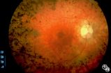 |
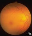
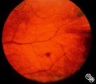
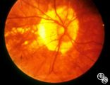
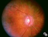
Optic Neuropathies | Rosa A. Tang, MD | Optic atrophy might be a false-localizing sign of optic nerve disease, as retinal disease may secondarily produce optic atrophy. In retinitis pigmentosa, for example, patients may exhibit a waxy disc palor. Fundus imaging. Anatomy: Retina. Disease/Diagnosis: Retinitis pigmentosa; Optic atrophy. |
| 302 |
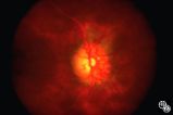 |
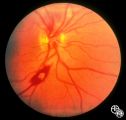
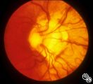
Acquired Disc Changes | Rosa A. Tang, MD | Optociliary shunt vessels are venous collaterals that form after chronic venous obstruction. The presence of optic atrophy, progressive visual loss, and optociliary shunt vessels may indicate a compressive optic nerve lesion such as meningioma. |
| 303 |
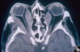 |
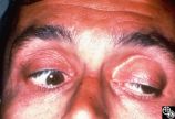
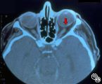
Ocular Manifestations of Systemic Disorders | Rosa A. Tang, MD | Thyroid eye disease may result in proptosis and restrictive external ophthalmoplegia. The extracoular muscles are often diffusely enlarged with sparing of the tendons. |
| 304 |
 |
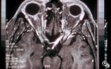
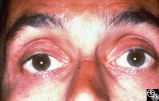
Ocular Manifestations of Systemic Disorders | Rosa A. Tang, MD | Thyroid eye disease may cause proptosis and extraocular muscle enlargement that may be seen on orbital imaging studies. In general, coronal images allow the best visualization of the extraocular muscle enlargement. Pair with 94_45 and 94_46. |
| 305 |
 |
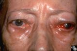
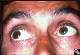
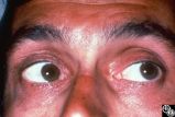
Ocular Manifestations of Systemic Disorders | Rosa A. Tang, MD | Thyroid eye disease can result in significant upper eyelid retraction and axial proptosis resulting in exposure keratopathy. |
| 306 |
 |
Isolated Congenital Optic Disc Anomalies | Rosa A. Tang, MD | Normally, there is no visible myelination of the nerve fiber layer in the retina. Occasionally, visible myelination occurs that takes on a characteristic white, arcuate, feathery appearance that follows the contour of the nerve fiber layer. Disease/Diagnosis: Myelinated Nerve Fiber. |
| 307 |
 |
Acquired Disc Changes | Rosa A. Tang, MD | Although optociliary shunt vessels are venous collaterals that form in response to chronic venous obstruction, they may occur in patients with chronic open-angle glaucoma. |
| 308 |
 |
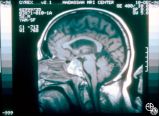
Neuro-Ophthalmic Imaging-MRI | Scott Forman, MD | This 23-year-old right-handed man had a history of idiopathic recurrent optic neuritis. The patient presented with acuity of 20/400 OD and 20/100 OS, with a central scotoma OD and a complete temporal defect OS. MRI with fat suppression and gadolinium revealed enhancement of the intracranial nerve an... |
| 309 |
 |
Neuro-Ophthalmic Imaging-MRI | Scott Forman, MD | This 23-year-old right-handed man had a history of idiopathic recurrent optic neuritis. The patient presented with acuity of 20/400 OD and 20/100 OS, with a central scotoma OD and a complete temporal defect OS. MRI with fat suppression and gadolinium revealed enhancement of the intracranial nerve an... |
| 310 |
 |
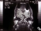
Neuro-Ophthalmic Imaging-MRI | Scott Forman, MD | This 23-year-old right-handed man had a history of idiopathic recurrent optic neuritis. The patient presented with acuity of 20/400 OD and 20/100 OS, with a central scotoma OD and a complete temporal defect OS. MRI with fat suppression and gadolinium revealed enhancement of the intracranial nerve an... |
| 311 |
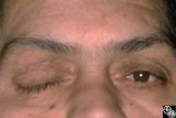 |
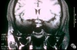
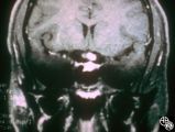
Chiasmal Syndromes | Shlomo A. Dotan, MD | A 52-year-old, morbidly obese man with a past medical history that included ischemic cardiac disease with a history of angioplasty, COPD, hypertension, and NIDDM, presented with a severe headache. The next day he had a frozen OD, complete right ptosis, and an unreactive middilated right pupil with V... |
| 312 |
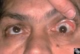 |
Chiasmal Syndromes | Shlomo A. Dotan, MD | A 52-year-old, morbidly obese man with a past medical history that included ischemic cardiac disease with a history of angioplasty, COPD, hypertension, and NIDDM, presented with a severe headache. The next day he had a frozen OD, complete right ptosis, and an unreactive middilated right pupil with V... |
| 313 |
 |
Chiasmal Syndromes | Shlomo A. Dotan, MD | A 52-year-old, morbidly obese man with a past medical history that included ischemic cardiac disease with a history of angioplasty, COPD, hypertension, and NIDDM, presented with a severe headache. The next day he had a frozen OD, complete right ptosis, and an unreactive middilated right pupil with V... |
| 314 |
 |
Chiasmal Syndromes | Shlomo A. Dotan, MD | A 52-year-old, morbidly obese man with a past medical history that included ischemic cardiac disease with a history of angioplasty, COPD, hypertension, and NIDDM, presented with a severe headache. The next day he had a frozen OD, complete right ptosis, and an unreactive middilated right pupil with V... |
| 315 |
 |
Chiasmal Syndromes | Shlomo A. Dotan, MD | A 52-year-old, morbidly obese man with a past medical history that included ischemic cardiac disease with a history of angioplasty, COPD, hypertension, and NIDDM, presented with a severe headache. The next day he had a frozen OD, complete right ptosis, and an unreactive middilated right pupil with V... |
| 316 |
 |
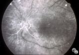
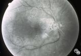
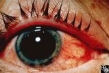
Neuro-Ophthalmic Vascular Disease | Steven A. Newman, MD | Occlusion of a branch or central retinal artery may result in acute visual loss. The ophthalmoscopic findings are retinal whitening due to ischemic retina in the distribution of the occluded artery. Sparing or selective involvement of cilioretinal artery branches may occur. Patients with a central r... |
| 317 |
 |
Neuro-Ophthalmic Vascular Disease | Steven A. Newman, MD | Occlusion of a branch or central retinal artery may result in acute visual loss. The ophthalmoscopic findings are retinal whitening due to ischemic retina in the distribution of the occluded artery. Sparing or selective involvement of cilioretinal artery branches may occur. Patients with a central r... |
| 318 |
 |
Neuro-Ophthalmic Vascular Disease | Steven A. Newman, MD | Occlusion of a branch or central retinal artery may result in acute visual loss. The ophthalmoscopic findings are retinal whitening due to ischemic retina in the distribution of the occluded artery. Sparing or selective involvement of cilioretinal artery branches may occur. Patients with a central r... |
| 319 |
 |
Neuro-Ophthalmic Vascular Disease | Steven A. Newman, MD | Occlusion of a branch or central retinal artery may result in acute visual loss. The ophthalmoscopic findings are retinal whitening due to ischemic retina in the distribution of the occluded artery. Sparing or selective involvement of cilioretinal artery branches may occur. Patients with a central r... |
| 320 |
 |
Traumatic Internal Carotid Artery Dissection | Steven Galetta, MD | Traumatic dissection of the carotid artery may result in neck pain, an ipsilateral Horner's syndrome (disruption of the pericarotid sympathetic fibers), or ipsilateral arterial occlusions from embolic disease. Pair with images 91_18 and 91_20. |
| 321 |
 |
Traumatic Internal Carotid Atery Dissection | Steven Galetta, MD | Traumatic dissection of the carotid artery may result in neck pain, an ipsilateral Horner's syndrome (disruption of the pericarotid sympathetic fibers), or ipsilateral arterial occlusions from embolic disease. Pair with images 91_19 and 91_20. |
| 322 |
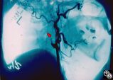 |
Traumatic Internal Carotid Artery Dissection | Steven Galetta, MD | Traumatic dissection of the carotid artery may result in neck pain, an ipsilateral Horner's syndrome (disruption of the pericarotid sympathetic fibers), or ipsilateral arterial occlusions from embolic disease. Pair with images 91_18 and 91_19. |
| 323 |
 |
Ocular Manifestations of Congenital/Inherited Diseases | Steven Galetta, MD | This patient is a 40-year-old man with a history of abetalipoproteinemia (Bassen-Kornweig syndrome), diagnosed at age 9. Neurologic complications have included ataxia, retinal degeneration, peripheral neuropathy, progressive leg weakness, dysarthria, and intermittent bladder incontinence. On his neu... |
| 324 |
 |
Ocular Manifestations of Congenital/Inherited Diseases | Steven Galetta, MD | This patient is a 40-year-old man with a history of abetalipoproteinemia (Bassen-Kornweig syndrome), diagnosed at age 9. Neurologic complications have included ataxia, retinal degeneration, peripheral neuropathy, progressive leg weakness, dysarthria, and intermittent bladder incontinence. On his neu... |
| 325 |
 |
Ocular Manifestations of Congenital/Inherited Diseases | Steven Galetta, MD | This patient is a 40-year-old man with a history of abetalipoproteinemia (Bassen-Kornweig syndrome), diagnosed at age 9. Neurologic complications have included ataxia, retinal degeneration, peripheral neuropathy, progressive leg weakness, dysarthria, and intermittent bladder incontinence. On his neu... |
| 326 |
 |
Ocular Manifestations of Congenital/Inherited Diseases | Steven Galetta, MD | This patient is a 40-year-old man with a history of abetalipoproteinemia (Bassen-Kornweig syndrome), diagnosed at age 9. Neurologic complications have included ataxia, retinal degeneration, peripheral neuropathy, progressive leg weakness, dysarthria, and intermittent bladder incontinence. On his neu... |
| 327 |
 |
Ocular Manifestations of Congenital/Inherited Diseases | Steven Galetta, MD | This patient is a 40-year-old man with a history of abetalipoproteinemia (Bassen-Kornweig syndrome), diagnosed at age 9. Neurologic complications have included ataxia, retinal degeneration, peripheral neuropathy, progressive leg weakness, dysarthria, and intermittent bladder incontinence. On his neu... |
| 328 |
 |
Ocular Manifestations of Congenital/Inherited Diseases | Steven Galetta, MD | This patient is a 40-year-old man with a history of abetalipoproteinemia (Bassen-Kornweig syndrome), diagnosed at age 9. Neurologic complications have included ataxia, retinal degeneration, peripheral neuropathy, progressive leg weakness, dysarthria, and intermittent bladder incontinence. On his neu... |
| 329 |
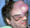 |
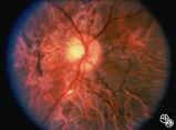
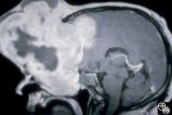
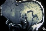
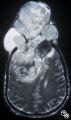
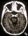
Neuro-Ophthalmic Manifestations of Brain Tumors | Steven Galetta, MD | The patient is a 45-year-old recluse found to harbor this frontal lobe mass. Remarkably, this patient had only mild bilateral optic neuropathies with visual acuities in the 20/25 range. This right disc was mildly swollen and the left mildly pale. He could not fit into the fundus camera for disc phot... |
| 330 |
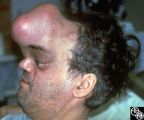 |
Neuro-Ophthalmic Manifestations of Brain Tumors | Steven Galetta, MD | The patient is a 45-year-old recluse found to harbor this frontal lobe mass. Remarkably, this patient had only mild bilateral optic neuropathies with visual acuities in the 20/25 range. This right disc was mildly swollen and the left mildly pale. He could not fit into the fundus camera for disc phot... |
| 331 |
 |
Ocular Manifestations of Congenital/Inherited Diseases | Steven Galetta, MD | This 21-year-old woman had a 2-year history of blurred vision. A computerized visual field demonstrated a temporal defect OS. MRI confirmed a chiasmal mass lesion. The pathology was consistent with hemangioblastoma. Further workup revealed retinal angiomas and multiple other hemangioblastomas of the... |
| 332 |
 |
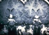
Neuro-Ophthalmic Manifestations of Brain Tumors | Steven Galetta, MD | The patient is a 45-year-old recluse found to harbor this frontal lobe mass. Remarkably, this patient had only mild bilateral optic neuropathies with visual acuities in the 20/25 range. This right disc was mildly swollen and the left mildly pale. He could not fit into the fundus camera for disc phot... |
| 333 |
 |
Neuro-Ophthalmic Manifestations of Brain Tumors | Steven Galetta, MD | The patient is a 45-year-old recluse found to harbor this frontal lobe mass. Remarkably, this patient had only mild bilateral optic neuropathies with visual acuities in the 20/25 range. This right disc was mildly swollen and the left mildly pale. He could not fit into the fundus camera for disc phot... |
| 334 |
 |
Neuro-Ophthalmic Manifestations of Brain Tumors | Steven Galetta, MD | The patient is a 45-year-old recluse found to harbor this frontal lobe mass. Remarkably, this patient had only mild bilateral optic neuropathies with visual acuities in the 20/25 range. This right disc was mildly swollen and the left mildly pale. He could not fit into the fundus camera for disc phot... |
| 335 |
 |
Neuro-Ophthalmic Manifestations of Brain Tumors | Steven Galetta, MD | The patient is a 45-year-old recluse found to harbor this frontal lobe mass. Remarkably, this patient had only mild bilateral optic neuropathies with visual acuities in the 20/25 range. This right disc was mildly swollen and the left mildly pale. He could not fit into the fundus camera for disc phot... |
| 336 |
 |
Ocular Manifestations of Congenital/Inherited Diseases | Steven Galetta, MD | This 21-year-old woman had a 2-year history of blurred vision. A computerized visual field demonstrated a temporal defect OS. MRI confirmed a chiasmal mass lesion. The pathology was consistent with hemangioblastoma. Further workup revealed retinal angiomas and multiple other hemangioblastomas of the... |
| 337 |
 |
Ocular Manifestations of Congenital/Inherited Diseases | Steven Galetta, MD | This 21-year-old woman had a 2-year history of blurred vision. A computerized visual field demonstrated a temporal defect OS. MRI confirmed a chiasmal mass lesion. The pathology was consistent with hemangioblastoma. Further workup revealed retinal angiomas and multiple other hemangioblastomas of the... |
| 338 |
 |
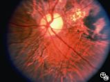
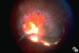
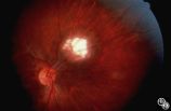
Systemic Disorders With Optic Nerve and Retinal Findings | Steven Galetta, MD | This fondus image shows a white-centered hemorrhage in a leukemia patient with orbital aspergillosis. |
| 339 |
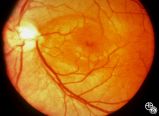 |
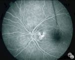
Isolated Congenital Optic Disc Anomalies | Thomas R. Wolf, MD | An optic pit is a small defect in the optic disc that may be asymptomatic in isolation. Patients may develop an associated serous detachment of the macula. The condition is usually unilateral but may be bilateral. A fluorescein angiogram may demonstrate the serous detachment, and laser photocoagulat... |
| 340 |
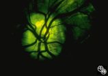 |
Optic Disc Drusen With Autofluorescence | Thomas R. Wolf, MD | This photograph of optic disc drusen demonstrates autoflourescence with flourescein barrier filters in place. Imaging: flourescein barrier filters. |
| 341 |
 |
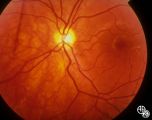
Isolated Congenital Optic Disc Anomalies | Thomas R. Wolf, MD | Benign tumors of blood vessels (hemangiomas) may occur on the optic nerve and may mimic optic disc edema. Disease/Diagnosis: Optic Nerve Hemangioma. |
| 342 |
 |
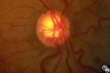
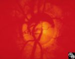
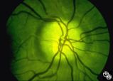
Isolated Congenital Optic Disc Anomalies | Thomas R. Wolf, MD | Patients with hypoplasia of the optic nerve may have normal or subnormal visual acuity or visual field. The condition may be unilateral or bilateral. Optic nerve hypoplasia is usually idiopathic, but maternal diabetes, or maternal use of anti-epileptic drugs or alcohol are predisposing factors. Opti... |
| 343 |
 |
Isolated Congenital Optic Disc Anomalies | Thomas R. Wolf, MD | Optociliary shunt vessels are venous collaterals that form in response to chronic venous obstruction, shunting the venous blood from the retinal circulation into the choroidal circulation. Although they may be congenital, they may occur in patients with chronic disc edema, following central retinal ... |
| 344 |
 |
Isolated Congenital Optic Disc Anomalies | Thomas R. Wolf, MD | This optic disc displays multiple drusen. Note the pseudopapilledema here. One can differentiate this from true papilledema in that there is no obscuration of the vessel by the peripapillary nerve fiber layer as they cross the disc margin. This photograph was taken with barrier filters in place, but... |
| 345 |
 |
Isolated Congenital Optic Disc Anomalies | Thomas R. Wolf, MD | An optic pit is a small defect in the optic disc that may be asymptomatic in isolation. The pit can be small or large, and central or peripheral. Disease/Diagnosis: Optic Pit. |
| 346 |
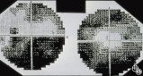 |
Optic Disc Drusen Visual Fields | Thomas R. Wolf, MD | This is the visual field of patient with optic nerve drusen. Whereas they typically do not cause central field loss, optic disc drusen may cause nerve fiber bundle layer defects and, thus, peripheral field defects, including altitudinal defects (seen inferiorly in the left eye) or arcuate defects (s... |
| 347 |
 |
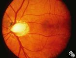
Retinal Coloboma Underneath a Relatively Normal Optic Nerve | Thomas R. Wolf, MD | Optic nerve colobomas appear as enlarged, white optic discs that are deeply excavated, often with some sapring of the superior rim. They result from an abnormal fusion of the proximal embryonic fissure. Optic nerve colobomas occur unilaterally or bilaterally with a similar frequency and can result i... |
| 348 |
 |
Isolated Congenital Optic Disc Anomalies | Thomas R. Wolf, MD | Patients with midline closure defects may exhibit abnormalities in the optic nerve, choroid, retinal pigment epithelium or retina. Anterior closure defects may result in colobomas of the structures of the anterior segment, such as the iris. Disease/Diagnosis: Coloboma. |
| 349 |
 |
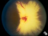
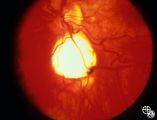
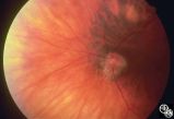
Peripapillary Staphyloma | Thomas R. Wolf, MD | Patients with ectasia of the outer layers of the eye may exhibit a posterior protrusion that appears on funduscopy as an area of deep excavation of the retina (posterior staphyloma). When it occurs around the optic disc, as in this case, it is termed a peripapillary staphyloma. This may occur in ass... |
| 350 |
 |
Motility Disturbances | Thomas R. Wolf, MD | The patient is a 53-year-old man with diplopia from right oculomotor nerve palsy and left hemiparesis (Weber's syndrome), with associated left lung hilar mass. The spinal tap showed pleocytosis consistent with carcinomatous meningitis. This image demonstrates oculomotor nerve metastatic carcinomatos... |
| 351 |
 |
Ocular Manifestations of Congenital/Inherited Diseases | William Fletcher Hoyt, MD | Tuberous sclerosis is an autosomal dominant phakomatosis characterized by astrocytic hamartomas of the retina or optic nerve, adenoma sebaceum of the face, periungual fibromas, and astrocytic hamartomas of the brain, with secondary seizures and mental retardation. Disease/Diagnosis: Tuberous Scleros... |
| 352 |
 |
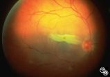
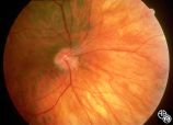
Isolated Congenital Optic Disc Anomalies | William Fletcher Hoyt, MD | This patient demonstrates bilateral tilted optic discs. Patients with this congenital optic disc anomaly may be asymptomatic or have bitemporal visual field defects that do not respect the vertical midline. Disease/Diagnosis: Tilted Discs. |
| 353 |
 |
Isolated Congenital Optic Disc Anomalies | William Fletcher Hoyt, MD | This patient demonstrates bilateral tilted optic discs. Patients with this congenital optic disc anomaly may be asymptomatic or have bitemporal visual field defects that do not respect the vertical midline. Disease/Diagnosis: Tilted Discs. |
| 354 |
 |
Isolated Congenital Optic Disc Anomalies | William Fletcher Hoyt, MD | This patient demonstrates bilateral tilted optic discs. Patients with this congenital optic disc anomaly may be asymptomatic or have bitemporal visual field defects that do not respect the vertical midline. Disease/Diagnosis: Tilted Discs. |
| 355 |
|
Isolated Congenital Optic Disc Anomalies | William Fletcher Hoyt, MD | This patient demonstrates bilateral tilted optic discs. Patients with this congenital optic disc anomaly may be asymptomatic or have bitemporal visual field defects that do not respect the vertical midline. Disease/Diagnosis: Tilted Discs. |
| 356 |
 |
Ocular Manifestations of Congenital/Inherited Diseases | William Fletcher Hoyt, MD | Image shows a patient with the Laurence-Moon-Biedl syndrome, an autosomal recessive syndrome characterized by obesity, mental retardation, retinitis pigmentosa (see Image 91_07), polydactyly, and hypogonadism. Pair with 91_07. |
| 357 |
 |
Ocular Disease With Retinal Findings | William Fletcher Hoyt, MD | Choroidal folds may result from choroidal tumors, compression on the eye wall from thyroid ophthalmopathy, orbital pseudotumor, orbital tumor, posterior scleritis, hypotony, scleral laceration, retinal detachment, marked hyperopia, or secondary to papilledema. Intraocular pressure measurements, refr... |
| 358 |
|
Isolated Congenital Optic Disc Anomalies | William Fletcher Hoyt, MD | This patient demonstrates bilateral tilted optic discs. Patients with this congenital optic disc anomaly may be asymptomatic or have bitemporal visual field defects that do not respect the vertical midline. Disease/Diagnosis: Tilted Discs. |
| 359 |
 |
Ocular Manifestations of Congenital/Inherited Diseases | William Fletcher Hoyt, MD | Image shows a patient with the Laurence-Moon_Biedl syndrome, an autosomal recessive syndrome characterized by obesity, mental retardation, retinitis pigmentosa, polydactyly, and hypogonadism. Pair with 91_06. |
| 360 |
|
Isolated Congenital Optic Disc Anomalies | William Fletcher Hoyt, MD | This patient demonstrates bilateral tilted optic discs. Patients with this congenital optic disc anomaly may be asymptomatic or have bitemporal visual field defects that do not respect the vertical midline. Disease/Diagnosis: Tilted Discs. |
| 361 |
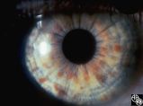 |
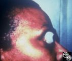
Ocular Manifestations of Congenital/Inherited Diseases | William Fletcher Hoyt, MD | Neurofibromatosis, type 1, is an autosomal dominant phakomatosis characterized by Lisch nodules of the iris (hamartomas) plexiform neurofibromas, café-au-lait spots on the skin, and axillary freckling. Intracranial tumors such as optic pathway gliomas may occur. Disease/Diagnosis: Neurofibromatosis... |
| 362 |
 |
Ocular Manifestations of Congenital/Inherited Diseases | William Fletcher Hoyt, MD | Tuberous sclerosis is an autosomal dominant phakomatosis characterized by astrocytic hamartomas of the retina or optic nerve, adenoma sebaceum of the face, periungual fibromas, and astrocytic hamartomas of the brain, with secondary seizures and mental retardation. Disease/Diagnosis: Tuberous Scleros... |