AAO-NANOS Neuro-Ophthalmology Clinical Collection: Derived from the AAO-NANOS Clinical Neuro-Ophthalmology collection produced on CD. The images are of selected cases from the NANOS teaching slide exchange, and the CD was produced under the direction of Larry Frohman, MD and Andrew Lee, MD.
The American Academy of Ophthalmology (AAO); The North American Neuro-Ophthalmology Association (NANOS).
NOVEL: https://novel.utah.edu/
TO
Filters: Collection: "ehsl_novel_aao_nanos"
| Title | Creator | Description | ||
|---|---|---|---|---|
| 201 |
 |
Systemic Disorders With Optic Nerve and Retinal Findings | Larry P. Frohman, MD | A 29-year-old African American woman presented with headaches, bilateral transient visual obscurations, blurred vision, numbness, and weakness of the lower extremities with myalgia and joint pains. She had an unplanned 12-pound weight loss over 2 months. A neurologist and internist diagnosed her wit... |
| 202 |
 |
Systemic Disorders With Optic Nerve and Retinal Findings | Larry P. Frohman, MD | A 35-year-old African-American woman had gradual bilateral painless visual loss over 3 months. When initially seen, the visual acuities were HM OD, NLP OS. The MRI showed diffused enhancement of the optic nerves and lacrimal glands. The evaluation strongly suggested sarcoidosis, with elevated angiot... |
| 203 |
 |
Systemic Disorders With Optic Nerve and Retinal Findings | Larry P. Frohman, MD | A 35-year-old African-American woman had gradual bilateral painless visual loss over 3 months. When initially seen, the visual acuities were HM OD, NLP OS. The MRI showed diffused enhancement of the optic nerves and lacrimal glands. The evaluation strongly suggested sarcoidosis, with elevated angiot... |
| 204 |
 |
Chiasmal Syndromes | Larry P. Frohman, MD | This 36-year-old woman presented in 1988 with 3 weeks of vertical binocular diplopia. She was a known amblyope OD. Her examination was notable for a right hyperdeviation of 1 PD present in right gaze and a subtle left noncongruous homonymous field defect. She was lost to follow-up, but 5 years later... |
| 205 |
 |
Chiasmal Syndromes | Larry P. Frohman, MD | This 36-year-old woman presented in 1988 with 3 weeks of vertical binocular diplopia. She was a known amblyope OD. Her examination was notable for a right hyperdeviation of 1 PD present in right gaze and a subtle left noncongruous homonymous field defect. She was lost to follow-up, but 5 years later... |
| 206 |
 |
Systemic Disorders With Optic Nerve and Retinal Findings | Larry P. Frohman, MD | Skin rashes occur in about 30 percent of patients with sarcoid. When seen, the rashes offer an accessible site for obtaining histologic material for confirmation of the clinical diagnosis. Pair with 91_68. |
| 207 |
 |
Ocular Manifestations of Congenital/Inherited Diseases | Larry P. Frohman, MD | On optic nerve CT scan, this patient with neurofibromatosis, type 1, shows the classic railroad-track sign of optic nerve meningioma and the kink sign of optic nerve glioma. Disease/Diagnosis: Neurofibromatosis, Type 1. |
| 208 |
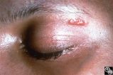 |
Systemic Disorders With Optic Nerve and Retinal Findings | Larry P. Frohman, MD | Skin rashes occur in about 30 percent of patients with sarcoid. When seen, the rashes offer an accessible site for obtaining histologic material for confirmation of the clinical diagnosis. Pair with 91_69. |
| 209 |
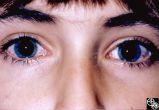 |
Ocular Manifestations of Congenital/Inherited Diseases | Larry P. Frohman, MD | This girl was 8 years old when these photographs were taken. She had been referred by pediatric neurology with ataxia. When seen, the findings of ataxia, poor convergence, early head thrusting maneuvers, and relatively preserved motility, in conjunction with the conjunctival findings as seen in the ... |
| 210 |
 |
Ocular Manifestations of Congenital/Inherited Diseases | Larry P. Frohman, MD | This girl was 8 years old when these photographs were taken. She had been referred by pediatric neurology with ataxia. When seen, the findings of ataxia, poor convergence, early head thrusting maneuvers, and relatively preserved motility, in conjunction with the conjunctival findings as seen in the ... |
| 211 |
 |
Systemic Disorders With Optic Nerve and Retinal Findings | Larry P. Frohman, MD | This is a 32-year-old HIV-positive man with anterior uveitis, vitritis, and bilateral papillitis from syphilis. With intravenous penicillin treatment, the optic discs and vision returned to normal. |
| 212 |
 |
Magnetic Resonance Imaging in Detection of Extracranial Internal Carotid Artery Dissection | Marilyn C. Kay, MD | This 28-year-old woman presented with a 4-week history of bilateral visual loss. She had a known history of multiple sclerosis. Her vision was 20/60 OD and 20/40 OS, with an RAPD OS and optic pallor OU. Her fields and MRI are shown. Optic tract lesions usually result in an incongruous homonymous hem... |
| 213 |
 |
Neuro-Ophthalmic Case With Notable Field Changes | Marilyn C. Kay, MD | This 28-year-old woman presented with a 4-week history of bilateral visual loss. She had a known history of multiple sclerosis. Her vision was 20/60 OD and 20/40 OS, with an RAPD OS and optic pallor OU. Her fields and MRI are shown. Optic tract lesions usually result in an incongruous homonymous hem... |
| 214 |
 |
Neuro-Ophthalmic Imaging-Cerebral Angiography | Mark J. Kupersmith, MD | Ehlers-Danlos syndrome is a connective tissue disorder that may affect blood vessels and predispose some affected patients to development of carotid cavernous fistula. Most patients with high-flow direct carotid cavernous sinus fistulas have suffered acute traumatic tears in the internal carotid art... |
| 215 |
 |
Neuro-Ophthalmic Consequences of Therapy | Mark J. Kupersmith, MD | radiation retinopathy may mimic diabetic or hypertensive optic neuropathy. A history of irradiation to the eye, orbit, or head is mandatory. Radiation retinopathy usually occurs many months after radiation therapy. |
| 216 |
 |
Neuro-Ophthalmic Consequences of Therapy | Mark J. Kupersmith, MD | Radiation causes a vascular retinopathy that may mimic diabetic or hypertensive retinopathy. It does not develop until many months or several years after radiation therapy to the eye, orbit or head. |
| 217 |
 |
Isolated Optic Neuritis/Neuropathy | Mark J. Kupersmith, MD | Papilledema may produce visual loss due to chronic atrophic papilledema, secondary macular hemorrhage, exudate or edema, secondary ischemic optic neuropathy, or secondary subretinal neovascular membrane formation. Patients with papilledema and visual loss should be suspected of harboring one of thes... |
| 218 |
 |
Isolated Optic Neuritis/Neuropathy | Mark J. Kupersmith, MD | Papilledema may produce visual loss due to chronic atrophic papilledema, secondary macular hemorrhage, exudate or edema, secondary ischemic optic neuropathy, or secondary subretinal neovascular membrane formation. Patients with papilledema and visual loss should be suspected of harboring one of thes... |
| 219 |
 |
Systemic Disorders With Optic Nerve and Retinal Findings | Mark J. Kupersmith, MD | Sarcoidosis is an inflammatory granulomatous disease that may result in inflammatory or infiltrative optic neuropathology or retinal vasculitis. Pair with 91_34. |
| 220 |
 |
Systemic Disorders With Optic Nerve and Retinal Findings | Mark J. Kupersmith, MD | Sarcoidosis is an inflammatory granulomatous disease that may result in inflammatory or infiltrative optic neuropathology or retinal vasculitis. Pair with 91_35. |
| 221 |
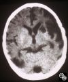 |
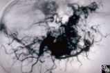
Neuro-Ophthalmic Vascular Disease | Mark J. Kupersmith, MD | A 9-year-old boy had recurrent ischemic episodes that had begun 2 years prior to evaluation. A significant right hemiparesis and a significant speech, learning, and memory disorder were present. His noncontrast axial view CT scan demonstrated multiple cerebral infarcts. Cerebral angiography revealed... |
| 222 |
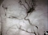 |
Neuro-Ophthalmic Vascular Disease | Mark J. Kupersmith, MD | A 9-year-old boy had recurrent ischemic episodes that had begun 2 years prior to evaluation. A significant right hemiparesis and a significant speech, learning, and memory disorder were present. His noncontrast axial view CT scan demonstrated multiple cerebral infarcts. Cerebral angiography revealed... |
| 223 |
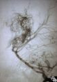 |
Neuro-Ophthalmic Vascular Disease | Mark J. Kupersmith, MD | A 9-year-old boy had recurrent ischemic episodes that had begun 2 years prior to evaluation. A significant right hemiparesis and a significant speech, learning, and memory disorder were present. His noncontrast axial view CT scan demonstrated multiple cerebral infarcts. Cerebral angiography revealed... |
| 224 |
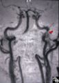 |
Neuro-Ophthalmic Vascular Disease | Mark J. Kupersmith, MD | MR angiography was performed on this 33-year-old woman, who complained of the onset of a bad taste in her mouth followed by pain along the left forehead and development of the left third-order Horner's syndrome during pregnancy. Except for the Horner's syndrome, the patient was neurologically intact... |
| 225 |
 |
Ocular Manifestations of Congenital/Inherited Diseases | Mark J. Kupersmith, MD | This 9-year-old girl, who had complained of recurrent spontaneous bleeding from the palate and slight swelling and increased warmth over the left cheek, was found to have Wyburn-Mason syndrome. Image 1993_16 shows a small area of arteriovenous shunt on the left optic disc in this patient, who has no... |
| 226 |
 |
Ocular Manifestations of Congenital/Inherited Diseases | Mark J. Kupersmith, MD | This 9-year-old girl, who had complained of recurrent spontaneous bleeding from the palate and slight swelling and increased warmth over the left cheek, was found to have Wyburn-Mason syndrome. Image 1993_16 shows a small area of arteriovenous shunt on the left optic disc in this patient, who has no... |
| 227 |
 |
Ocular Manifestations of Congenital/Inherited Diseases | Mark J. Kupersmith, MD | This 9-year-old girl, who had complained of recurrent spontaneous bleeding from the palate and slight swelling and increased warmth over the left cheek, was found to have Wyburn-Mason syndrome. Image 1993_16 shows a small area of arteriovenous shunt on the left optic disc in this patient, who has no... |
| 228 |
 |
Ocular Manifestations of Congenital/Inherited Diseases | Mark J. Kupersmith, MD | This 9-year-old girl, who had complained of recurrent spontaneous bleeding from the palate and slight swelling and increased warmth over the left cheek, was found to have Wyburn-Mason syndrome. Image 1993_16 shows a small area of arteriovenous shunt on the left optic disc in this patient, who has no... |
| 229 |
 |
Ocular Manifestations of Congenital/Inherited Diseases | Mark J. Kupersmith, MD | This 9-year-old girl, who had complained of recurrent spontaneous bleeding from the palate and slight swelling and increased warmth over the left cheek, was found to have Wyburn-Mason syndrome. Image 1993_16 shows a small area of arteriovenous shunt on the left optic disc in this patient, who has no... |
| 230 |
 |
Neuro-Ophthalmic Consequences of Therapy | Mark J. Kupersmith, MD | radiation retinopathy may mimic diabetic or hypertensive optic neuropathy. A history of irradiation to the eye, orbit, or head is mandatory. Radiation retinopathy usually occurs many months after radiation therapy. |
| 231 |
 |
Neuro-Ophthalmic Consequences of Therapy | Mark J. Kupersmith, MD | radiation retinopathy may mimic diabetic or hypertensive optic neuropathy. A history of irradiation to the eye, orbit, or head is mandatory. Radiation retinopathy usually occurs many months after radiation therapy. |
| 232 |
 |
Neuro-Ophthalmic Vascular Disease | Mark J. Kupersmith, MD | Aneurysms of the intracranial circulation may act as mass lesions and compress the afferent of efferent visual pathway. Ophthalmic artery aneurysms may compress the optic nerve and result in an optic neuropathy (ie, visual loss, afferent pupillary defect, optic atrophy). Treatment includes endovascu... |
| 233 |
 |
Neuro-Ophthalmic Vascular Disease | Mark J. Kupersmith, MD | Aneurysms of the intracranial circulation may act as mass lesions and compress the afferent of efferent visual pathway. Ophthalmic artery aneurysms may compress the optic nerve and result in an optic neuropathy (ie, visual loss, afferent pupillary defect, optic atrophy). Treatment includes endovascu... |
| 234 |
 |
Systemic Disorders With Optic Nerve and Retinal Findings | Mark J. Kupersmith, MD | Sarcoidosis is an inflammatory multisystem granulomatous disease that may result in an inflammatory or infiltrative optic neuropathy, papilledema from increased intracranial pressure due to meningeal inflammation or intracranial granuloma, or may present with an optic disc granuloma. Pair with 91_31... |
| 235 |
 |
Systemic Disorders With Optic Nerve and Retinal Findings | Mark J. Kupersmith, MD | Sarcoidosis is an inflammatory multisystem granulomatous disease that may result in an inflammatory or infiltrative optic neuropathy, papilledema from increased intracranial pressure due to meningeal inflammation or intracranial granuloma, or may present with an optic disc granuloma. Pair with 91_32... |
| 236 |
 |
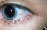
Neuro-Ophthalmic Vascular Disease | Mark L. Moster, MD | Neovascularization of the iris may form in response to an ischemic disease of the retina, such as diabetic retinopathy. Carotid artery occlusion may result in ocular ischemia that may induce neovascularization.This is a dramatic image of iris neovascularization. |
| 237 |
 |
Isolated Optic Neuritis/Neuropathy | Michael Wall, MD | This 36-year-old man noticed blurry vision in his right eye when attempting to sight a gun. He also reported bifrontal headaches responsive to aspirin. Acuity was 20/50 OD, 20/13 OS. His right eye could be refracted to 20/20 with a +2.50 sphere. A 0.3 right relative afferent pupillary defect was pre... |
| 238 |
 |
Optic Neuropathies | Michael Wall, MD | Optic disc edema with a macular star figure may occur in infectious diseases (eg, cat-scratch disease, syphilis, tuberculosis, Lyme disease), inflammatory diseases (eg, sarcoid), ischemic diseases (anterior ischemic optic neuropathy), and in papilledema. Infectious causes should be sought in patient... |
| 239 |
 |
Neuro-Ophthalmic Vascular Disease | Mitchell J. Wolin, MD | Carotid cavernous fistulas (CCFs) are connections between the arterial blood flow from the carotid artery system and the cavernous sinus. CCFs may be direct high-flow fistulas or indirect low-flow fistulas. Most direct CCFs are due to trauma. Enlargement of the superior ophthalmic vein may be demons... |
| 240 |
 |
Motility Disturbances | Mitchell J. Wolin, MD | The trochlear nerve (fourth nerve) runs from the midbrain, exits dorsally, crosses in the anterior medullary velum, enters the subarachnoid space, travels within the lateral wall of the cavernous sinus, and enters the orbit through the superior orbital fissure. A trochlear nerve palsy may be due to ... |
| 241 |
 |
Motility Disturbances | Mitchell J. Wolin, MD | The trochlear nerve (fourth nerve) runs from the midbrain, exits dorsally, crosses in the anterior medullary velum, enters the subarachnoid space, travels within the lateral wall of the cavernous sinus, and enters the orbit through the superior orbital fissure. A trochlear nerve palsy may be due to ... |
| 242 |
 |
Neuro-Ophthalmic Vascular Disease | Mitchell J. Wolin, MD | Carotid cavernous fistulas (CCFs) are connections between the arterial blood flow from the carotid artery system and the cavernous sinus. CCFs may be direct high-flow fistulas or indirect low-flow fistulas. Most direct CCFs are due to trauma. Enlargement of the superior ophthalmic vein may be demons... |
| 243 |
 |
Chiasmal Syndromes | Mitchell J. Wolin, MD | The patient is a 60-year-old woman with a chief complaint of decreased vision. In 1986 she was diagnosed with a poorly differentiated breast cancer in her left breast. She underwent mastectomy, and all nodes were negative. She did well until 1991, when she was found to have a chest wall mass. This m... |
| 244 |
 |
Chiasmal Syndromes | Mitchell J. Wolin, MD | The patient is a 60-year-old woman with a chief complaint of decreased vision. In 1986 she was diagnosed with a poorly differentiated breast cancer in her left breast. She underwent mastectomy, and all nodes were negative. She did well until 1991, when she was found to have a chest wall mass. This m... |
| 245 |
 |
Chiasmal Syndromes | Mitchell J. Wolin, MD | The patient is a 60-year-old woman with a chief complaint of decreased vision. In 1986 she was diagnosed with a poorly differentiated breast cancer in her left breast. She underwent mastectomy, and all nodes were negative. She did well until 1991, when she was found to have a chest wall mass. This m... |
| 246 |
 |
Chiasmal Syndromes | Mitchell J. Wolin, MD | The patient is a 60-year-old woman with a chief complaint of decreased vision. In 1986 she was diagnosed with a poorly differentiated breast cancer in her left breast. She underwent mastectomy, and all nodes were negative. She did well until 1991, when she was found to have a chest wall mass. This m... |
| 247 |
 |
Orbital Tumors | Mitchell J. Wolin, MD | Cavernous hemangiomas of the orbit usually result in painless orbital signs such as proptosis or visual loss. Orbital imaging of the lesion, which usually is a well-defined orbital mass, is demonstrated in this study. The lesion is benign and usually occurs in young to middle-aged adults. Surgical e... |
| 248 |
 |
Ocular Manifestations of Congenital/Inherited Diseases | Mitchell J. Wolin, MD | Patients with olivopontocerebellar atrophy may exhibit signs of ocular motor deficits, such as ocular motor apraxia or cerebellar eye signs, and peripheral pigmentary retinopathy and optic atrophy. Pair with 94_55. |
| 249 |
 |
Ocular Manifestations of Congenital/Inherited Diseases | Mitchell J. Wolin, MD | Patients with olivopontocerebellar atrophy may exhibit signs of ocular motor deficits, such as ocular motor apraxia or cerebellar eye signs, and peripheral pigmentary retinopathy and optic atrophy. Pair with 94_54. |
| 250 |
 |
Orbital Tumors | Mitchell J. Wolin, MD | Cavernous hemangiomas of the orbit usually result in painless orbital signs such as proptosis or visual loss. Orbital imaging of the lesion, which usually is a well-defined orbital mass, is demonstrated in this study. The lesion is benign and usually occurs in young to middle-aged adults. Surgical e... |
| 251 |
 |
Orbital Tumors | Mitchell J. Wolin, MD | Cavernous hemangiomas of the orbit usually result in painless orbital signs such as proptosis or visual loss. Orbital imaging of the lesion, which usually is a well-defined orbital mass, is demonstrated in this study. The lesion is benign and usually occurs in young to middle-aged adults. Surgical e... |
| 252 |
 |
Neuro-Ophthalmic Imaging-CT Scan | Mitchell J. Wolin, MD | Idiopathic orbital pseudotumor is an inflammatory disorder that may effect any part of the ocular anatomy. The site of inflammation determines the nomenclature. For example, involvement of the sclera is referred to as scleritis. And involvement of one or more of the extraocular muscles is referred t... |
| 253 |
 |
Neuro-Ophthalmic Imaging-CT Scan | Mitchell J. Wolin, MD | Idiopathic orbital pseudotumor is an inflammatory disorder that may effect any part of the ocular anatomy. The site of inflammation determines the nomenclature. For example, involvement of the sclera is referred to as scleritis. And involvement of one or more of the extraocular muscles is referred t... |
| 254 |
 |
Ocular Manifestations of Systemic Disorders | Mitchell J. Wolin, MD | Thyroid eye disease is the most common cause of unilateral or bilateral proptosis in the adult patient. Other signs of thyroid eye disease should be sought, including lid retraction, inferior scleral show, and lid lag. Patients with markedly asymmetric or strictly unilateral proptosis should probabl... |
| 255 |
 |
Ocular Manifestations of Systemic Disorders | Mitchell J. Wolin, MD | Thyroid eye disease is the most common cause of unilateral or bilateral proptosis in the adult patient. Other signs of thyroid eye disease should be sought, including lid retraction, inferior scleral show, and lid lag. Patients with markedly asymmetric or strictly unilateral proptosis should probabl... |
| 256 |
 |
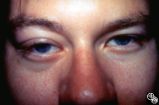
Migraine Syndrome | Mitchell J. Wolin, MD | The image shows a patient with cluster headache and eye displaying Horner's syndrome. |
| 257 |
 |
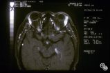
Neuro-Ophthalmic Imaging-MRI | Mitchell J. Wolin, MD | Axial view of Arnold-Chiari malformation on a patient with downbeat nystagmus. Note the presence of the cerebellar tonsils posterior to the caudal medulla. In addition to downbeat nystagmus, Arnold-Chiari malformations can sometimes lead to increased intracranial pressure and papilledema. |
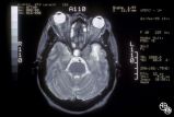
| 258 |
 |
Neuro-Ophthalmic Imaging-MRI | Mitchell J. Wolin, MD | Sagittal view of Arnold-Chiari malformation on a patient with downbeat nystagmus. The compression of the cervicomedullary junction is clearly depicted in the sagittal view. |
| 259 |
 |
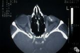
Neuro-Ophthalmic Imaging-CT Scan | Mitchell J. Wolin, MD | This is a patient with trauma leading to enucleation, with swelling years later over the implant. This is a presumed chronic abscess between orbit and dura. |
| 260 |
 |
Isolated Optic Neuritis/Neuropathy | Ralph A. Sawyer, MD | Papilledema usually results in bilateral optic disc edema without visual loss. The blind spot may enlarge initially, but progressive visual field loss may occur with chronic optic disc edema. Asymmetric or frankly unilateral optic disc edema may occur due to structural disc fractures that prevent th... |
| 261 |
 |
Isolated Optic Neuritis/Neuropathy | Ralph A. Sawyer, MD | Papilledema usually results in bilateral optic disc edema without visual loss. The blind spot may enlarge initially, but progressive visual field loss may occur with chronic optic disc edema. Asymmetric or frankly unilateral optic disc edema may occur due to structural disc fractures that prevent th... |
| 262 |
 |
Optic Neuropathies | Ralph A. Sawyer, MD | Optic disc edema with a macular star figure has been referred to as neuroretinitis. |
| 263 |
 |
Isolated Optic Neuritis/Neuropathy | Richard H. Legge, MD | Papilledema is a term reserved for optic disc edema related to increased intracranial pressure (eg. Papilledema, sixth nerve palsy, headache), a normal neuroimaging study, and an elevated opening pressure with normal cerebrospinal fluid contents. |
| 264 |
 |
Systemic Disorders With Optic Nerve and Retinal Findings | Robert F. Saul, MD | This patent has known pseudoxanthoma elasticum (an uncommon elastic tissue disorder characterized by plaque-like skin folds [plucked chicken skin], and degeneration of collagen fibers involving multiple systems, including the GI tract and heart), angioid streaks, and optic disc drusen. |
| 265 |
 |
Systemic Disorders With Optic Nerve and Retinal Findings | Robert F. Saul, MD | This patent has known pseudoxanthoma elasticum (an uncommon elastic tissue disorder characterized by plaque-like skin folds [plucked chicken skin], and degeneration of collagen fibers involving multiple systems, including the GI tract and heart), angioid streaks, and optic disc drusen. |
| 266 |
 |
Systemic Disorders With Optic Nerve and Retinal Findings | Robert F. Saul, MD | This patent has known pseudoxanthoma elasticum (an uncommon elastic tissue disorder characterized by plaque-like skin folds [plucked chicken skin], and degeneration of collagen fibers involving multiple systems, including the GI tract and heart), angioid streaks, and optic disc drusen. |
| 267 |
 |
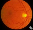
Neuro-Ophthalmic Vascular Disease | Robert F. Saul, MD | In image 93_29, taken during the episode, note the change in caliber of the blood vessels. |
| 268 |
 |
Neuro-Ophthalmic Vascular Disease | Robert F. Saul, MD | Image 93_30 is immediately after the attack, note the slight redness to the macula. |
| 269 |
 |
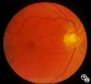
Neuro-Ophthalmic Vascular Disease | Robert F. Saul, MD | Image 93_28 shows the fundus before the attack. |
| 270 |
 |
Systemic Disorders With Optic Nerve and Retinal Findings | Robert F. Saul, MD | This patent has known pseudoxanthoma elasticum (an uncommon elastic tissue disorder characterized by plaque-like skin folds [plucked chicken skin], and degeneration of collagen fibers involving multiple systems, including the GI tract and heart), angioid streaks, and optic disc drusen. Imaging of a... |
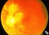
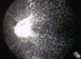
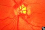
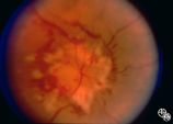
| 271 |
 |
Systemic Disorders With Optic Nerve and Retinal Findings | Robert F. Saul, MD | This patent has known pseudoxanthoma elasticum (an uncommon elastic tissue disorder characterized by plaque-like skin folds [plucked chicken skin], and degeneration of collagen fibers involving multiple systems, including the GI tract and heart), angioid streaks, and optic disc drusen. Imaging of a... |
| 272 |
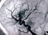
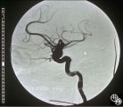
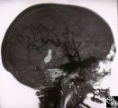
 |
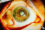
Ocular Manifestations of Systemic Disorders | Robert F. Saul, MD | Wilson's disease (hepatolenticular degeneration) is a progressive autosomal recessive multisystem disease that may result in cirrhosis of the liver, renal dysfunction, and motor neurologic disease. A Kayser-Fleischer ring may occur as a green-brown band at the level of Descemet's membrane in the cor... |
| 273 |
 |
Systemic Disorders With Optic Nerve and Retinal Findings | Robert L. Lesser, MD | Intraocular lymphoma may present with an unexplained vitritis, optic disc infiltration, or choroidal infiltration. One unusual manifestation of large-cell lymphoma is this leopard-spot appearance. Pair with 94_32, 94_33, and 94_35. This is a fundus photo. |
| 274 |
 |
Systemic Disorders With Optic Nerve and Retinal Findings | Robert L. Lesser, MD | Intraocular lymphoma may present with an unexplained vitritis, optic disc infiltration, or choroidal infiltration. One unusual manifestation of large-cell lymphoma is this leopard-spot appearance. Pair with 94_32, 94_34, and 94_35. This is a fundus photo. |
| 275 |
 |
Systemic Disorders With Optic Nerve and Retinal Findings | Robert L. Lesser, MD | Intraocular lymphoma may present with an unexplained vitritis, optic disc infiltration, or choroidal infiltration. One unusual manifestation of large-cell lymphoma is this leopard-spot appearance. Pair with 94_33, 94_34, and 94_35. This is a fundus photo. |
| 276 |
 |
Systemic Disorders With Optic Nerve and Retinal Findings | Robert L. Lesser, MD | Intraocular lymphoma may present with an unexplained vitritis, optic disc infiltration, or choroidal infiltration. One unusual manifestation of large-cell lymphoma is this leopard-spot appearance. Pair with 94_32, 94_33, and 94_34. This is a fluorescein angiogram. |
| 277 |
 |
Isolated Congenital Optic Disc Anomalies | Roger Turbin, MD | Shown are the fundi of a - year old child with dominant optic atrophy, 20/200 OU. Pair with 1996_59. Disease/Diagnosis: Congenital optic atrophy. |
| 278 |
 |
Isolated Congenital Optic Disc Anomalies | Roger Turbin, MD | Shown are the fundi of a - year old child with dominant optic atrophy, 20/200 OU. Pair with 1996_60. Disease/Diagnosis: Congenital optic atrophy. |
| 279 |
 |
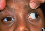
Ocular Manifestations of Systemic Disorders | Rosa A. Tang, MD | Myasthenia gravis should be considered in any patient with painless, pupil-spared, nonapoptotic ophthalmoplegia. It may mimic any ophthalmoparesis. Involvement of the medical rectus may result in a pseudointernuclear ophthalmoplegia. Pair with 96_24 and 96_25. |
| 280 |
 |
Motility Disturbances | Rosa A. Tang, MD | Traumatic damage to the third cranial nerve may result in aberrant regeneration of fibers that innervate the eyelid, pupil, or extraocular muscles. For instance, there may be lid retraction in attempted downgaze. Any combination of aberrant activation of third nerve-innervated structures may occur, ... |
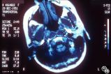
| 281 |
 |
Ocular Manifestations of Systemic Disorders | Rosa A. Tang, MD | Myasthenia gravis should be considered in any patient with painless, pupil-spared, nonapoptotic ophthalmoplegia. It may mimic any ophthalmoparesis. Involvement of the medical rectus may result in a pseudointernuclear ophthalmoplegia. Pair with 96_23 and 96_25. |
| 282 |
 |
Ocular Manifestations of Systemic Disorders | Rosa A. Tang, MD | Myasthenia gravis should be considered in any patient with painless, pupil-spared, nonapoptotic ophthalmoplegia. It may mimic any ophthalmoparesis. Involvement of the medical rectus may result in a pseudointernuclear ophthalmoplegia. Pair with 96_23 and 96_24. |
| 283 |
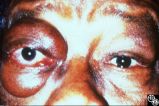
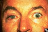
 |
Isolated Optic Neuritis/Neuropathy | Rosa A. Tang, MD | Papilledema may produce visual loss due to chronic atrophic papilledema, secondary macular hemorrhage, exudate or edema, secondary ischemic optic neuropathy, or secondary subretinal neovascular membrane formation. |
| 284 |
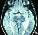
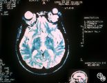
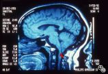
 |
Isolated Optic Neuritis/Neuropathy | Rosa A. Tang, MD | Papilledema in pseudotumor cerebri may result in adjacent choroidal or retinal folds. |
| 285 |
 |
Isolated Congenital Optic Disc Anomalies | Rosa A. Tang, MD | This patient has optic disc drusen and evidence of a superimposed optic neuropathy, including loss of visual field, an ipsilateral afferent pupillary defect, and optic atrophy. Although optic disc drusen typically causes visual field loss without visual acuity loss superimposed, ischemic optic neuro... |
| 286 |
 |
Motility Disturbances | Rosa A. Tang, MD | Skew deviation is a vertical deviation that is not localized to any one muscle or muscle group. The deviation may be comitant or not, and intermittent or constant. Skew deviation is often defined by the company it keeps, that is, skew usually occurs in association with other brain-stem signs, and is... |
| 287 |
 |
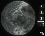
Motility Disturbances | Rosa A. Tang, MD | Skew deviation is a vertical deviation that is not localized to any one muscle or muscle group. The deviation may be comitant or not, and intermittent or constant. Skew deviation is often defined by the company it keeps, that is, skew usually occurs in association with other brain-stem signs, and is... |
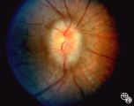
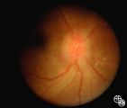
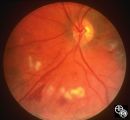
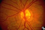
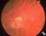
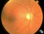
| 288 |
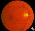
 |
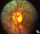
Acquired Disc Changes | Rosa A. Tang, MD | Although optociliary shunt vessels are venous collaterals that typically form in response to chronic venous obstruction, they may occur on a congenital basis as seen here. |
| 289 |
 |
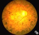
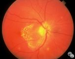
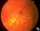
Isolated Optic Neuritis/Neuropathy | Rosa A. Tang, MD | Papilledema is a term reserved for optic disc edema related to increased intracranial pressure. Fluid within the optic nerve sheath or elevation of the intraocular optic nerve head may be visible on magnetic resonance imaging studies of the head and orbit. |
| 290 |
 |
Neuro-Ophthalmic Imaging-MRI | Rosa A. Tang, MD | Aneurisms may result in neuro-ophthalmologic sign and symptoms by direct compression of the afferent or efferent systems or by the secondary effects of hemorrhage. Basilar aneurisms may result in ocular motor deficits such as a unilateral or bilateral third nerve palsy. |
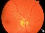
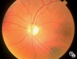
| 291 |
 |
Acquired Disc Changes | Rosa A. Tang, MD | Optociliary shunt vessels are venous collaterals that form in response to chronic venous obstruction. They may occur in patients following central retinal vein occlusion. |
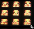
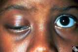
| 292 |
 |
Motility Disturbances | Rosa A. Tang, MD | Cyclical oculomotor paresis may occur in patients as an intermittent phenomenon, with a paretic phase and diplopia and intervals that are nonparetic. The history and examination are classic for the disorder. Pair with Images 95_19 and 95_20. |
| 293 |
 |
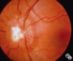
Motility Disturbances | Rosa A. Tang, MD | Cyclical oculomotor paresis may occur in patients as an intermittent phenomenon, with a paretic phase and diplopia and intervals that are nonparetic. The history and examination are classic for the disorder. Pair with Images 95_18 and 95_19. |
| 294 |
 |
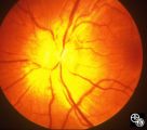
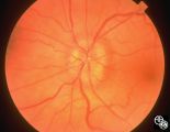
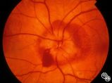
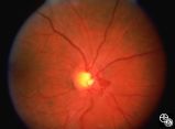
Ocular Manifestations of Systemic Disorders | Rosa A. Tang, MD | Systemic lymphoma may occur in the orbit and may involve the lacrimal gland. Patients usually present with a painless, progressive proptosis or a mass. CT scan usually demonstrates an irregularly shaped lesion conforming to the globe or lacrimal fossa, and bone erosion is not usually found. Pair wit... |
| 295 |
 |
Ocular Manifestations of Systemic Disorders | Rosa A. Tang, MD | Systemic lymphoma may occur in the orbit and may involve the lacrimal gland. Patients usually present with a painless, progressive proptosis or a mass. CT scan usually demonstrates an irregularly shaped lesion conforming to the globe or lacrimal fossa, and bone erosion is not usually found. Pair wit... |
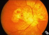
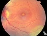
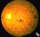
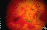
| 296 |
 |
Systemic Disorders With Optic Nerve and Retinal Findings | Rosa A. Tang, MD | Neoplasms may result in an optic neuropathy by direct metastatic involvement. In this patient, a lung adenocarcinoma was metastatic to the optic nerve.This is a fundus photo. |
| 297 |
 |
Motility Disturbances | Rosa A. Tang, MD | Cyclical oculomotor paresis may occur in patients as an intermittent phenomenon, with a paretic phase and diplopia and intervals that are nonparetic. The history and examination are classic for the disorder. Pair with Images 95_18 and 95_20. |
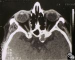
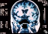
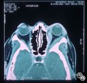
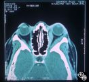
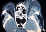
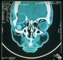
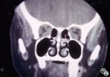
| 298 |
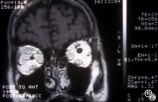 |
Ocular Manifestations of Systemic Disorders | Rosa A. Tang, MD | Thyroid eye disease may cause proptosis and extraocular muscle enlargement that may be seen on orbital imaging studies. In general, coronal images allow the best visualization of the extraocular muscle enlargement. Pair with 94_44 and 94_46. |
| 299 |
 |
Ocular Manifestations of Systemic Disorders | Rosa A. Tang, MD | Thyroid eye disease may cause proptosis and extraocular muscle enlargement that may be seen on orbital imaging studies. In general, coronal images allow the best visualization of the extraocular muscle enlargement. Pair with 94_44 and 94_45. |
| 300 |
 |
Neuro-Ophthalmic Imaging-MRI | Rosa A. Tang, MD | Aneurisms may result in neuro-ophthalmologic sign and symptoms by direct compression of the afferent or efferent systems or by the secondary effects of hemorrhage. Basilar aneurisms may result in ocular motor deficits such as a unilateral or bilateral third nerve palsy. |