AAO-NANOS Neuro-Ophthalmology Clinical Collection: Derived from the AAO-NANOS Clinical Neuro-Ophthalmology collection produced on CD. The images are of selected cases from the NANOS teaching slide exchange, and the CD was produced under the direction of Larry Frohman, MD and Andrew Lee, MD.
The American Academy of Ophthalmology (AAO); The North American Neuro-Ophthalmology Association (NANOS).
NOVEL: https://novel.utah.edu/
TO
Filters: Collection: "ehsl_novel_aao_nanos"
| Title | Creator | Description | ||
|---|---|---|---|---|
| 251 |
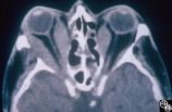 |
Ocular Manifestations of Systemic Disorders | Rosa A. Tang, MD | Thyroid eye disease may result in proptosis and restrictive external ophthalmoplegia. The extracoular muscles are often diffusely enlarged with sparing of the tendons. |
| 252 |
 |
Ocular Manifestations of Systemic Disorders | Rosa A. Tang, MD | Thyroid eye disease may cause proptosis and extraocular muscle enlargement that may be seen on orbital imaging studies. In general, coronal images allow the best visualization of the extraocular muscle enlargement. Pair with 94_45 and 94_46. |
| 253 |
 |
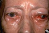
Ocular Manifestations of Systemic Disorders | Rosa A. Tang, MD | Thyroid eye disease can result in significant upper eyelid retraction and axial proptosis resulting in exposure keratopathy. |
| 254 |
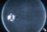 |
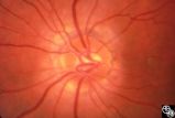
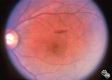
Optic Disc Drusen Autofluorescence | Anthony C. Arnold, MD | This case of optic disc drusen demonstrates the typical autofluorescence. Pair with 92_68. Imaging: Autoflourescence? |
| 255 |
 |
Optic Disc Drusen Visual Fields | Thomas R. Wolf, MD | This is the visual field of patient with optic nerve drusen. Whereas they typically do not cause central field loss, optic disc drusen may cause nerve fiber bundle layer defects and, thus, peripheral field defects, including altitudinal defects (seen inferiorly in the left eye) or arcuate defects (s... |
| 256 |
 |
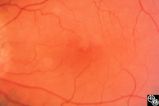
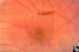
Optic Disc Drusen With Autofluorescence | Thomas R. Wolf, MD | This photograph of optic disc drusen demonstrates autoflourescence with flourescein barrier filters in place. Imaging: flourescein barrier filters. |
| 257 |
 |
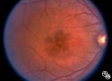
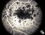
Optic Disc Drusen, Fluorescein Angiogram | Anthony C. Arnold, MD | Images 92_64 and 92_67 demonstrate the characteristics of optic disc drusen on flourescein angiography. This image shows the early arteriovenous phase, with irregular dye uptake and focal hypoflourescence superotemporally. Pair with 92_63 and 92_67 |
| 258 |
 |
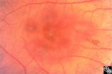
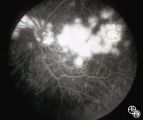
Optic Nerve Drusen, Late Fluorescein Angiogram | Anthony C. Arnold, MD | Images 92_64 and 92_67 demonstrate the characteristics of optic disc drusen on flourescein angiography. This image displays the nodular staining of the drusen without leakage. Pair with 92_63 and 92_64. |
| 259 |
 |
Optic Neuropathies | Larry P. Frohman, MD | This healthy 29-year-old man with dense amblyopia OS presented with a foreign-body sensation OS and further visual loss in his amblyopic eye. He was noted to have bilateral disc edema and lesions in the left eye consistent with unilateral acute multifocal placoid pigment epitheliopathy (AMPPE). He r... |
| 260 |
 |
Optic Neuropathies | Larry P. Frohman, MD | This healthy 29-year-old man with dense amblyopia OS presented with a foreign-body sensation OS and further visual loss in his amblyopic eye. He was noted to have bilateral disc edema and lesions in the left eye consistent with unilateral acute multifocal placoid pigment epitheliopathy (AMPPE). He r... |
| 261 |
 |
Optic Neuropathies | Larry P. Frohman, MD | This healthy 29-year-old man with dense amblyopia OS presented with a foreign-body sensation OS and further visual loss in his amblyopic eye. He was noted to have bilateral disc edema and lesions in the left eye consistent with unilateral acute multifocal placoid pigment epitheliopathy (AMPPE). He r... |
| 262 |
 |
Optic Neuropathies | Larry P. Frohman, MD | This healthy 29-year-old man with dense amblyopia OS presented with a foreign-body sensation OS and further visual loss in his amblyopic eye. He was noted to have bilateral disc edema and lesions in the left eye consistent with unilateral acute multifocal placoid pigment epitheliopathy (AMPPE). He r... |
| 263 |
 |
Optic Neuropathies | Daniel M. Jacobson MD | This 28-year-old otherwise-healthy woman was referred to for treatment of what was thought to be optic neuritis OD. Three weeks earlier she had noted a dark inferior scotoma OD that progressed to involve fixation over the next 10-12 days. She experienced photopsias OD at the onset. She had no viral ... |
| 264 |
 |
Optic Neuropathies | Daniel M. Jacobson MD | This 28-year-old otherwise-healthy woman was referred to for treatment of what was thought to be optic neuritis OD. Three weeks earlier she had noted a dark inferior scotoma OD that progressed to involve fixation over the next 10-12 days. She experienced photopsias OD at the onset. She had no viral ... |
| 265 |
 |
Optic Neuropathies | Daniel M. Jacobson MD | This 34-year-old otherwise-healthy woman was referred for a neuro-ophthalmologic consultation for unexplained loss of vision OD following head trauma. Two months earlier, she was involved in a motor vehicle accident that resulted in a closed head injury complicated by a spontaneously resolving small... |
| 266 |
 |
Optic Neuropathies | Daniel M. Jacobson MD | This 34-year-old otherwise-healthy woman was referred for a neuro-ophthalmologic consultation for unexplained loss of vision OD following head trauma. Two months earlier, she was involved in a motor vehicle accident that resulted in a closed head injury complicated by a spontaneously resolving small... |
| 267 |
 |
Optic Neuropathies | Daniel M. Jacobson MD | This 34-year-old otherwise-healthy woman was referred for a neuro-ophthalmologic consultation for unexplained loss of vision OD following head trauma. Two months earlier, she was involved in a motor vehicle accident that resulted in a closed head injury complicated by a spontaneously resolving small... |
| 268 |
 |
Optic Neuropathies | Daniel M. Jacobson MD | This 34-year-old otherwise-healthy woman was referred for a neuro-ophthalmologic consultation for unexplained loss of vision OD following head trauma. Two months earlier, she was involved in a motor vehicle accident that resulted in a closed head injury complicated by a spontaneously resolving small... |
| 269 |
 |
Optic Neuropathies | Larry P. Frohman, MD | This healthy 29-year-old man with dense amblyopia OS presented with a foreign-body sensation OS and further visual loss in his amblyopic eye. He was noted to have bilateral disc edema and lesions in the left eye consistent with unilateral acute multifocal placoid pigment epitheliopathy (AMPPE). He r... |
| 270 |
 |
Optic Neuropathies | Larry P. Frohman, MD | This healthy 29-year-old man with dense amblyopia OS presented with a foreign-body sensation OS and further visual loss in his amblyopic eye. He was noted to have bilateral disc edema and lesions in the left eye consistent with unilateral acute multifocal placoid pigment epitheliopathy (AMPPE). He r... |
| 271 |
 |
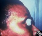
Optic Neuropathies | Larry P. Frohman, MD | The patient is a 66-year-old man with a history of ethanol abuse. He presented with 3 months of right-sided headache and a few days of progressive visual loss OD to hand motions only. When seen by the orbital service, he had nearly complete ophthalmoplegia and ptosis. Sinus biopsy showed fungus, whi... |
| 272 |
 |
Optic Neuropathies | Larry P. Frohman, MD | The patient is a 66-year-old man with a history of ethanol abuse. He presented with 3 months of right-sided headache and a few days of progressive visual loss OD to hand motions only. When seen by the orbital service, he had nearly complete ophthalmoplegia and ptosis. Sinus biopsy showed fungus, whi... |
| 273 |
 |
Optic Neuropathies | Larry P. Frohman, MD | The patient is a 66-year-old man with a history of ethanol abuse. He presented with 3 months of right-sided headache and a few days of progressive visual loss OD to hand motions only. When seen by the orbital service, he had nearly complete ophthalmoplegia and ptosis. Sinus biopsy showed fungus, whi... |
| 274 |
 |
Optic Neuropathies | Larry P. Frohman, MD | The patient is a 66-year-old man with a history of ethanol abuse. He presented with 3 months of right-sided headache and a few days of progressive visual loss OD to hand motions only. When seen by the orbital service, he had nearly complete ophthalmoplegia and ptosis. Sinus biopsy showed fungus, whi... |
| 275 |
 |
Optic Neuropathies | Rosa A. Tang, MD | Optic atrophy might be a false-localizing sign of optic nerve disease, as retinal disease may secondarily produce optic atrophy. In retinitis pigmentosa, for example, patients may exhibit a waxy disc palor. Fundus imaging. Anatomy: Retina. Disease/Diagnosis: Retinitis pigmentosa; Optic atrophy. |