AAO-NANOS Neuro-Ophthalmology Clinical Collection: Derived from the AAO-NANOS Clinical Neuro-Ophthalmology collection produced on CD. The images are of selected cases from the NANOS teaching slide exchange, and the CD was produced under the direction of Larry Frohman, MD and Andrew Lee, MD.
The American Academy of Ophthalmology (AAO); The North American Neuro-Ophthalmology Association (NANOS).
NOVEL: https://novel.utah.edu/
TO
| Title | Creator | Description | ||
|---|---|---|---|---|
| 1 |
 |
Neuro-Ophthalmic Imaging-Cerebral Angiography | Mark J. Kupersmith, MD | Ehlers-Danlos syndrome is a connective tissue disorder that may affect blood vessels and predispose some affected patients to development of carotid cavernous fistula. Most patients with high-flow direct carotid cavernous sinus fistulas have suffered acute traumatic tears in the internal carotid art... |
| 2 |
 |
Optic Neuropathies | Daniel M. Jacobson MD | This 34-year-old otherwise-healthy woman was referred for a neuro-ophthalmologic consultation for unexplained loss of vision OD following head trauma. Two months earlier, she was involved in a motor vehicle accident that resulted in a closed head injury complicated by a spontaneously resolving small... |
| 3 |
 |
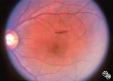
Neuro-Ophthalmic Consequences of Therapy | Don Bienfang, MD | An elderly woman was first seen in 1991, after being on Plaquenil (Hydroxychloroquine Sulfate) for 5 to 6 years, with a new symptomatic disturbance in her reading. Because her blue/yellow color perception was disturbed, and because she had symptoms, the medication was stopped. However the fundi and ... |
| 4 |
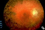 |
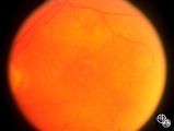
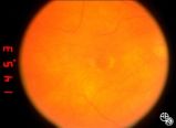
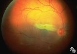
Optic Neuropathies | Rosa A. Tang, MD | Optic atrophy might be a false-localizing sign of optic nerve disease, as retinal disease may secondarily produce optic atrophy. In retinitis pigmentosa, for example, patients may exhibit a waxy disc palor. Fundus imaging. Anatomy: Retina. Disease/Diagnosis: Retinitis pigmentosa; Optic atrophy. |
| 5 |
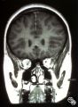 |
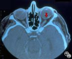
Ocular Manifestations of Congenital/Inherited Diseases | Jacqueline A. Leavitt, MD | This 22-year-old woman has neurofibromatosis, type 2. Acuity, color plates, pupillary responses, slit-lamp examination, IOP, fields, and funduscopy are all normal. There is a 3 mm proptosis OS. The patient has recently undergone gamma knife for the acoustic tumor, and she has residual facial nerve p... |
| 6 |
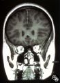 |
Ocular Manifestations of Congenital/Inherited Diseases | Jacqueline A. Leavitt, MD | This 22-year-old woman has neurofibromatosis, type 2. Acuity, color plates, pupillary responses, slit-lamp examination, IOP, fields, and funduscopy are all normal. There is a 3 mm proptosis OS. The patient has recently undergone gamma knife for the acoustic tumor, and she has residual facial nerve p... |
| 7 |
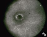 |
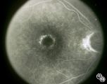
Neuro-Ophthalmic Consequences of Therapy | Don Bienfang, MD | An elderly woman was first seen in 1991, after being on Plaquenil (Hydroxychloroquine Sulfate) for 5 to 6 years, with a new symptomatic disturbance in her reading. Because her blue/yellow color perception was disturbed, and because she had symptoms, the medication was stopped. However the fundi and ... |
| 8 |
 |
Neuro-Ophthalmic Consequences of Therapy | Don Bienfang, MD | An elderly woman was first seen in 1991, after being on Plaquenil (Hydroxychloroquine Sulfate) for 5 to 6 years, with a new symptomatic disturbance in her reading. Because her blue/yellow color perception was disturbed, and because she had symptoms, the medication was stopped. However the fundi and ... |
| 9 |
 |
Neuro-Ophthalmic Consequences of Therapy | Don Bienfang, MD | An elderly woman was first seen in 1991, after being on Plaquenil (Hydroxychloroquine Sulfate) for 5 to 6 years, with a new symptomatic disturbance in her reading. Because her blue/yellow color perception was disturbed, and because she had symptoms, the medication was stopped. However the fundi and ... |
| 10 |
 |
Neuro-Ophthalmic Consequences of Therapy | Don Bienfang, MD | An elderly woman was first seen in 1991, after being on Plaquenil (Hydroxychloroquine Sulfate) for 5 to 6 years, with a new symptomatic disturbance in her reading. Because her blue/yellow color perception was disturbed, and because she had symptoms, the medication was stopped. However the fundi and ... |
| 11 |
 |
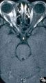
Neuro-Ophthalmic Manifestations of Brain Tumors | Jacqueline A. Leavitt, MD | Chordomas of the clivus may result in diplopia due to a sixth nerve palsy. The sixth nerve runs up the clivus and may be the presenting manifestation of the lesion. Pair with Images 97_33, 97_34, and 97_36. |
| 12 |
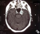 |
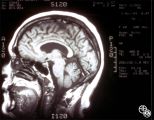
Neuro-Ophthalmic Manifestations of Brain Tumors | Jacqueline A. Leavitt, MD | Chordomas of the clivus may result in diplopia due to a sixth nerve palsy. The sixth nerve runs up the clivus and may be the presenting manifestation of the lesion. Imaging: MRI, T-1 axial with contrast. Pair with Images 97_33, 97_34, and 97_36. Anatomy: Clivus, Sixth nerve. Pathology: Sixth nerve p... |
| 13 |
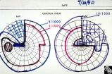 |
Chiasmal Syndromes | Larry P. Frohman, MD | This woman was 61 years old when she underwent initial neuro-ophthalmologic evaluation. Twenty-three years earlier, she had undergone removal of a pituitary adenoma followed by radiation therapy. At that time, she had noted a preoperative visual field defect that improved somewhat after the surgery.... |
| 14 |
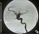 |
Neuro-Ophthalmic Vascular Disease | Mark J. Kupersmith, MD | Aneurysms of the intracranial circulation may act as mass lesions and compress the afferent of efferent visual pathway. Ophthalmic artery aneurysms may compress the optic nerve and result in an optic neuropathy (ie, visual loss, afferent pupillary defect, optic atrophy). Treatment includes endovascu... |
| 15 |
 |
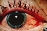
Systemic Disorders With Optic Nerve and Retinal Findings | Larry P. Frohman, MD | A 42-year old woman presented with a history of severe brow pain and 4 days of progressive visual loss OD. There was no increased pain on ocular rotation. Aside from heavy menses, she denied any significant past medical history. Her examination revealed acuity NLP OD, 20/25 OS; color vision 9/10 OS;... |
| 16 |
 |
Systemic Disorders With Optic Nerve and Retinal Findings | Larry P. Frohman, MD | A 42-year old woman presented with a history of severe brow pain and 4 days of progressive visual loss OD. There was no increased pain on ocular rotation. Aside from heavy menses, she denied any significant past medical history. Her examination revealed acuity NLP OD, 20/25 OS; color vision 9/10 OS;... |
| 17 |
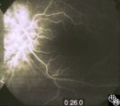 |
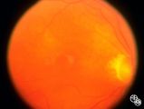
Systemic Disorders With Optic Nerve and Retinal Findings | Larry P. Frohman, MD | A 42-year old woman presented with a history of severe brow pain and 4 days of progressive visual loss OD. There was no increased pain on ocular rotation. Aside from heavy menses, she denied any significant past medical history. Disease/Diagnosis: Syphilitic optic neuritis/perineuritis, fundus, OS -... |
| 18 |
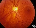 |
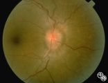
Isolated Congenital Optic Disc Anomalies | Thomas R. Wolf, MD | Patients with hypoplasia of the optic nerve may have normal or subnormal visual acuity or visual field. The condition may be unilateral or bilateral. Optic nerve hypoplasia is usually idiopathic, but maternal diabetes, or maternal use of anti-epileptic drugs or alcohol are predisposing factors. Opti... |
| 19 |
 |
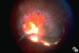
Ocular Manifestations of Congenital/Inherited Diseases | Larry P. Frohman, MD | This 14-year-old boy presented with sudden visual loss of the right eye that occurred 3 weeks before and due to a central retinal vein occlusion. His ocular history was quite complicated. He had had a resection of a lymphangioma of the left upper lid at age 7 months and underwent left orbitotomy for... |
| 20 |
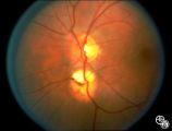 |
Bridge Coloboma | Anthony C. Arnold, MD | The patient is a 35-year-old asymptomatic woman with a bridge coloboma, also known as double optic disc. Disease/Diagnosis: Coloboma. |
| 21 |
 |
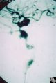
Ocular Manifestations of Congenital/Inherited Diseases | William Fletcher Hoyt, MD | Tuberous sclerosis is an autosomal dominant phakomatosis characterized by astrocytic hamartomas of the retina or optic nerve, adenoma sebaceum of the face, periungual fibromas, and astrocytic hamartomas of the brain, with secondary seizures and mental retardation. Disease/Diagnosis: Tuberous Scleros... |
| 22 |
 |
Isolated Congenital Optic Disc Anomalies | William Fletcher Hoyt, MD | This patient demonstrates bilateral tilted optic discs. Patients with this congenital optic disc anomaly may be asymptomatic or have bitemporal visual field defects that do not respect the vertical midline. Disease/Diagnosis: Tilted Discs. |
| 23 |
 |
Traumatic Internal Carotid Artery Dissection | Steven Galetta, MD | Traumatic dissection of the carotid artery may result in neck pain, an ipsilateral Horner's syndrome (disruption of the pericarotid sympathetic fibers), or ipsilateral arterial occlusions from embolic disease. Pair with images 91_18 and 91_20. |
| 24 |
 |
Traumatic Internal Carotid Atery Dissection | Steven Galetta, MD | Traumatic dissection of the carotid artery may result in neck pain, an ipsilateral Horner's syndrome (disruption of the pericarotid sympathetic fibers), or ipsilateral arterial occlusions from embolic disease. Pair with images 91_19 and 91_20. |
| 25 |
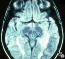 |
Magnetic Resonance Imaging in Detection of Extracranial Internal Carotid Artery Dissection | Marilyn C. Kay, MD | This 28-year-old woman presented with a 4-week history of bilateral visual loss. She had a known history of multiple sclerosis. Her vision was 20/60 OD and 20/40 OS, with an RAPD OS and optic pallor OU. Her fields and MRI are shown. Optic tract lesions usually result in an incongruous homonymous hem... |