AAO-NANOS Neuro-Ophthalmology Clinical Collection: Derived from the AAO-NANOS Clinical Neuro-Ophthalmology collection produced on CD. The images are of selected cases from the NANOS teaching slide exchange, and the CD was produced under the direction of Larry Frohman, MD and Andrew Lee, MD.
The American Academy of Ophthalmology (AAO); The North American Neuro-Ophthalmology Association (NANOS).
NOVEL: https://novel.utah.edu/
TO
Filters: Collection: "ehsl_novel_aao_nanos"
1 - 25 of 19
| Title | Curriculum | Description | Subject | Collection | ||
|---|---|---|---|---|---|---|
| 1 |
 |
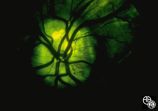
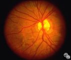
Isolated Congenital Optic Disc Anomalies | This patient has optic disc drusen and evidence of a superimposed optic neuropathy, including loss of visual field, an ipsilateral afferent pupillary defect, and optic atrophy. Although optic disc drusen typically causes visual field loss without visual acuity loss superimposed, ischemic optic neuro... | Optic Disc Drusen; Optic Nerve Drusen; Optic Neuropathy | Neuro-Ophthalmology Virtual Education Library: NOVEL http://NOVEL.utah.edu | |
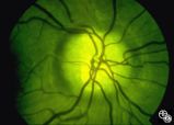
| 2 |
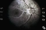
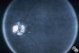 |
Optic Disc Drusen Autofluorescence | curriculum_fellow; IC-D1hvi-optic-nerve-drusen | This case of optic disc drusen demonstrates the typical autofluorescence. Pair with 92_68. Imaging: Autoflourescence? | Optic Disc Drusen; Optic Nerve Drusen; Autofluorescence | Neuro-Ophthalmology Virtual Education Library: NOVEL http://NOVEL.utah.edu |
| 3 |
 |
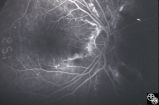
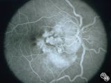
Optic Nerve Drusen, Late Fluorescein Angiogram | Images 92_64 and 92_67 demonstrate the characteristics of optic disc drusen on flourescein angiography. This image displays the nodular staining of the drusen without leakage. Pair with 92_63 and 92_64. | Optic Disc Drusen; Optic Nerve Drusen; Fluorescein Angiogram | Neuro-Ophthalmology Virtual Education Library: NOVEL http://NOVEL.utah.edu | |
| 4 |
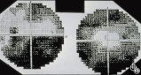 |
Optic Disc Drusen Visual Fields | This is the visual field of patient with optic nerve drusen. Whereas they typically do not cause central field loss, optic disc drusen may cause nerve fiber bundle layer defects and, thus, peripheral field defects, including altitudinal defects (seen inferiorly in the left eye) or arcuate defects (s... | Optic Disc Drusen; Optic Nerve Drusen; Visual Fields | Neuro-Ophthalmology Virtual Education Library: NOVEL http://NOVEL.utah.edu | |
| 5 |
 |
Isolated Congenital Optic Disc Anomalies | curriculum_fellow; IC-D1hvi-optic-nerve-drusen | This is a photograph of peripheral drusen. The paired image 92_69 demonstrates the typical autofluorescence. | Optic Disc Drusen; Optic Nerve Drusen; Autofluorescence | Neuro-Ophthalmology Virtual Education Library: NOVEL http://NOVEL.utah.edu |
| 6 |
 |
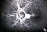
Optic Disc Drusen, Fluorescein Angiogram | Images 92_64 and 92_67 demonstrate the characteristics of optic disc drusen on flourescein angiography. This image shows the early arteriovenous phase, with irregular dye uptake and focal hypoflourescence superotemporally. Pair with 92_63 and 92_67 | Optic Disc Drusen; Optic Nerve Drusen; Fluorescein Angiogram | Neuro-Ophthalmology Virtual Education Library: NOVEL http://NOVEL.utah.edu | |
| 7 |
 |
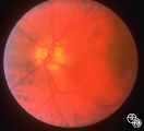
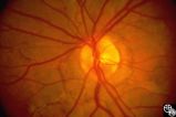
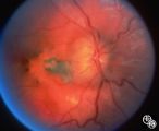
Isolated Congenital Optic Disc Anomalies | This image shows drusen that are especially prominent superotemporally. Pair with 92_64 and 92_67. | Optic Disc Drusen; Optic Nerve Drusen; Pseudopapilledema | Neuro-Ophthalmology Virtual Education Library: NOVEL http://NOVEL.utah.edu | |
| 8 |
 |
Optic Disc Drusen With Autofluorescence | This photograph of optic disc drusen demonstrates autoflourescence with flourescein barrier filters in place. Imaging: flourescein barrier filters. | Optic Disc Drusen; Autofluorescence | Neuro-Ophthalmology Virtual Education Library: NOVEL http://NOVEL.utah.edu | |
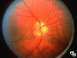
| 9 |
 |
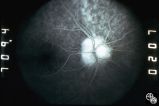
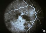
Isolated Congenital Optic Disc Anomalies | This optic disc displays multiple drusen. Note the pseudopapilledema here. One can differentiate this from true papilledema in that there is no obscuration of the vessel by the peripapillary nerve fiber layer as they cross the disc margin. This photograph was taken with barrier filters in place, but... | Optic Disc Drusen; Optic Nerve Drusen; Pseudopapilledema | Neuro-Ophthalmology Virtual Education Library: NOVEL http://NOVEL.utah.edu | |
| 10 |
 |
Isolated Congenital Optic Disc Anomalies | Retinaldystrophy | This patient has known pseudoxanthoma elasticum (an uncommon elastic tissue disorder characterized by plaque-like skin folds [plucked chicken skin] and degeneration of collagen fibers involving multiple systems, including the GI tract and heart), angioid streaks, and optic disc drusen. | Angioid Streaks; Pseudoxanthoma Elasticum; Retinal Dystrophy | Neuro-Ophthalmology Virtual Education Library: NOVEL http://NOVEL.utah.edu |
| 11 |
 |
Systemic Disorders With Optic Nerve and Retinal Findings | This patent has known pseudoxanthoma elasticum (an uncommon elastic tissue disorder characterized by plaque-like skin folds [plucked chicken skin], and degeneration of collagen fibers involving multiple systems, including the GI tract and heart), angioid streaks, and optic disc drusen. | Pseudoxanthoma Elasticum | Neuro-Ophthalmology Virtual Education Library: NOVEL http://NOVEL.utah.edu | |
| 12 |
 |
Systemic Disorders With Optic Nerve and Retinal Findings | This patent has known pseudoxanthoma elasticum (an uncommon elastic tissue disorder characterized by plaque-like skin folds [plucked chicken skin], and degeneration of collagen fibers involving multiple systems, including the GI tract and heart), angioid streaks, and optic disc drusen. | Pseudoxanthoma Elasticum | Neuro-Ophthalmology Virtual Education Library: NOVEL http://NOVEL.utah.edu | |
| 13 |
 |
Systemic Disorders With Optic Nerve and Retinal Findings | This patent has known pseudoxanthoma elasticum (an uncommon elastic tissue disorder characterized by plaque-like skin folds [plucked chicken skin], and degeneration of collagen fibers involving multiple systems, including the GI tract and heart), angioid streaks, and optic disc drusen. | Pseudoxanthoma Elasticum | Neuro-Ophthalmology Virtual Education Library: NOVEL http://NOVEL.utah.edu | |
| 14 |
 |
Systemic Disorders With Optic Nerve and Retinal Findings | This patent has known pseudoxanthoma elasticum (an uncommon elastic tissue disorder characterized by plaque-like skin folds [plucked chicken skin], and degeneration of collagen fibers involving multiple systems, including the GI tract and heart), angioid streaks, and optic disc drusen. Imaging of a... | Pseudoxanthoma Elasticum | Neuro-Ophthalmology Virtual Education Library: NOVEL http://NOVEL.utah.edu | |
| 15 |
 |
Systemic Disorders With Optic Nerve and Retinal Findings | This patent has known pseudoxanthoma elasticum (an uncommon elastic tissue disorder characterized by plaque-like skin folds [plucked chicken skin], and degeneration of collagen fibers involving multiple systems, including the GI tract and heart), angioid streaks, and optic disc drusen. Imaging of a... | Pseudoxanthoma Elasticum | Neuro-Ophthalmology Virtual Education Library: NOVEL http://NOVEL.utah.edu | |
| 16 |
 |
Isolated Congenital Optic Disc Anomalies | This 8-year-old boy presented with a 2-week history of decreased vision in the right eye. He had undergone a normal MRI and CSF examination, including intracranial pressure, before neuro-ophthalmologic assessment. The fundus photographs and fluorescein angiograms show subretinal neovascularization a... | Pseudopapilledema; Edema; Papilledema; Retinal Neurovascularization | Neuro-Ophthalmology Virtual Education Library: NOVEL http://NOVEL.utah.edu | |
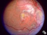
| 17 |
 |
Isolated Congenital Optic Disc Anomalies | This 8-year-old boy presented with a 2-week history of decreased vision in the right eye. He had undergone a normal MRI and CSF examination, including intracranial pressure, before neuro-ophthalmologic assessment. The fundus photographs and fluorescein angiograms show subretinal neovascularization a... | Pseudopapilledema; Edema; Papilledema; Retinal Neurovascularization | Neuro-Ophthalmology Virtual Education Library: NOVEL http://NOVEL.utah.edu | |
| 18 |
 |
Pseudopapilledema With Retinal Neovascularization, Fluorescein Angiogram | This 8-year-old boy presented with a 2-week history of decreased vision in the right eye. He had undergone a normal MRI and CSF examination, including intracranial pressure, before neuro-ophthalmologic assessment. The fundus photographs and fluorescein angiograms show subretinal neovascularization a... | Pseudopapilledema; Edema; Papilledema; Retinal Neurovascularization; Fluorescein Angiogram | Neuro-Ophthalmology Virtual Education Library: NOVEL http://NOVEL.utah.edu | |
| 19 |
 |
Pseudopapilledema With Subretinal Neovascularization on Fluorescein Angiogram | This 8-year-old boy presented with a 2-week history of decreased vision in the right eye. He had undergone a normal MRI and CSF examination, including intracranial pressure, before neuro-ophthalmologic assessment. The fundus photographs and fluorescein angiograms show subretinal neovascularization a... | Pseudopapilledema; Edema; Papilledema; Retinal Neurovascularization; Fluorescein Angiogram | Neuro-Ophthalmology Virtual Education Library: NOVEL http://NOVEL.utah.edu |
1 - 25 of 19