Collection of materials relating to neuro-ophthalmology as part of the Neuro-Ophthalmology Virtual Education Library.
NOVEL: https://novel.utah.edu/
TO
- NOVEL230
| Title | Creator | Description | Subject | ||
|---|---|---|---|---|---|
| 1 |
 |
Clinical Characteristics of Ocular Lateropulsion | Jorge C Kattah, MD | A discussion of the normal mechanism that maintain the eyes in normal horizontal position. | Ocular Lateropulsion; Unilateral Gaze Palsy; Radiographic h-CGD |
| 2 |
 |
Cogan's Lid Twitch Sign | Raed Behbehani, MD | Cogan's lid twitch sign is a twitch sign of he upper lid upon looking straight from a sustained downgaze position. It is associated with Ocular Myasthenia Gavis. | Myasthenia; Ptosis; Lid Twitch |
| 3 |
 |
Common Patterns of Visual Field Defects | Sean Gratton, MD; Sarah Lam, 6th year BA/MD | Lecture covering common visual field defects, including those of the retina, optic nerve, chiasm, and retrochiasmal. | Visual Field Defects |
| 4 |
 |
Complications of Strabismus Surgery and Botox | W. Walker Motley, MD | A narrated video slideshow outlining complications associated with strabismus surgery. | Strabismus; Surgery; Surgical Complications; Botox |
| 5 |
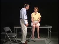 |
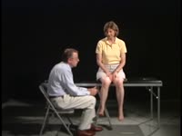
Coordination Exam: Normal Exam: Finger-to-nose (includes Spanish audio & captions) | Paul D. Larsen, MD | The patient moves her pointer finger from her nose to the examiner's finger as the examiner moves his finger to new positions and tests accuracy at the furthest outreach of the arm. NeuroLogic Exam has been supported by a grant from the Slice of Life Development Fund at the University of Utah, the D... | Coordination Examination; Finger-to-nose Test |
| 6 |
 |
Coordination Exam: Normal Exam: Heel-to-shin (includes Spanish audio & captions) | Paul D. Larsen, MD | The patient places her heel on the opposite knee then runs the heel down the shin to the ankle and back to the knee in a smooth coordinated fashion. NeuroLogic Exam has been supported by a grant from the Slice of Life Development Fund at the University of Utah, the Department of Pediatrics and the O... | Coordination Examination; Heel-shin Test |
| 7 |
 |
Cranial Nerves: Neuroanatomy Video Lab - Brain Dissections | Suzanne S. Stensaas, PhD | The approach is to learn to associate the cranial nerves with their brainstem level and blood supply. Emphasis is given to the midbrain (3, 4), pons (5, 6, 7, 8), medulla (9, 10, 11, 12) and their most important functions. | Cranial Nerves; Brain; Dissection |
| 8 |
 |
Direct and Consensual Response | Karl C. Golnik, MD | Explanation of direct and consensual response testing. | Direct and Consensual Response |
| 9 |
 |
Distance Visual Acuity Testing | Sean Gratton, MD | Demonstration of measuring distance visual accuity. | Visual Acuity Testing |
| 10 |
 |
Pediatric Visual Acuity Strategies: Induced Tropia Test | Anat Bachar Zipori, MD; Nasrin Najm-Tehrani, FRCS Ed (Ophth), FRCSC | Induced Tropia Test. Using a 10-20 base-down prism, this exam demonstrates equal vision in both eyes. If there is evidence of ocular misalignment, prisms can be used to neutralize the movement and thereby measure the deviation, whether it is a heterotropia or heterophoria. Prisms are placed in front... | Induced Tropia; Prism Test |
| 11 |
 |
Vertical optokinetic nystagmus | Anat Bachar Zipori, MD; Nasrin Najm-Tehrani, FRCS Ed (Ophth), FRCSC | Visual depiction of the vertical OKN drum test. The vertical optokinetic drum test is an exam used to elicit vertical optokinetic nystagmus (OKN) when there is horizontal nystagmus. The hand-held "optokinetic" drums or tapes that are used to elicit smooth movements primarily test the pursuit system.... | Vertical Optokinetic Nystagmus; OKN Drum Test |
| 12 |
 |
Horizontal Right Optokinetic Nystagmus | Anat Bachar Zipori, MD; Nasrin Najm-Tehrani, FRCS Ed (Ophth), FRCSC | Visual depiction of the horizontal OKN drum test. The hand-held "optokinetic" drums or tapes that are used to elicit smooth movements primarily test the pursuit system. This video supplements Paediatric Neuro-ophthalmology: Visual Acuity Assessment Strategies: https://collections.lib.utah.edu/ark:/... | Horizontal Right Optokinetic Nystagmus; OKN Drum Test |
| 13 |
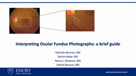 |
Interpreting Ocular Fundus Photographs: A Brief Guide | Gabriele Berman; Sachin Kedar; Nancy J. Newman; Valerie Biousse | This is a brief guide to the interpretation of the ocular fundus photograph. In this presentation we will describe the structures that comprise the normal ocular fundus followed the abnormalities that can be detected on fundus photographs. By the end of the presentation, learners should be able to d... | Fundus Photograph; Glaucoma; Papilledema; Retinal Detachment |
| 14 |
 |
John Cunningham Virus | Alison Gibbons; Amanda D. Henderson, MD | This learning object is a narrated Power Point presentation describing the features of, risk factors for, and clinical presentations of the John Cunningham, or JC, virus. It includes a discussion of various immunosuppressed states, including HIV, use of natalizumab (a disease-modifying therapy that ... | John Cunningham Virus; JC Virus; Natalizumab |
| 15 |
 |
Panoptic Ophthalmoscope | Amrita D. Vuppala, MD | Demonstration of using the panoptic ophthalmoscope in examinations. | Panoptic Ophthalmoscope |
| 16 |
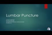 |
Lumbar Puncture | Jamison Hofer, DO; Sachin Kedar, MD | Explanation of lumbar puncture procedure. | Lumbar Puncture |
| 17 |
 |
A Five-Minute, Intravenous Edrophonium -Window to Observe the CNS Response to Myasthenic Ophthalmoplegia | Jorge C Kattah, MD | A video showing how ocular motor adaptation may be observed. | Ocular Motor Adaptation |
| 18 |
 |
Indirect Ophthalmoscopy | Jonathan Micieli, MD, CM | Demonstration of using the indirect ophthalmoscope in examinations. | Indirect Ophthalmoscopy |
| 19 |
 |
Nystagmus Elicitation Techniques | Jorge C. Kattah, MD | An examination of the patient days or weeks after the acute event requires fixation block, and a variety of techniques, known as nystagmus elicitation maneuvers to detect the recent vestibular imbalance. | Nystagmus |
| 20 |
 |
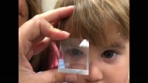
Pupil Exam | Carleigh N. Bruce, MD; Eric D. Habbe, MD; Ryan D. Walsh, MD | This video demonstrates how to conduct a pupillary exam on a patient. Specifically, pupils are evaluated in light and dark conditions, reactivity is assessed, and a swinging flashlight test is used to evaluate for a relative afferent pupillary defect. This video will be most helpful to early ophthal... | Pupil; Examination; Afferent Pupillary Defect; Swinging Flashlight Test; Reactivity |
| 21 |
 |
Introduction to Examination of the Pediatric Patient | Jason H. Peragallo, MD | Introduction to examination of the pediatric patient. | Pediatric Patient |
| 22 |
 |
Introduction to Examination of the Pupil in NANOS NOTE | Clare Fraser, MBBS, MMed | Introduction to Examination of the Pupil | Pupil; Examination |
| 23 |
 |
Vascular Headache Disorders | Salman Ali Mahmood, MD; Sean Gratton, MD | This is a PowerPoint with embedded audio presenting a detailed summary of the evaluation and management of an important cause of secondary headaches: vascular headache disorders. Topics covered include intracranial hemorrhage, arterial dissection, giant cell arteritis, cerebral venous sinus thrombos... | Vascular Headache Disorders; Epidural Hematoma; Subdural Hematoma; Subarachnoid Hemorrhage; Intracranial Hemorrhage; Arterial Dissection; Giant Cell Arteritis; Cerebral Venous Sinus Thrombosis; Pituitary Apoplexy |
| 24 |
 |
Natural Language Processing in Neuro-Ophthalmology | Areeba Abid, BS; Sachin Kedar, MD | This video provides an overview of natural language processing (NLP) techniques, applications, and limitations in neuro-ophthalmology. NLP, a branch of artificial intelligence, enables computers to understand and analyze human language. This video discusses three types of NLP techniques: sentiment a... | Artificial Intelligence; Machine Learning; Natural Language Processing; Word Prediction; Word Embeddings; Sentiment Analysis |
| 25 |
 |
Thiamine (Vitamin B1) Deficiency (Video) | Nirupama Devanathan; Devin D. Mackay, MD | 33-year-old female who initially presented with heat stroke that was thought to provoke cyclical vomiting over the course of 6 weeks, perpetuated by anxiety, resulting in a 15- pound weight loss. She presented to the ED with bilateral vision loss, bilateral lower extremity weakness and vertical nyst... | Thiamine; Upbeat Nystagmus; Nutritional Optic Neuropathy; Wernicke Encephalopathy |