A collection of videos relating to the diagnosis and treatment of eye movement disorders. This collection includes many demonstrations of examination techniques.
Dan Gold, D.O., Associate Professor of Neurology, Ophthalmology, Neurosurgery, Otolaryngology - Head & Neck Surgery, Emergency Medicine, and Medicine, The Johns Hopkins School of Medicine.
A collection of videos relating to the diagnosis and treatment of eye movement disorders.
NOVEL: https://novel.utah.edu/
TO
| Title | Description | Type | ||
|---|---|---|---|---|
| 1 |
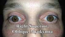 |
Superior Oblique Myokymia (SOM) | 𝗢𝗿𝗶𝗴𝗶𝗻𝗮𝗹 𝗗𝗲𝘀𝗰𝗿𝗶𝗽𝘁𝗶𝗼𝗻: This is a patient with transient monocular oscillopsia OD and vertical diplopia noted to have many episodes of SOM in the office. There was not only myokymia OD, but also a 4 prism diopter left hypertropia during episodes... | Image/MovingImage |
| 2 |
 |
Evaluation of Convergence | 𝗢𝗿𝗶𝗴𝗶𝗻𝗮𝗹 𝗗𝗲𝘀𝗰𝗿𝗶𝗽𝘁𝗶𝗼𝗻: The assessment of convergence includes measuring alignment at near versus distance (see video, https://collections.lib.utah.edu/details?id=187677), near point of convergence and convergence amplitude. Near point of conve... | Image/MovingImage |
| 3 |
 |
Paroxysmal Ocular Tilt Reaction | 𝗢𝗿𝗶𝗴𝗶𝗻𝗮𝗹 𝗗𝗲𝘀𝗰𝗿𝗶𝗽𝘁𝗶𝗼𝗻: This is a 60-year-old woman who 2 years prior experienced a left sided hypertensive hemorrhagic stroke, resulting in right hemiparesis, dysarthria and vertical diplopia. The initial vertical diplopia resolved completely a... | Image/MovingImage |
| 4 |
 |
Range of Motion (Ductions) | Range of motion (ductions): check the range of each individual eye (ductions) if there is diplopia or if a motility deficit is suspected. Instructing the patient to hold their head 20o to the right or to the left may provide a better view of the range of horizontal gaze, if there is diplopia or if a... | Image/MovingImage |
| 5 |
 |
Bilateral INOs Due to Stroke | This is a 65-year-old man with multiple vascular risk factors who experienced the abrupt onset of diplopia 6 months prior to this video. MRI done within 24 hours of onset was unremarkable. Examination demonstrated subtle bilateral adduction lag with horizontal saccades. There was very mild abducting... | Image/MovingImage |
| 6 |
 |
Mild 6th Nerve Palsy Due to Pontine Stroke | This is a 70-year-old woman with HTN and diabetes who presented with horizontal diplopia for several weeks, worse in right gaze. There was a very subtle abduction paresis OD with full motility elsewhere. With cover-uncover testing, there was a small esotropia in right gaze (esodeviation seen with al... | Image/MovingImage |
| 7 |
 |
Measuring Divergence Amplitude | Divergence insufficiency should be suspected in patients with binocular horizontal diplopia at distance (but not near) who lack abduction deficits. There should be an esodeviation greater at distance, and in older patients with levator dehiscence (or previous ptosis surgery) and prominent superior s... | Image/MovingImage |
| 8 |
 |
Wall-eyed Bilateral Internuclear Ophthalmoplegia (WEBINO) in MS | This is a young woman with a years-long history of multiple sclerosis who presented 2 years prior to this examination with complaints of oscillopsia (which was due to spontaneous upbeat nystagmus), as well as diplopia (which was due to bilateral internuclear ophthalmoplegia, INO). ; ; At the time of... | Image/MovingImage |
| 9 |
|
Spontaneous Torsional Nystagmus and Ocular Tilt Reaction | 𝗢𝗿𝗶𝗴𝗶𝗻𝗮𝗹 𝗗𝗲𝘀𝗰𝗿𝗶𝗽𝘁𝗶𝗼𝗻: This is a 70-year-old man who experienced "a delay in focusing" with "some twisting movement" that began about 18 months prior to this video with mild progression over days or weeks. For the same period of time, he experi... | Image/MovingImage |
| 10 |
 |
Ocular Motor Signs in Progressive Supranuclear Palsy (PSP) | 𝗢𝗿𝗶𝗴𝗶𝗻𝗮𝗹 𝗗𝗲𝘀𝗰𝗿𝗶𝗽𝘁𝗶𝗼𝗻: This is a 65-yo-woman complaining of imbalance and double vision. She had significant convergence insufficiency (and would close her right eye with near viewing), providing an explanation for her diplopia. Convergence ins... | Image/MovingImage |
| 11 |
 |
Typical Features of Duane Syndrome Type 1 | 𝗢𝗿𝗶𝗴𝗶𝗻𝗮𝗹 𝗗𝗲𝘀𝗰𝗿𝗶𝗽𝘁𝗶𝗼𝗻: This is a patient seen for vestibular complaints, who on exam, was found to have (unrelated to her vestibular symptoms) impaired abduction OS. In adduction, there was narrowing of the palpebral fissure OS, a result of glo... | Image/MovingImage |
| 12 |
 |
Mesodiencephalic Stroke Causing Unilateral riMLF and INC Ocular Motor Syndromes | 𝗢𝗿𝗶𝗴𝗶𝗻𝗮𝗹 𝗗𝗲𝘀𝗰𝗿𝗶𝗽𝘁𝗶𝗼𝗻: This is a 65-year-old man who experienced the sudden onset of diplopia (with horizontal and vertical components), dysarthria and imbalance. An MRI performed the following day showed a left mesodiencephalic stroke. The pat... | Image/MovingImage |
| 13 |
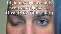 |
Cavernous Sinus Mass Causing Right 3rd and 4th Nerve Palsies | 𝗢𝗿𝗶𝗴𝗶𝗻𝗮𝗹 𝗗𝗲𝘀𝗰𝗿𝗶𝗽𝘁𝗶𝗼𝗻: 25-yo-man who complained of diplopia and was initially found to have right 4th and 6th nerve palsies in the setting of a right cavernous sinus mass (subsequently diagnosed as Ewing's sarcoma). When seen in follow-up (this... | Image/MovingImage |
| 14 |
 |
Leukemic Leptomeningeal Carcinomatosis Causing 4th and 6th Nerve Palsies | 𝗢𝗿𝗶𝗴𝗶𝗻𝗮𝗹 𝗗𝗲𝘀𝗰𝗿𝗶𝗽𝘁𝗶𝗼𝗻: This is a 55-yo-man with CML that recurred as AML. Diagonal diplopia developed, and on examination he was found to have a partial right 6th nerve palsy, in addition to a left hypertropia that increased in right gaze, down... | Image/MovingImage |
| 15 |
 |
Wallenberg Syndrome in MS | 𝗢𝗿𝗶𝗴𝗶𝗻𝗮𝗹 𝗗𝗲𝘀𝗰𝗿𝗶𝗽𝘁𝗶𝗼𝗻: 30-yo-woman with MS presenting with acute vertigo and vertical diplopia. Examination demonstrated several aspects of the Wallenberg syndrome (her acute demyelinating lesion was in the left lateral medulla): ipsilesional (... | Image/MovingImage |
| 16 |
 |
Miller Fisher Syndrome - Ophthalmoplegia and Hyperreflexia | 𝗢𝗿𝗶𝗴𝗶𝗻𝗮𝗹 𝗗𝗲𝘀𝗰𝗿𝗶𝗽𝘁𝗶𝗼𝗻: This is a 45-yo-woman who presented with mild imbalance and diplopia. There had been a preceding viral illness several weeks prior. Examination demonstrated horizontal gaze paresis (sparing unilateral adduction), mild gai... | Image/MovingImage |
| 17 |
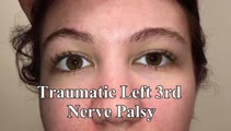 |
Traumatic 3rd Nerve Palsy with Aberrant Regeneration | 𝗢𝗿𝗶𝗴𝗶𝗻𝗮𝗹 𝗗𝗲𝘀𝗰𝗿𝗶𝗽𝘁𝗶𝗼𝗻: This is a 20-yo-woman who experienced severe head trauma and diplopia upon awakening from a coma several weeks after the injury. She had a partial left 3rd nerve palsy (adduction spared), and when she looked to the right ... | Image/MovingImage |
| 18 |
 |
Complete Microvascular 6th Nerve Palsy with Slow Abducting Saccade | 𝗢𝗿𝗶𝗴𝗶𝗻𝗮𝗹 𝗗𝗲𝘀𝗰𝗿𝗶𝗽𝘁𝗶𝗼𝗻: This is a 90-year-man with HTN, HLD, DM who woke up with horizontal diplopia. Two years prior, he was diagnosed with a microvascular right 6th nerve palsy that resolved over several months. There was little concern for gi... | Image/MovingImage |
| 19 |
 |
Slow Volitional Saccades and Poor Fast Phases to an Optokinetic Stimulus, with Preserved Head Impulse Testing | This is a 67-year-old woman presenting with imbalance and binocular horizontal diplopia at near. On examination there were frequent square wave jerks, limited supraduction OU and convergence insufficiency, which explained her diplopia. Pursuit and suppression of the vestibulo-ocular reflex were sa... | Image/MovingImage |
| 20 |
 |
Central HINTS (With an Abnormal Head Impulse Sign) in the Acute Vestibular Syndrome Due to Lateral Pontine/Middle Cerebellar Peduncle Demyelination | 𝗢𝗿𝗶𝗴𝗶𝗻𝗮𝗹 𝗗𝗲𝘀𝗰𝗿𝗶𝗽𝘁𝗶𝗼𝗻: This is a 30-year-old man presenting with vertigo, diplopia and mild left facial weakness (not seen in the video). On exam, there was right-beating nystagmus (RBN) in primary gaze that increased in right gaze (in accordan... | Image/MovingImage |
| 21 |
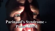 |
Parinaud's Syndrome in a Man with GBM of the Pineal Gland | 𝗢𝗿𝗶𝗴𝗶𝗻𝗮𝗹 𝗗𝗲𝘀𝗰𝗿𝗶𝗽𝘁𝗶𝗼𝗻: This is a 60-yo-man who presented with diplopia, headaches, and difficulty looking up, and was found to have a mass involving the pineal gland. Biopsy was diagnostic of a GBM. Major features of Parinaud's (dorsal midbrain... | Image/MovingImage |
| 22 |
|
Saccadic Hypermetria and Ipsipulsion (Behind Closed Eyelids and with Vertical Saccades) | 𝗢𝗿𝗶𝗴𝗶𝗻𝗮𝗹 𝗗𝗲𝘀𝗰𝗿𝗶𝗽𝘁𝗶𝗼𝗻: This is a 40-year-old woman who experienced oscillopsia and vertical diplopia, due to spontaneous torsional nystagmus and a skew deviation (right hypotropia), respectively. The symptom onset was 7 months prior to these vi... | Image/MovingImage |
| 23 |
 |
Oculopalatal Tremor and Internuclear Ophthalmoplegia Due to Hemorrhagic Pontine Cavernoma | 𝗢𝗿𝗶𝗴𝗶𝗻𝗮𝗹 𝗗𝗲𝘀𝗰𝗿𝗶𝗽𝘁𝗶𝗼𝗻: This is a 60-year-old woman who experienced 2 episodes of vertigo, nausea and vomiting, which was felt to be related to recurrent hemorrhage of a pontine cavernoma that was adjacent to the fourth ventricle. The cavernoma ... | Image/MovingImage |
| 24 |
|
Superior Oblique Myokymia - Three Patients with Recorded Attacks Using VOG and Frenzel Goggles | 𝗢𝗿𝗶𝗴𝗶𝗻𝗮𝗹 𝗗𝗲𝘀𝗰𝗿𝗶𝗽𝘁𝗶𝗼𝗻: Patients with superior oblique myokymia (SOM) commonly present with complaints of monocular oscillopsia and/or vertical diplopia, which are related to the primary and secondary actions of the SO (incycloduction and depres... | Image/MovingImage |
| 25 |
 |
Wall-eyed Bilateral INO in Caudal Midbrain Lesion | This is a 30-yo-woman with the relatively acute onset of diplopia. There was a large angle exotropia, very subtle lag of the adducting saccades OD>OS, suggestive of bilateral INOs. This was best seen with rapid horizontal saccades, and a lesion involving bilateral MLFs in the caudal midbrain was dem... | Image/MovingImage |