A collection of videos relating to the diagnosis and treatment of eye movement disorders. This collection includes many demonstrations of examination techniques.
Dan Gold, D.O., Associate Professor of Neurology, Ophthalmology, Neurosurgery, Otolaryngology - Head & Neck Surgery, Emergency Medicine, and Medicine, The Johns Hopkins School of Medicine.
A collection of videos relating to the diagnosis and treatment of eye movement disorders.
NOVEL: https://novel.utah.edu/
TO
| Title | Description | Type | ||
|---|---|---|---|---|
| 201 |
 |
Ocular Bobbing Due to Hepatic Encephalopathy | 𝗢𝗿𝗶𝗴𝗶𝗻𝗮𝗹 𝗗𝗲𝘀𝗰𝗿𝗶𝗽𝘁𝗶𝗼𝗻: This is a 55-year-old man presented with hepatic encephalopathy, and found to have ocular bobbing. Head CT did not show any acute changes. Ocular bobbing almost always localizes to the pons, although cerebellar pathology ... | Image/MovingImage |
| 202 |
 |
Ocular Motor & Vestibular Features of the MLF Syndrome | 𝗢𝗿𝗶𝗴𝗶𝗻𝗮𝗹 𝗗𝗲𝘀𝗰𝗿𝗶𝗽𝘁𝗶𝗼𝗻: This 61-year-old woman with HTN and DM presented for evaluation of acute onset diagonal diplopia. Adduction OS was about 60% of normal while medialization OS improved with convergence. In right gaze, dissociated abducti... | Image/MovingImage |
| 203 |
 |
Oculogyric Crisis | 𝗢𝗿𝗶𝗴𝗶𝗻𝗮𝗹 𝗗𝗲𝘀𝗰𝗿𝗶𝗽𝘁𝗶𝗼𝗻: This is a patient with neuroleptic-induced oculogyric crisis. 𝗡𝗲𝘂𝗿𝗼-𝗼𝗽𝗵𝘁𝗵𝗮𝗹𝗺𝗼𝗹𝗼𝗴𝘆 𝗮𝗻𝗱 𝗡𝗲𝘂𝗿𝗼-𝗼𝘁𝗼𝗹𝗼𝗴𝘆 𝗧𝗲𝘅𝘁𝗯�... | Image/MovingImage |
| 204 |
 |
Oculopalatal Tremor and Internuclear Ophthalmoplegia Due to Hemorrhagic Pontine Cavernoma | 𝗢𝗿𝗶𝗴𝗶𝗻𝗮𝗹 𝗗𝗲𝘀𝗰𝗿𝗶𝗽𝘁𝗶𝗼𝗻: This is a 60-year-old woman who experienced 2 episodes of vertigo, nausea and vomiting, which was felt to be related to recurrent hemorrhage of a pontine cavernoma that was adjacent to the fourth ventricle. The cavernoma ... | Image/MovingImage |
| 205 |
 |
Oculopalatal Tremor and One-and-a-Half Syndrome Due to Pontine Hemorrhage | 𝗢𝗿𝗶𝗴𝗶𝗻𝗮𝗹 𝗗𝗲𝘀𝗰𝗿𝗶𝗽𝘁𝗶𝗼𝗻: This is a 65-year-old man who was put on a blood thinner, and shortly thereafter experienced a midline pontine hemorrhage, which was more dense on the left side. Immediately afterwards, right hemiparesis and hemi-anesthes... | Image/MovingImage |
| 206 |
 |
Oculopalatal Tremor with Prominent Nystagmus, Bilateral Horizontal Gaze Palsy, and Bilateral Facial Palsies | 𝗢𝗿𝗶𝗴𝗶𝗻𝗮𝗹 𝗗𝗲𝘀𝗰𝗿𝗶𝗽𝘁𝗶𝗼𝗻: This is a 50-year-old woman who experienced the acute onset of right sixth and seventh nerve palsies and left hemiparesis. Two cavernomas within the right pons (one in the region of the facial colliculus) were demonstrat... | Image/MovingImage |
| 207 |
 |
Optokinetic Nystagmus | 𝗢𝗿𝗶𝗴𝗶𝗻𝗮𝗹 𝗗𝗲𝘀𝗰𝗿𝗶𝗽𝘁𝗶𝗼𝗻: During the bedside evaluation of optokinetic nystagmus (OKN), the patient is instructed to look at each red (or white) square as it moves past. Because this is not a full-field visual stimuli, using an optokinetic flag m... | Image/MovingImage |
| 208 |
 |
Pendular, Gaze-Evoked and Abducting Nystagmus in MS | This is a 40-year-old woman with a history of multiple sclerosis who presented for oscillopsia. On examination, she had bilateral internuclear ophthalmoplegia (INO-adduction lag OU and abducting nystagmus OU), with a corresponding exotropia that increased in right and left gaze. She also had horiz... | Image/MovingImage |
| 209 |
 |
Penlight Cover Test (Partial Removal of Fixation) | Penlight cover test (partial removal of fixation): during in-person clinical encounters, the maneuvers below are best tested with complete (or near complete) removal of fixation (e.g., Frenzel or video Frenzel goggles). Removal of fixation is more challenging during virtual evaluations but can be ap... | Image/MovingImage |
| 210 |
 |
Peripheral (Vestibular) and Central (Gaze-Evoked) Patterns of Nystagmus in a Single Patient | A 55-year-old man experienced episodic vertigo and was diagnosed with Meniere's disease affecting the left ear (based on audiograms and his clinical course) about 1 year prior to presentation. About 6 months prior to presentation, intratympanic (IT) gentamicin was injected into the left ear, at whic... | Image/MovingImage |
| 211 |
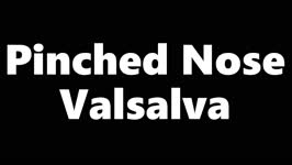 |
Pinched Nose Valsalva | Valsalva (closed glottis or pinched nose): instruct the patient to take a deep breath and ‘bear down' (closed glottis) or take a deep breath and ‘try to pop their ears' (pinched nose). Assess for nystagmus. In superior canal dehiscence, pressure changes may be transmitted to the superior canal, ... | Image/MovingImage |
| 212 |
 |
Pressure Testing for Superior Canal Dehiscence Syndrome | 𝗢𝗿𝗶𝗴𝗶𝗻𝗮𝗹 𝗗𝗲𝘀𝗰𝗿𝗶𝗽𝘁𝗶𝗼𝗻: Superior semicircular canal dehiscence syndrome (SCDS) is caused by a third mobile window in the inner ear. This allows for transmission of sound or pressure to the superior canal. Tragal compression and/or glottic and ... | Image/MovingImage |
| 213 |
 |
Provocative Maneuvers (Removal of Fixation, Vibration, Head-Shaking) to Accentuate Peripheral Vestibular Nystagmus) | 𝗢𝗿𝗶𝗴𝗶𝗻𝗮𝗹 𝗗𝗲𝘀𝗰𝗿𝗶𝗽𝘁𝗶𝗼𝗻: With an acute destructive process like vestibular neuritis that causes significant unilateral vestibular loss, spontaneous nystagmus is always present. However, over days to months, spontaneous nystagmus should resolve co... | Image/MovingImage |
| 214 |
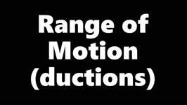 |
Range of Motion (Ductions) | Range of motion (ductions): check the range of each individual eye (ductions) if there is diplopia or if a motility deficit is suspected. Instructing the patient to hold their head 20o to the right or to the left may provide a better view of the range of horizontal gaze, if there is diplopia or if a... | Image/MovingImage |
| 215 |
 |
Saccades | Saccades: instruct the patient to make rapid movements of their eyes in each gaze direction, noting the speed, conjugacy, latency, and accuracy. First have the patient look between an eccentric target and the camera horizontally and vertically, making assessment of accuracy easier - e.g., overshooti... | Image/MovingImage |
| 216 |
 |
Skew Deviation and Spontaneous Nystagmus Due to Posterior Fossa Lesions | This is a 50-year-old woman who reported the abrupt onset of imbalance, right upper extremity incoordination and binocular vertical diplopia several months prior to her presentation to our clinic. On examination, she had a left hypertropia that was fairly comitant (measuring 5 prism diopters) assoc... | Image/MovingImage |
| 217 |
 |
Slow Saccades Due to Unilateral Paramedian Pontine Reticular Formation (PPRF) Injury with Preserved Movements Using the Vestibulo-Ocular Reflex | 𝗢𝗿𝗶𝗴𝗶𝗻𝗮𝗹 𝗗𝗲𝘀𝗰𝗿𝗶𝗽𝘁𝗶𝗼𝗻: This is a 60-year-old man who presented for imbalance and oscillopsia 10 months after surgery and 8 months after radiation for Merkel cell carcinoma of the neck. He developed imbalance after surgery and diplopia and osci... | Image/MovingImage |
| 218 |
 |
Slow Volitional Saccades and Poor Fast Phases to an Optokinetic Stimulus, with Preserved Head Impulse Testing | This is a 67-year-old woman presenting with imbalance and binocular horizontal diplopia at near. On examination there were frequent square wave jerks, limited supraduction OU and convergence insufficiency, which explained her diplopia. Pursuit and suppression of the vestibulo-ocular reflex were sa... | Image/MovingImage |
| 219 |
 |
Smooth Pursuit | Smooth pursuit: instruct the patient to hold their head steady, fix their eyes on the camera and slowly move the camera in the horizontal and vertical planes. Or, have the patient focus on their outstretched thumbnail (or other small fixation target), while following the slowly moving object horizon... | Image/MovingImage |
| 220 |
 |
Spontaneous Upbeat Nystagmus in Acute Wernicke's Encephalopathy | 𝗢𝗿𝗶𝗴𝗶𝗻𝗮𝗹 𝗗𝗲𝘀𝗰𝗿𝗶𝗽𝘁𝗶𝗼𝗻: This is a 40-year-old woman presenting with imbalance, confusion and oscillopsia. Exam demonstrated upbeat nystagmus (UBN) in primary gaze that remained UB in all directions of gaze, with a slight torsional component (top... | Image/MovingImage |
| 221 |
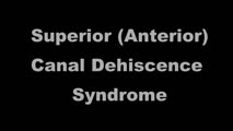 |
Superior Canal Dehiscence | 𝗢𝗿𝗶𝗴𝗶𝗻𝗮𝗹 𝗗𝗲𝘀𝗰𝗿𝗶𝗽𝘁𝗶𝗼𝗻: This is a 60-yo-man who complained of autophony (eg, hearing his own heartbeat, noting that his own voice sounded too loud) and dizziness triggered with loud noises and straining at times. With pinched-nose Valsalva maneu... | Image/MovingImage |
| 222 |
 |
Test Your Knowledge - Bilateral 4th Nerve Palsies | Watch the video until instructed to stop. Which of the following features is likely to be present given her exam findings? A. Gaze-evoked nystagmus and impaired smooth pursuit B. History of traumatic brain injury C. History of blepharoplasty or brow lift surgery and prominence of superior sulcus on ... | Image/MovingImage |
| 223 |
|
Test Your Knowledge - Central and Peripheral Vestibular and Ocular Motor Signs Due to a Large Vestibular Schwannoma | Which of the following is least likely to be the correct localization or etiology given the findings seen in the video? 1) Acute right 8th cranial neuropathy 2) Right-sided vestibular schwannoma 3) Right vestibular nucleus infarction 4) Right anterior inferior cerebellar artery distribution stroke A... | Image/MovingImage |
| 224 |
|
Test Your Knowledge - Monocular Oscillopsia | 𝗢𝗿𝗶𝗴𝗶𝗻𝗮𝗹 𝗗𝗲𝘀𝗰𝗿𝗶𝗽𝘁𝗶𝗼𝗻: Which of the following associated signs is most likely to be seen in this patient presenting with oscillopsia? A. Optic nerve pallor B. Palatal tremor C. Severe unilateral cataract D. Head bobbing E. Neurovascular contact... | Image/MovingImage |
| 225 |
|
Test Your Knowledge - Optokinetic Nystagmus with a Parietal Lesion | 𝗢𝗿𝗶𝗴𝗶𝗻𝗮𝗹 𝗗𝗲𝘀𝗰𝗿𝗶𝗽𝘁𝗶𝗼𝗻: Given the finding seen in the first part of the video, which of the following associated features are most likely? (more than one answer may be correct) A. Left homonymous visual field defect B. Right homonymous visual fi... | Image/MovingImage |