Best known for his world-renowned neuro-ophthalmology unit based at the University of California, San Francisco, William Hoyt, MD collected here more than 850 of his best images covering a wide range of disorders.
William F. Hoyt, MD, Professor Emeritus of Ophthalmology, Neurology and Neurosurgery, Department of Ophthalmology, University of California, San Francisco.
NOVEL: https://novel.utah.edu/
TO
| Title | Description | Type | ||
|---|---|---|---|---|
| 1 |
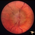 |
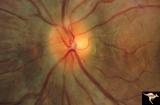
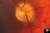
Bilateral Chronic Papilledema | Left eye. Frisen's stage 5. Patient with long standing aqueductal stenosis. Bilateral Chronic Papilledema. Man. Anatomy: Optic disc. Pathology: Papilledema. Disease/Diagnosis: Papilledema from aqueductal stenosis. | Image |
| 2 |
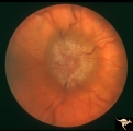 |
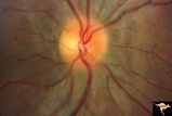
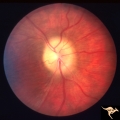
Bilateral Chronic Papilledema | Right eye. Frisen's stage 5. Patient with long standing aqueductal stenosis. Bilateral Chronic Papilledema. Man. Anatomy: Optic disc. Pathology: Papilledema. Disease/Diagnosis: Papilledema from aqueductal stenosis. | Image |
| 3 |
 |
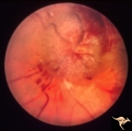
Chronic Atrophic Papilledema | Left eye. Left eye blind. Chronic Atrophic Papilledema. Obese woman (300 lbs) with large tentorial meningioma. "Pseudo Pseudotumor". Anatomy: Optic disc. Pathology: Papilledema. Disease/Diagnosis: Papilledema from large tentorial meningioma. | Image |
| 4 |
 |
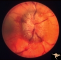
Chronic Atrophic Papilledema | Right eye. Chronic Atrophic Papilledema. Obese woman (300 lbs) with large tentorial meningioma. "Pseudo Pseudotumor" Anatomy: Optic disc. Pathology: Papilledema. Disease/Diagnosis: Papilledema from large tentorial meningioma. | Image |
| 5 |
 |
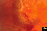
Early Papilledema due to Brain Tumor - Resolving | Left eye. Same eye as P_34a. One month post op, papilledema resolving. Boy. Anatomy: Optic disc. Pathology: Papilledema. Disease/Diagnosis: Papilledema from posterior fossa hemangioblastoma. | Image |
| 6 |
 |
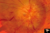
Early Papilledema due to Tumor | Left eye. Asymmetric Papilledema with posterior fossa hemangioblastoma. Left - moderate papilledema. Blurring of disc. Young man. Anatomy: Optic disc. Pathology: Papilledema. Disease/Diagnosis: Papilledema from posterior fossa hemangioblastoma. | Image |
| 7 |
 |
Papilledema due to Brain Tumor - Natural History | Left eye. 3.5 years after presentation. Atrophy appears about the same. Note especially the narowing of retinal arterioles. Visual loss is profound in both eyes. Note the horizontal retinal folds. Papilledema due to brain tumor - 3 year natural history. Patient refused treatment. Man. | Image |
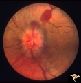
| 8 |
 |
Progression of Papilledema due to Metastatic Melanoma | Right eye at presentation. Early stage bilateral papilledema in a man. Note increased papilledema. Rapid progression of papilledema due to occipital metastatic melanoma. Anatomy: Optic disc. Pathology: Papilledema. Disease/Diagnosis: Early stage bilateral papilledema. | Image |
| 9 |
 |
Progression of Papilledema due to Metastatic Melanoma | Right eye at presentation. Early stage bilateral papilledema in a man. Rapid progression of bilateral papilledema due to metastatic occipital melanoma. Anatomy: Optic disc. Pathology: Papilledema. Disease/Diagnosis: Papilledema. | Image |
| 10 |
 |
Chronic Papilledema due to Brain Tumor | Right eye. Chronic papilledema wth white centrally located exudates in a man with hemispheric glioma. Anatomy: Optic disc. Pathology: Papilledema. Disease/Diagnosis: Chronic papilledema. | Image |
| 11 |
 |
Chronic Papilledema due to Brain Tumor | Left eye. Chronic papilledema with white centrally located exudates in a man with hemispheric glioma. Anatomy: Optic disc. Pathology: Papilledema. Disease/Diagnosis: Chronic papilledema. | Image |
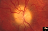
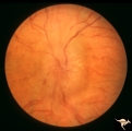
| 12 |
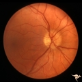 |
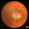
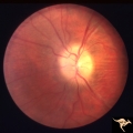
P43a Asymmetrical Papilledema due to Brain Tumor | Right eye. Early papilledema. Incipient papilledema barely recognizable. Early papilledema due to posterior fossa meningioma in a boy. Anatomy: Optic disc. Pathology: Papilledema. Disease/ Diagnosis: Asymmetrical papilledema due to posterior fossa meningioma. | Image |
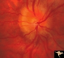
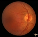
| 13 |
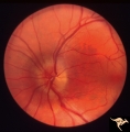 |
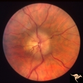
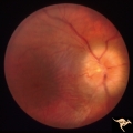
P43b Asymmetrical Papilledema due to Brain Tumor | Left eye. Early papilledema. Clearly has papilledema. Early papilledema to posterior fossa meningioma in a boy. | Image |
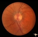
| 14 |
 |
Papilledema with Choroidal Folds | Chronic papilledema with choroidal folds. Frontal astrocytoma. Man. Anatomy: Optic disc. Pathology: Papilledema. Disease/Diagnosis: Chronic papilledema. | Image |
| 15 |
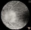 |
Papilledema with Choroidal Folds | Chronic papilledema with choroidal folds. Frontal astrocytoma. Flouroscein angiogram of choroidal folds. Man. | Image |
| 16 |
 |
Post Papilledema | Right eye. Post Papilledema with minimal optic disc changes after treatment for temporal lobe glioma. Minimal optic disc haze. Optic disc. Pathology: Papilledema. Disease/Diagnosis: Post Papilledema due to temporal lobe glioma. | Image |
| 17 |
 |
Post Papilledema | Left eye. Post Papilledema with minimal optic disc changes after treatment for temporal lobe glioma. Minimal optic disc haze. | Image |
| 18 |
 |
Post Papilledema Disc Blurring | Left eye. 8 year old boy. Post papilledema due to brain tumor. Note the entire peripapillary nerve fiber is blurred but the optic discs are barely elevated. Anatomy: Optic disc. Pathology: Brain tumor. Disease/Diagnosis: Papilledema. Clinical: Post papilledema due to brain tumor. | Image |
| 19 |
 |
Post Papilledema Disc Blurring | Right eye. 8 year old boy. Post papilledema due to brain tumor. Note the entire peripapillary nerve fiber is blurred but the optic discs are barely elevated. Anatomy: Optic disc. Pathology: Post papilledema. Disease/Diagnosis: Post papilledema due to brain tumor. | Image |
| 20 |
 |
Bilateral Papilledema from Occipital Tumor | Left eye. Bilateral hemorrhagic papilledema. Occipital glioma. Woman. Anatomy: Optic disc. Pathology: Papilledema. Disease/Diagnosis: Hemorrhagic papilledema from occipital glioma. | Image |
| 21 |
 |
Bilateral Papilledema from Occipital Tumor | Right eye. Bilateral hemorrhagic papilledema. Occipital glioma. Right hemianopia. Woman. Anatomy: Optic disc. Pathology: Papilledema. Disease/Diagnosis: Hemorrhagic papilledema from occipital glioma. | Image |
| 22 |
 |
Papilledema due to Brain Tumor - Natural History | Right eye 33 months after presentation. Atrophy appears about the same. Notice especially the narrowing of retinal arterioles. Visual loss is severe in both eyes. Papilledema due to brain tumor - 3 year natural history. Patient refused treatment. Man. Anatomy: Optic disc. Pathology: Papilledema. Dis... | Image |
| 23 |
 |
Papilledema due to Brain Tumor - Natural History | Right eye at 27 months after presentation. Atrophy is more profound in both eyes. Papilledema due to brain tumor - 3 year natural history. Patient refused treatment. Man. Anatomy: Optic disc. Pathology: Papilledema. Disease/Diagnosis: Papilledema from acoustic neuronoma. | Image |
| 24 |
 |
Papilledema due to Brain Tumor - Natural History | Left eye at 25 months after presentation. Atrophy is more profound in both eyes. Papilledema due to brain tumor - 3 year natural history. Patient refused treatment. Man. Anatomy: Optic disc. Pathology: Papilledema. Disease/Diagnosis: Papilledema from acoustic neuronoma. | Image |
| 25 |
 |
Papilledema due to Brain Tumor - Natural History | Right eye 23 months after presentation. Atrophy is replacing papilledema in both eyes. Papilledema due to brain tumor - 3 year natural history. Patient refused treatment. Man. Anatomy: Optic disc. Pathology: Papilledema. Disease/Diagnosis: Papilledema from acoustic neuronoma. | Image |