AAO-NANOS Neuro-Ophthalmology Clinical Collection: Derived from the AAO-NANOS Clinical Neuro-Ophthalmology collection produced on CD. The images are of selected cases from the NANOS teaching slide exchange, and the CD was produced under the direction of Larry Frohman, MD and Andrew Lee, MD.
The American Academy of Ophthalmology (AAO); The North American Neuro-Ophthalmology Association (NANOS).
NOVEL: https://novel.utah.edu/
TO
| Title | Description | Subject | ||
|---|---|---|---|---|
| 301 |
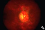 |
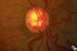
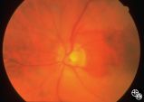
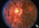
Acquired Disc Changes | Optociliary shunt vessels are venous collaterals that form after chronic venous obstruction. The presence of optic atrophy, progressive visual loss, and optociliary shunt vessels may indicate a compressive optic nerve lesion such as meningioma. | Shunt Vessels (Meningioma) |
| 302 |
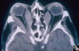 |
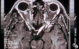
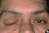
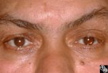
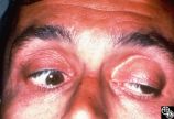
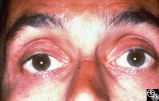
Ocular Manifestations of Systemic Disorders | Thyroid eye disease may result in proptosis and restrictive external ophthalmoplegia. The extracoular muscles are often diffusely enlarged with sparing of the tendons. | Thyroid Disease; Restriction Syndromes; Thyroid Eye Disease; Thyroid-Associated Ophthalmopathy; Blow-Out Fracture; Thyroid Orbitopathy; Thyroid Eye Disease; Myopathy; Proptosis; Chemosis; Periorbital Edema; Lid Retraction; CT |
| 303 |
 |
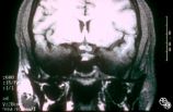
Ocular Manifestations of Systemic Disorders | Thyroid eye disease may cause proptosis and extraocular muscle enlargement that may be seen on orbital imaging studies. In general, coronal images allow the best visualization of the extraocular muscle enlargement. Pair with 94_45 and 94_46. | Thyroid Disease; Thyroid Orbitopathy; Thyroid Eye Disease; Myopathy; Proptosis; Chemosis; Periorbital Edema; Lid Retraction |
| 304 |
 |
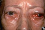
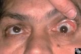
Ocular Manifestations of Systemic Disorders | Thyroid eye disease can result in significant upper eyelid retraction and axial proptosis resulting in exposure keratopathy. | Thyroid Disease; Thyroid Orbitopathy; Thyroid Eye Disease; Myopathy; Proptosis; Chemosis; Periorbital Edema; Lid Retraction |
| 305 |
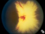 |
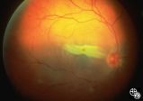
Isolated Congenital Optic Disc Anomalies | Normally, there is no visible myelination of the nerve fiber layer in the retina. Occasionally, visible myelination occurs that takes on a characteristic white, arcuate, feathery appearance that follows the contour of the nerve fiber layer. Disease/Diagnosis: Myelinated Nerve Fiber. | Myelinated Nerve Fiber Layer |
| 306 |
 |
Acquired Disc Changes | Although optociliary shunt vessels are venous collaterals that form in response to chronic venous obstruction, they may occur in patients with chronic open-angle glaucoma. | Shunt Vessels (Glaucoma) |
| 307 |
 |
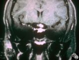
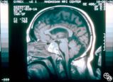
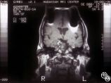
Neuro-Ophthalmic Imaging-MRI | This 23-year-old right-handed man had a history of idiopathic recurrent optic neuritis. The patient presented with acuity of 20/400 OD and 20/100 OS, with a central scotoma OD and a complete temporal defect OS. MRI with fat suppression and gadolinium revealed enhancement of the intracranial nerve an... | Chiasmal Optic Neuritis |
| 308 |
 |
Neuro-Ophthalmic Imaging-MRI | This 23-year-old right-handed man had a history of idiopathic recurrent optic neuritis. The patient presented with acuity of 20/400 OD and 20/100 OS, with a central scotoma OD and a complete temporal defect OS. MRI with fat suppression and gadolinium revealed enhancement of the intracranial nerve an... | Chiasmal Optic Neuritis |
| 309 |
 |
Neuro-Ophthalmic Imaging-MRI | This 23-year-old right-handed man had a history of idiopathic recurrent optic neuritis. The patient presented with acuity of 20/400 OD and 20/100 OS, with a central scotoma OD and a complete temporal defect OS. MRI with fat suppression and gadolinium revealed enhancement of the intracranial nerve an... | Chiasmal Optic Neuritis |
| 310 |
 |
Chiasmal Syndromes | A 52-year-old, morbidly obese man with a past medical history that included ischemic cardiac disease with a history of angioplasty, COPD, hypertension, and NIDDM, presented with a severe headache. The next day he had a frozen OD, complete right ptosis, and an unreactive middilated right pupil with V... | Pituitary Apoplexy |
| 311 |
 |
Chiasmal Syndromes | A 52-year-old, morbidly obese man with a past medical history that included ischemic cardiac disease with a history of angioplasty, COPD, hypertension, and NIDDM, presented with a severe headache. The next day he had a frozen OD, complete right ptosis, and an unreactive middilated right pupil with V... | Pituitary Apoplexy |
| 312 |
 |
Chiasmal Syndromes | A 52-year-old, morbidly obese man with a past medical history that included ischemic cardiac disease with a history of angioplasty, COPD, hypertension, and NIDDM, presented with a severe headache. The next day he had a frozen OD, complete right ptosis, and an unreactive middilated right pupil with V... | Pituitary Apoplexy |
| 313 |
 |
Chiasmal Syndromes | A 52-year-old, morbidly obese man with a past medical history that included ischemic cardiac disease with a history of angioplasty, COPD, hypertension, and NIDDM, presented with a severe headache. The next day he had a frozen OD, complete right ptosis, and an unreactive middilated right pupil with V... | Pituitary Apoplexy |
| 314 |
 |
Chiasmal Syndromes | A 52-year-old, morbidly obese man with a past medical history that included ischemic cardiac disease with a history of angioplasty, COPD, hypertension, and NIDDM, presented with a severe headache. The next day he had a frozen OD, complete right ptosis, and an unreactive middilated right pupil with V... | Pituitary Apoplexy |
| 315 |
 |
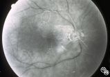
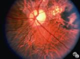
Neuro-Ophthalmic Vascular Disease | Occlusion of a branch or central retinal artery may result in acute visual loss. The ophthalmoscopic findings are retinal whitening due to ischemic retina in the distribution of the occluded artery. Sparing or selective involvement of cilioretinal artery branches may occur. Patients with a central r... | Central/Branch Retinal Artery Occlusion |
| 316 |
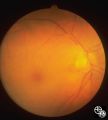 |
Neuro-Ophthalmic Vascular Disease | Occlusion of a branch or central retinal artery may result in acute visual loss. The ophthalmoscopic findings are retinal whitening due to ischemic retina in the distribution of the occluded artery. Sparing or selective involvement of cilioretinal artery branches may occur. Patients with a central r... | Central/Branch Retinal Artery Occlusion |
| 317 |
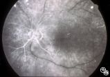 |
Neuro-Ophthalmic Vascular Disease | Occlusion of a branch or central retinal artery may result in acute visual loss. The ophthalmoscopic findings are retinal whitening due to ischemic retina in the distribution of the occluded artery. Sparing or selective involvement of cilioretinal artery branches may occur. Patients with a central r... | Central/Branch Retinal Artery Occlusion |
| 318 |
 |
Neuro-Ophthalmic Vascular Disease | Occlusion of a branch or central retinal artery may result in acute visual loss. The ophthalmoscopic findings are retinal whitening due to ischemic retina in the distribution of the occluded artery. Sparing or selective involvement of cilioretinal artery branches may occur. Patients with a central r... | Central/Branch Retinal Artery Occlusion |
| 319 |
 |
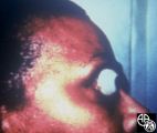
Traumatic Internal Carotid Artery Dissection | Traumatic dissection of the carotid artery may result in neck pain, an ipsilateral Horner's syndrome (disruption of the pericarotid sympathetic fibers), or ipsilateral arterial occlusions from embolic disease. Pair with images 91_18 and 91_20. | Carotid Dissection |
| 320 |
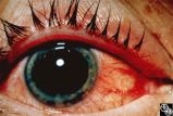 |
Traumatic Internal Carotid Atery Dissection | Traumatic dissection of the carotid artery may result in neck pain, an ipsilateral Horner's syndrome (disruption of the pericarotid sympathetic fibers), or ipsilateral arterial occlusions from embolic disease. Pair with images 91_19 and 91_20. | Carotid Dissection |
| 321 |
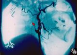 |
Traumatic Internal Carotid Artery Dissection | Traumatic dissection of the carotid artery may result in neck pain, an ipsilateral Horner's syndrome (disruption of the pericarotid sympathetic fibers), or ipsilateral arterial occlusions from embolic disease. Pair with images 91_18 and 91_19. | Carotid Dissection |
| 322 |
 |
Ocular Manifestations of Congenital/Inherited Diseases | This patient is a 40-year-old man with a history of abetalipoproteinemia (Bassen-Kornweig syndrome), diagnosed at age 9. Neurologic complications have included ataxia, retinal degeneration, peripheral neuropathy, progressive leg weakness, dysarthria, and intermittent bladder incontinence. On his neu... | Bassen-Kornzweig Syndrome |
| 323 |
 |
Ocular Manifestations of Congenital/Inherited Diseases | This patient is a 40-year-old man with a history of abetalipoproteinemia (Bassen-Kornweig syndrome), diagnosed at age 9. Neurologic complications have included ataxia, retinal degeneration, peripheral neuropathy, progressive leg weakness, dysarthria, and intermittent bladder incontinence. On his neu... | Bassen-Kornzweig Syndrome |
| 324 |
 |
Ocular Manifestations of Congenital/Inherited Diseases | This patient is a 40-year-old man with a history of abetalipoproteinemia (Bassen-Kornweig syndrome), diagnosed at age 9. Neurologic complications have included ataxia, retinal degeneration, peripheral neuropathy, progressive leg weakness, dysarthria, and intermittent bladder incontinence. On his neu... | Bassen-Kornzweig Syndrome |
| 325 |
 |
Ocular Manifestations of Congenital/Inherited Diseases | This patient is a 40-year-old man with a history of abetalipoproteinemia (Bassen-Kornweig syndrome), diagnosed at age 9. Neurologic complications have included ataxia, retinal degeneration, peripheral neuropathy, progressive leg weakness, dysarthria, and intermittent bladder incontinence. On his neu... | Bassen-Kornzweig Syndrome |