AAO-NANOS Neuro-Ophthalmology Clinical Collection: Derived from the AAO-NANOS Clinical Neuro-Ophthalmology collection produced on CD. The images are of selected cases from the NANOS teaching slide exchange, and the CD was produced under the direction of Larry Frohman, MD and Andrew Lee, MD.
The American Academy of Ophthalmology (AAO); The North American Neuro-Ophthalmology Association (NANOS).
NOVEL: https://novel.utah.edu/
TO
| Title | Description | Subject | ||
|---|---|---|---|---|
| 251 |
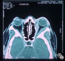 |
Neuro-Ophthalmic Imaging-CT Scan | Idiopathic orbital pseudotumor is an inflammatory disorder that may effect any part of the ocular anatomy. The site of inflammation determines the nomenclature. For example, involvement of the sclera is referred to as scleritis. And involvement of one or more of the extraocular muscles is referred t... | Myositis; Orbital Inflammation |
| 252 |
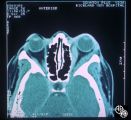 |
Neuro-Ophthalmic Imaging-CT Scan | Idiopathic orbital pseudotumor is an inflammatory disorder that may effect any part of the ocular anatomy. The site of inflammation determines the nomenclature. For example, involvement of the sclera is referred to as scleritis. And involvement of one or more of the extraocular muscles is referred t... | Myositis |
| 253 |
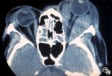 |
Ocular Manifestations of Systemic Disorders | Thyroid eye disease is the most common cause of unilateral or bilateral proptosis in the adult patient. Other signs of thyroid eye disease should be sought, including lid retraction, inferior scleral show, and lid lag. Patients with markedly asymmetric or strictly unilateral proptosis should probabl... | Thyroid Disease; Thyroid Eye Disease; Myopathy; Proptosis; Periorbital Edema; Lid Retraction; CT |
| 254 |
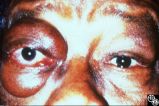 |
Ocular Manifestations of Systemic Disorders | Thyroid eye disease is the most common cause of unilateral or bilateral proptosis in the adult patient. Other signs of thyroid eye disease should be sought, including lid retraction, inferior scleral show, and lid lag. Patients with markedly asymmetric or strictly unilateral proptosis should probabl... | Thyroid Disease; Thyroid Eye Disease; Myopathy; Proptosis; Periorbital Edema; Lid Retraction |
| 255 |
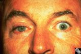 |
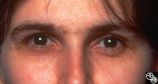
Migraine Syndrome | The image shows a patient with cluster headache and eye displaying Horner's syndrome. | Cluster Headache |
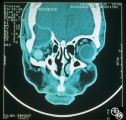
| 256 |
 |
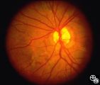
Neuro-Ophthalmic Imaging-MRI | Axial view of Arnold-Chiari malformation on a patient with downbeat nystagmus. Note the presence of the cerebellar tonsils posterior to the caudal medulla. In addition to downbeat nystagmus, Arnold-Chiari malformations can sometimes lead to increased intracranial pressure and papilledema. | Arnold-Chiari Malformation; Chiari Malformation; Inferior Tonsillar Herniation; Downbeat Nystagmus |
| 257 |
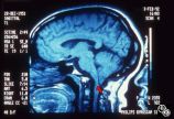 |
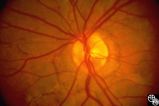
Neuro-Ophthalmic Imaging-MRI | Sagittal view of Arnold-Chiari malformation on a patient with downbeat nystagmus. The compression of the cervicomedullary junction is clearly depicted in the sagittal view. | Arnold-Chiari Malformation; Chiari Malformation; Inferior Tonsillar Herniation; Downbeat Nystagmus |
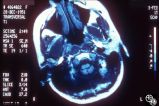
| 258 |
 |
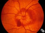
Neuro-Ophthalmic Imaging-CT Scan | This is a patient with trauma leading to enucleation, with swelling years later over the implant. This is a presumed chronic abscess between orbit and dura. | Orbital Abscess |
| 259 |
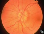 |
Isolated Optic Neuritis/Neuropathy | Papilledema usually results in bilateral optic disc edema without visual loss. The blind spot may enlarge initially, but progressive visual field loss may occur with chronic optic disc edema. Asymmetric or frankly unilateral optic disc edema may occur due to structural disc fractures that prevent th... | Pseudotumor Cerebri/Papilledema; Edema |
| 260 |
 |
Isolated Optic Neuritis/Neuropathy | Papilledema usually results in bilateral optic disc edema without visual loss. The blind spot may enlarge initially, but progressive visual field loss may occur with chronic optic disc edema. Asymmetric or frankly unilateral optic disc edema may occur due to structural disc fractures that prevent th... | Pseudotumor Cerebri/Papilledema; Edema |
| 261 |
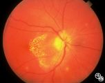 |
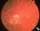
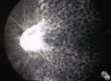
Optic Neuropathies | Optic disc edema with a macular star figure has been referred to as neuroretinitis. | Neuroretinitis |
| 262 |
 |
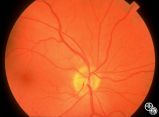
Isolated Optic Neuritis/Neuropathy | Papilledema is a term reserved for optic disc edema related to increased intracranial pressure (eg. Papilledema, sixth nerve palsy, headache), a normal neuroimaging study, and an elevated opening pressure with normal cerebrospinal fluid contents. | Pseudotumor Cerebri/Papilledema; Edema |
| 263 |
 |
Systemic Disorders With Optic Nerve and Retinal Findings | This patent has known pseudoxanthoma elasticum (an uncommon elastic tissue disorder characterized by plaque-like skin folds [plucked chicken skin], and degeneration of collagen fibers involving multiple systems, including the GI tract and heart), angioid streaks, and optic disc drusen. | Pseudoxanthoma Elasticum |
| 264 |
 |
Systemic Disorders With Optic Nerve and Retinal Findings | This patent has known pseudoxanthoma elasticum (an uncommon elastic tissue disorder characterized by plaque-like skin folds [plucked chicken skin], and degeneration of collagen fibers involving multiple systems, including the GI tract and heart), angioid streaks, and optic disc drusen. | Pseudoxanthoma Elasticum |
| 265 |
 |
Systemic Disorders With Optic Nerve and Retinal Findings | This patent has known pseudoxanthoma elasticum (an uncommon elastic tissue disorder characterized by plaque-like skin folds [plucked chicken skin], and degeneration of collagen fibers involving multiple systems, including the GI tract and heart), angioid streaks, and optic disc drusen. | Pseudoxanthoma Elasticum |
| 266 |
 |
Neuro-Ophthalmic Vascular Disease | In image 93_29, taken during the episode, note the change in caliber of the blood vessels. | Vasospastic Amaurosis Fugax |
| 267 |
 |
Neuro-Ophthalmic Vascular Disease | Image 93_30 is immediately after the attack, note the slight redness to the macula. | Vasospastic Amaurosis Fugax |
| 268 |
 |
Neuro-Ophthalmic Vascular Disease | Image 93_28 shows the fundus before the attack. | Vasospastic Amaurosis Fugax |
| 269 |
 |
Systemic Disorders With Optic Nerve and Retinal Findings | This patent has known pseudoxanthoma elasticum (an uncommon elastic tissue disorder characterized by plaque-like skin folds [plucked chicken skin], and degeneration of collagen fibers involving multiple systems, including the GI tract and heart), angioid streaks, and optic disc drusen. Imaging of a... | Pseudoxanthoma Elasticum |
| 270 |
 |
Systemic Disorders With Optic Nerve and Retinal Findings | This patent has known pseudoxanthoma elasticum (an uncommon elastic tissue disorder characterized by plaque-like skin folds [plucked chicken skin], and degeneration of collagen fibers involving multiple systems, including the GI tract and heart), angioid streaks, and optic disc drusen. Imaging of a... | Pseudoxanthoma Elasticum |
| 271 |
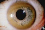 |
Ocular Manifestations of Systemic Disorders | Wilson's disease (hepatolenticular degeneration) is a progressive autosomal recessive multisystem disease that may result in cirrhosis of the liver, renal dysfunction, and motor neurologic disease. A Kayser-Fleischer ring may occur as a green-brown band at the level of Descemet's membrane in the cor... | Wilson's Disease; Hepatolenticular Degeneration; Kayser-Fleisher Rings; Slit Lamp |
| 272 |
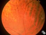 |
Systemic Disorders With Optic Nerve and Retinal Findings | Intraocular lymphoma may present with an unexplained vitritis, optic disc infiltration, or choroidal infiltration. One unusual manifestation of large-cell lymphoma is this leopard-spot appearance. Pair with 94_32, 94_33, and 94_35. This is a fundus photo. | Lymphoma; Leopard Spot Fundus; Lymphoma; Optic Nerve |
| 273 |
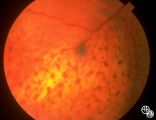 |
Systemic Disorders With Optic Nerve and Retinal Findings | Intraocular lymphoma may present with an unexplained vitritis, optic disc infiltration, or choroidal infiltration. One unusual manifestation of large-cell lymphoma is this leopard-spot appearance. Pair with 94_32, 94_34, and 94_35. This is a fundus photo. | Lymphoma; Leopard Spot Fundus; Lymphoma; Optic Nerve |
| 274 |
 |
Systemic Disorders With Optic Nerve and Retinal Findings | Intraocular lymphoma may present with an unexplained vitritis, optic disc infiltration, or choroidal infiltration. One unusual manifestation of large-cell lymphoma is this leopard-spot appearance. Pair with 94_33, 94_34, and 94_35. This is a fundus photo. | Lymphoma; Leopard Spot Fundus; Lymphoma; Optic Nerve |
| 275 |
 |
Systemic Disorders With Optic Nerve and Retinal Findings | Intraocular lymphoma may present with an unexplained vitritis, optic disc infiltration, or choroidal infiltration. One unusual manifestation of large-cell lymphoma is this leopard-spot appearance. Pair with 94_32, 94_33, and 94_34. This is a fluorescein angiogram. | Lymphoma; Leopard Spot Fluorescein Angiogram; Lymphoma; Optic Nerve |