Best known for his world-renowned neuro-ophthalmology unit based at the University of California, San Francisco, William Hoyt, MD collected here more than 850 of his best images covering a wide range of disorders.
William F. Hoyt, MD, Professor Emeritus of Ophthalmology, Neurology and Neurosurgery, Department of Ophthalmology, University of California, San Francisco.
NOVEL: https://novel.utah.edu/
TO
| Title | Description | Type | ||
|---|---|---|---|---|
| 1 |
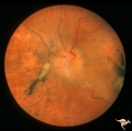 |
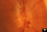
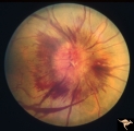
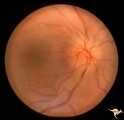
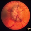
A301a Disc Swelling, Chorioretinal Disease | a and b same eye. Bad chorioretinal scars with disc swelling. Anatomy: Optic disc. Pathology: Unknown. | Image |
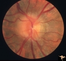
| 2 |
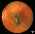 |
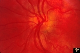
A302b Disc Swelling, Chorioretinal Disease | Bad chorioretinal scars with disc swelling. Temporal extent of chorioretinal scarring. A and B are the same eye. Anatomy: Optic disc. Pathology: Unknown. | Image |
| 3 |
 |
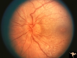
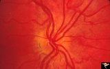
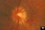
B101 Disc Swelling, Ischemic Papillopathies, AION | Pallid swelling in course of acute AION. 48 year old man who developed disc swelling after a flu like illness, then developed AION. Anatomy: Optic disc. Pathology: Axoplasmic stasis due to ischemia. Disease/ Diagnosis: AION. Clinical: Visual loss after flu-like illness. | Image |
| 4 |
 |
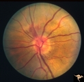
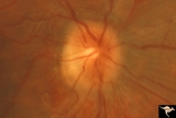
B203 Disc Swelling, Diabetic Papillopathy | Disc swelling in a diabetic woman. Recovered without visual loss. Right eye. Pair with B2_04. Anatomy: Optic disc. Pathology: Axoplasmic stasis due to ischemia. Disease/ Diagnosis: Diabetic papillopathy. Clinical: Visual loss with recovery. | Image |
| 5 |
 |
B204 Disc Swelling, Diabetic Papillopathy | Disc swelling in a diabetic. Recovered without visual loss. Left eye. Pair with B2_03. Anatomy: Optic disc. Pathology: Axoplasmic stasis due to ischemia. Disease/ Diagnosis: Diabetic papillopathy. Clinical: Visual loss with recovery. | Image |
| 6 |
 |
B301 Disc Swelling, Giant Cell Arteritis | Disc swelling. Giant Cell Arteritis. Temporal. Ischemic swelling. Blind eye with pallid swelling and marked dilation of central retinal vein. | Image |
| 7 |
 |
B403 Disc Swelling, Radiation Papillopathy | Man with blind eye. Ischemic hemorrhages. Vitreous haze. Anatomy: Optic disc. Pathology: Axoplasmic stasis due to ischemia. Disease/ Diagnosis: Radiation papillopathy; optic neuropathy. Clinical: Visual loss after radiation therapy. | Image |
| 8 |
 |
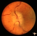
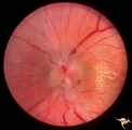
Bilateral Crowded Discs | Left eye. Bilateral crowded discs with congenital blurring. Blurred disc margins are not from edema. Note optic cup is absent. Pair with right eye in PP_1a, and brother in PP_2. Mother has drusen of the optic disc in PP_11aa & b. Sister has drusen in PP_11c. Anatomy: Optic disc. Pathology: Normal va... | Image |
| 9 |
 |
Bilateral Crowded Discs (Family) | Right eye. Bilateral crowded discs with congenital blurring. Blurred disc margins are not from edema. Note optic cup is absent. Pair with left eye in PP_1b, and brother in PP_2. Mother has drusen of the optic disc in PP_11a & b. Sister has drusen in PP_11c. Anatomy: Optic disc. Pathology: Normal var... | Image |
| 10 |
 |
Bilateral Hemorrhagic Papilledema | Left eye. Bilateral Hemorrhagic Papilledema from cardio-respiratory disease. Woman. Anatomy: Optic disc. Pathology: Bilateral papilledema, hemorrhagic. Disease/Diagnosis: Pseudotumor due to cardio-respiratory disease. Clinical notes: Woman with headache, shortness of breath. | Image |
| 11 |
 |
Bilateral Hemorrhagic Papilledema | Bilateral Hemorrhagic Papilledema from cardio-respiratory disease. Woman. Anatomy: Optic disc. Pathology: Bilateral papilledema, hemorrhagic. Disease/Diagnosis: Pseudotumor due to cardio-respiratory disease. Clinical notes: Woman with headache, shortness of breath. | Image |
| 12 |
 |
Bilateral Papilledema | Left eye. Has intra-retinal exudate and unusual vascular changes in the optic disc. Pre-pubertal girl. Anatomy: Optic disc. Pathology: Bilateral papilledema; exudative deposits in macula. Disease/Diagnosis: Pseudotumor. Clinical: Pubertal girl; headaches. | Image |
| 13 |
 |
Bilateral Papilledema | Right eye. Bilateral Papilledema in a patient with hyperthyroidism. Woman. Anatomy: Optic disc. Pathology: Papilledema. Disease/Diagnosis: Bilateral papilledema with hyperthyroidism. | Image |
| 14 |
 |
Bilateral Papilledema | Left eye. Bilateral Papilledema with hypoparathyroidism. Woman. Anatomy: Optic disc. Pathology: Papilledema. Papilledema with hypopararthyroidism. | Image |
| 15 |
 |
Bilateral Papilledema | Right eye. Bilateral Papilledema with hypoparathyroidism. Woman. Anatomy: Optic disc. Pathology: Papilledema. Disease/Diagnosis: Papilledema with hypoparathyroidism. | Image |
| 16 |
 |
Bilateral Papilledema | Left eye. Bilateral Papilledema in a patient with hyperthyroidism. Woman. Anatomy: Optic disc. Pathology: Papilledema. Disease/Diagnosis: Bilateral papilledema. | Image |
| 17 |
 |
Bilateral Papilledema | Right eye. Pre-pubertal girl. Anatomy: Optic disc. Pathology: Bilateral papilledema; exudative deposits in macula. Disease/Diagnosis: Pseudotumor. Clinical notes: Pubertal girl; headaches. | Image |
| 18 |
 |
Bilateral Papilledema from Occipital Tumor | Left eye. Bilateral hemorrhagic papilledema. Occipital glioma. Woman. Anatomy: Optic disc. Pathology: Papilledema. Disease/Diagnosis: Hemorrhagic papilledema from occipital glioma. | Image |
| 19 |
 |
Bilateral Papilledema from Occipital Tumor | Right eye. Bilateral hemorrhagic papilledema. Occipital glioma. Right hemianopia. Woman. Anatomy: Optic disc. Pathology: Papilledema. Disease/Diagnosis: Hemorrhagic papilledema from occipital glioma. | Image |
| 20 |
 |
Bilateral Papilledema from Pseudotumor | Left eye. Atrophic changes in left optic disc. Chronic papilledema with involution to atrophy on the left. Woman. Anatomy: Optic disc. Pathology: Bilateal papilledema; atrophic papilledema. Disease/Diagnosis: Pseudotumor. Clinical: Headache. | Image |
| 21 |
 |
Bilateral Papilledema from Pseudotumor | Right eye. Chronic papilledema. Woman. Anatomy: Optic disc. Pathology: Bilateral papilledema; atrophic papilledema. Disease/Diagnosis: Pseudotumor. Clinical notes: Headache. | Image |
| 22 |
 |
Bilateral Severe Hemorrhagic Papilledema | Left eye. Two months later, resolving Bilateral Severe Hemorrhagic Papilledema. Same eye as P_32b | Image |
| 23 |
 |
Bilateral Severe Hemorrhagic Papilledema | Right eye. 2 months later, resolving Bilateral Severe Hemorrhagic Papilledema. Same eye as P_32a | Image |
| 24 |
 |
Bilateral Severe Hemorrhagic Papilledema | Right eye. Bilateral Severe Hemorrhagic Papilledema in a woman with hyperthyroidism and dural sinus occlusion. | Image |
| 25 |
 |
Bilateral Severe Hemorrhagic Papilledema | Left eye. Bilateral Severe Hemorrhagic Papilledema in a woman with hyperthyroidism and dural sinus occlusion. | Image |