Best known for his world-renowned neuro-ophthalmology unit based at the University of California, San Francisco, William Hoyt, MD collected here more than 850 of his best images covering a wide range of disorders.
William F. Hoyt, MD, Professor Emeritus of Ophthalmology, Neurology and Neurosurgery, Department of Ophthalmology, University of California, San Francisco.
NOVEL: https://novel.utah.edu/
TO
1 - 25 of 13
| Title | Description | Type | ||
|---|---|---|---|---|
| 1 |
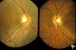 |
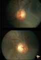
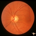
H80 Chiasmal Hemioptic Hypoplasia | Discs show striking nasal hypoplasia and band atrophy. DeMorsier synrome. Congenital bitemporal hemianopia with see-saw nystagmus. Note vertically oral shape of these hypoplastic nerves. The CT scan showed the median bar of the chiasm in this patient is totally hypoplastic. Anatomy: Optic disc. Path... | Image |
| 2 |
 |
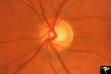
H82 Chiasmal Hemioptic Hypoplasia | De Morsier synrome with congenital bitemporal hemianopia. Left eye. Same patient as H_81. Anatomy: Optic disc. Pathology: Chiasmal hemioptic hypoplasia. Disease/ Diagnosis: Congenital anomaly involving chiasm. | Image |
| 3 |
 |
H88 Chiasmal Hemioptic Hypoplasia | Nasal hypoplasia with temporal hemianopia from a congenital Rathke Pouch Cyst. Anatomy: Optic disc. Pathology: Chiasmal hemioptic hypoplasia. Disease/ Diagnosis: Congenital anomaly involving chiasm. | Image |
| 4 |
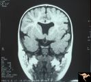 |
H07 Panhypoplasia | MRI Scan, coronal view showing absence of septum pellucidum. Hypoplastic chiasm. De Morsier's syndrome. Same patient as H_6. Anatomy: Optic disc. Pathology: Hypoplasia of the optic nerve. Disease/ Diagnosis: Hypoplasia. Imaging: MRI scan. | Image |
| 5 |
 |
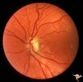
H81 Chiasmal Hemioptic Hypoplasia | De Morsier synrome with congenital bitemporal hemianopia. Right eye. Note nasal hypoplasia of the right optic disc. Same patient as H_82. Anatomy: Optic disc. Pathology: Chiasmal hemioptic hypoplasia. Disease/ Diagnosis: Congenital anomaly involving chiasm | Image |
| 6 |
 |
H83 Chiasmal Hemioptic Hypoplasia | De Morsier synrome with congenital bitemporal hemianopia. Note nasal hypoplasia of both optic discs. Left eye above, right eye below. Anatomy: Optic disc. Pathology: Chiasmal hemioptic hypoplasia. Disease/ Diagnosis: Congenital anomaly involving chiasm. | Image |
| 7 |
 |
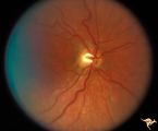
H84 Chiasmal Hemioptic Hypoplasia | Congenital bitemporal hemianopia with marked bi-nasal hypoplasia. Left eye. 17 year old male. Same patient as H_85. Anatomy: Optic disc. Pathology: Chiasmal hemioptic hypoplasia. Disease/ Diagnosis: Congenital anomaly involving chiasm. | Image |
| 8 |
 |
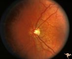
H85 Chiasmal Hemioptic Hypoplasia | Congenital bitemporal hemianopia with marked bi-nasal hypoplasia. Right eye. 17 year old male. Same patient as H_84. Anatomy: Optic disc. Pathology: Chiasmal hemioptic hypoplasia. Disease/ Diagnosis: Congenital anomaly involving chiasm. | Image |
| 9 |
 |
H86 Chiasmal Hemioptic Hypoplasia | Congenital bitemporal hemianopia with nasal hypoplasia. 24 year old man. Same patient as H_87. Anatomy: Optic disc. Pathology: Chiasmal hemioptic hypoplasia. Disease/ Diagnosis: Congenital anomaly involving chiasm. | Image |
| 10 |
 |
H87 Chiasmal Hemioptic Hypoplasia | Congenital bitemporal hemianopia with nasal hypoplasia. 24 year old man. Same patient as H_86. Anatomy: Optic disc. Pathology: Chiasmal hemioptic hypoplasia. Disease/ Diagnosis: Congenital anomaly involving chiasm. | Image |
| 11 |
 |
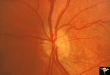
Segmental Atrophy - Hemianopic (Band) Atrophy | Segmental Atrophy - Band atrophy with temporal hemianopia. 1983. Anatomy: Optic disc. Pathology: Atrophy of the chiasm or left optic tract. Disease/Diagnosis: Segmental band atrophy. Clinical: Right temporal field defect. | Image |
| 12 |
 |
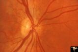
Segmental Atrophy - Hemianopic (Band) Atrophy | Segmental Atrophy - Band atrophy in an eye with temporal hemianopia. Wyburn-Mason Syndrome extending to the chiasm. Left eye 1975. Anatomy: Optic disc. Pathology: Right sided chiasmal AVM. Disease/Diagnosis: Band atrophy due to chiasmal AVM and Wyburn-Mason Syndrome. Clinical: Blind right eye, temp... | Image |
| 13 |
 |
Segmental Atrophy - Hemianopic (Band) Atrophy | Segmental Atrophy - Magnification of IIA2C_02a. Band atrophy in an eye with temporal hemianopia. Wyburn-Mason Syndrome extending to the chiasm. Left eye. 1975. Right eye in patient was blind. Anatomy: Optic disc. Pathology: Right sided chiasmal AVM. Disease/Diagnosis: Band atrophy due to chiasmal A... | Image |
1 - 25 of 13