Best known for his world-renowned neuro-ophthalmology unit based at the University of California, San Francisco, William Hoyt, MD collected here more than 850 of his best images covering a wide range of disorders.
William F. Hoyt, MD, Professor Emeritus of Ophthalmology, Neurology and Neurosurgery, Department of Ophthalmology, University of California, San Francisco.
NOVEL: https://novel.utah.edu/
TO
| Title | Description | Type | ||
|---|---|---|---|---|
| 151 |
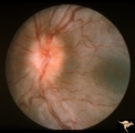 |
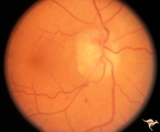
C301 Nodular Papillopathies (Sarcoid) | Disc swelling. Sarcoid papillopathy. Note infiltrative nodule at 9:00 on the disc.The patient had proven sarcoid. Perivenous inflammatory cuffing visible on image C3_02. Right eye. Pair with C3_02. Anatomy: Optic disc; Retina. Pathology: Axoplasmic stasis due to sarcoid infiltration. Disease/ Diagn... | Image |
| 152 |
 |
C302 Nodular Papillopathies (Sarcoid) | Perivenous Inflammatory Cuffing in a Patient with Proven Sarcoid. Left eye. Pair with C3_01. Anatomy: Retina. Pathology: Axoplasmic stasis due to sarcoid infiltration with retinal venous exudation? Disease/ Diagnosis: Sarcoid papillopathy with perivenous inflammatory disease. Clinical: Unknown? | Image |
| 153 |
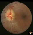 |
C303 Nodular Papillopathies (Sarcoid) | Lumpy infiltrative papillopathy in a patient with proven sarcoid. Anatomy: Optic disc. Pathology: Axoplasmic stasis due to sarcoid infiltration. Disease/ Diagnosis: Sarcoid papillopathy. Clinical: Unknown? | Image |
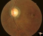
| 154 |
 |
C304 Nodular Papillopathies (Sarcoid) | Lumpy nodular disc infiltration from sarcoid. Anatomy: Optic disc. Pathology: Axoplasmic stasis due to sarcoid infiltration. Disease/ Diagnosis: Sarcoid papillopathy. Clinical: Unknown? | Image |
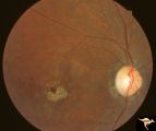
| 155 |
 |
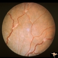
C305 Nodular Papillopathies (Sarcoid) | July 1984 shows multiple infiltrative nodules on the optic disc in addition to circumferential subretinal yellow exudates. 32 year old black woman. Same patient as C3_06 and C3_07. Anatomy: Optic disc; Retina. Pathology: Axoplasmic stasis due to sarcoid infiltration and retinal exudation. Disease/ ... | Image |
| 156 |
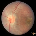 |
C306 Nodular Papillopathies (Sarcoid) | Lumpy disc swelling with retinal folds and a macular star in a patient with sarcoid. Presentation in October 1983. Same patient as C3_05 and C3_07. Anatomy: Optic disc; Retina. Pathology: Axoplasmic stasis due to sarcoid infiltration. Disease/ Diagnosis: Axoplasmic stasis due to sarcoid infiltration... | Image |
| 157 |
 |
C307 Nodular Papillopathies (Sarcoid) | Fluorescein angiogram shows striking staining of the sarcoid nodules. July 1984. Same patient as C3_05 and C3_06. Corresponds with July 1984 image, C3_06. Anatomy: Optic disc. Pathology: Axoplasmic stasis due to sarcoid infiltration. Disease/ Diagnosis: Sarcoid papillopathy. Clinical: Unknown? | Image |
| 158 |
 |
C308 Nodular Papillopathies (Sarcoid) | Nodular infiltrative papillopathy in a patient with sarcoid. Woman. Anatomy: Optic disc. Pathology: Axoplasmic stasis due to sarcoid infiltration. Disease/ Diagnosis: Sarcoid papillopahty. Clinical: Unknown? | Image |
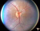
| 159 |
 |
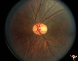
C33 Anomalous Pale Disc | Woman. Multiple cilioretinal arteries. Veins all empty into eye. Anomalous venous exit from nasal edge of optic disc. Visual function normal. Pair with C_36. Anatomy: Optic disc. | Image |
| 160 |
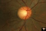 |
C35 Anomalous Pale Disc | Macro disc appears pale because of large diameter. Woman. Right eye. Anatomy: Optic disc. | Image |
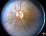
| 161 |
 |
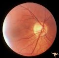
C36 Anomalous Pale Disc | Multiple cilioretinal arteries. Pale appearance. Normal optic nerve function. Good example of "empty disc". Pair with C_33. Anatomy: Optic disc. | Image |
| 162 |
 |
C37 Anomalous Pale Disc | "Watermelon" disc. Woman. Normal function. Left eye. Anatomy: Optic disc. | Image |
| 163 |
 |
C38 Anomalous Pale Disc | Megalopapilla in -8 myopic eye. Right eye. Anatomy: Optic disc. Clinical: High myope. | Image |
| 164 |
 |
C401 Luetic Papillopathy (Gumma of the Optic Disc) | Diffuse optic disc swelling with tortuous capillary dilations indicating inflammatory cellular infiltration. October 2001. Same eye as C4_02. Anatomy: Optic disc. Pathology: Axoplasmic stasis due to syphillitic infection. Luetic papillopathy (Syphyllis). Clinical: Visual loss. | Image |
| 165 |
 |
C402 Luetic Papillopathy (Gumma of the Optic Disc) | November 2001. Same eye as C4_01 after treatment with penicillin. Disc swelling went away and good visual function returned. Anatomy: Optic disc. Pathology: Axoplasmic stasis due to syphillitic infection. Disease/ Diagnosis: Luetic papillopathy (Syphillis). Clinical: Improving visual loss. | Image |
| 166 |
 |
Cerebellar Macular Degeneration | Cerebellar retinal degeneration with narrowed arterioles. Disc pallor. Granular retinal degeneration. 10 year old boy with mental degenerations and seizures. Anatomy: Retina. Pathology: Optic atrophy. Disease/Diagnosis: Congenital retinal cerebellar degeneration. Clinical: Severe mental retardation ... | Image |
| 167 |
 |
Cerebellar Macular Degeneration | Cerebellar retinal degenerative disease in a 12 year old boy who was blind and demented. His siblings were also blind. Was referred to as Voght-Spielmeyer Disease (Pair with R2_B1_3b shows granular retinal degeneration.) Anatomy: Retina. Pathology: Optic atrophy. Disease/Diagnosis: Congenital retina... | Image |
| 168 |
 |
Cerebellar Macular Degeneration | Cerebellar macular degeneration in a 7 year old boy with blindness. Rectal biopsy positive for storage material. Nature of cerebral degeneration was not defined in era when picture was taken. Sister also had similar findings. Anatomy: Retina. Pathology: Optic atrophy. Disease/Diagnosis: Congenital r... | Image |
| 169 |
 |
Cerebellar Macular Degeneration | Cerebellar retinal degenerative disease in a 12 year old boy who was blind and demented. His siblings were also blind. Was referred to as Vogt-Spielmeyer Disease. Pair with R2_B1_3a. Anatomy: Retina. Pathology: Optic atrophy. Disease/Diagnosis: Congenital retinal cerebral degeneration. Clinical: Sev... | Image |
| 170 |
 |
Cerebellar Macular Degenerative Disease | Ocular fundus shows prominent retinal degeneration in the region of the maculae, bilateral optic disc pallor with narrowed retinal arterioles. Interesting peripapillary halo of retinal pigment degeneration. Most consistent with Spinal Cerebellar Degeneration Type 7 (SCA-7). Anatomy: Retina. Patholog... | Image |
| 171 |
 |
Cerebellar Macular Degenerative Disease | Ocular fundus shows prominent retinal degeneration in the region of the maculae, bilateral optic disc pallor with narrowed retinal arterioles. Interesting peripapillary halo of retinal pigment degeneration. Most consistent with Spinal Cerebellar Degeneration Type 7 (SCA-7). Anatomy: Retina. Patholog... | Image |
| 172 |
 |
Cerebellar Macular Degenerative Disease | Cerebellar degeneration with granular maculae changes and bone spicules. Right eye. Anatomy: Retina. Pathology: Cerebellar macular degenerative disease. Disease/Diagnosis: Spinal Cerebellar Degeneration Type 7 (SCA-7). Clinical notes: Blindness and cerebellar degeneration. | Image |
| 173 |
 |
Cerebellar Macular Degenerative Disease | Cerebellar degeneration with granular maculae changes and bone spicules. Right eye. Anatomy: Retina. Pathology: Cerebellar macular degenerative disease. Disease/Diagnosis: Spinal Cerebellar Degeneration Type 7 (SCA-7). Clinical notes: Blindness and cerebellar degeneration. | Image |
| 174 |
 |
Cerebellar Macular Degenerative Disease | Cerebellar degeneration with granular maculae changes and bone spicules. Left eye. Anatomy: Retina. Pathology: Cerebellar macular degenerative disease. Disease/Diagnosis: Spinal Cerebellar Degeneration Type 7 (SCA-7). Clinical notes: Blindness and cerebellar degeneration. | Image |
| 175 |
 |
Cerebellar Macular Degenerative Disease | Cerebellar degeneration with granular maculae changes and bone spicules. Left eye. Anatomy: Retina. Pathology: Cerebellar macular degenerative disease. Disease/Diagnosis: Spinal Cerebellar Degeneration Type 7 (SCA-7). Clinical: Blindness and cerebellar degeneration. | Image |