Best known for his world-renowned neuro-ophthalmology unit based at the University of California, San Francisco, William Hoyt, MD collected here more than 850 of his best images covering a wide range of disorders.
William F. Hoyt, MD, Professor Emeritus of Ophthalmology, Neurology and Neurosurgery, Department of Ophthalmology, University of California, San Francisco.
NOVEL: https://novel.utah.edu/
TO
| Title | Description | Type | ||
|---|---|---|---|---|
| 26 |
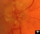 |
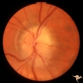
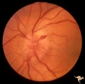
B103 Disc Swelling, Ischemic Papillopathies, AION | Ischemic swelling. 50 year old woman, 12 days after a viral illness. Nasal nerve fiber layer bundle visual field defect. Anatomy: Optic disc. Pathology: Axoplasmic stasis due to ischemia. Disease/ Diagnosis: AION. Clinical: Visual loss after viral illness. | Image |
| 27 |
 |
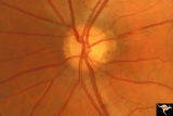
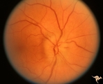
Segmental Atrophy - Temporal | Segmental Atrophy - Temporal - Nutritional Amblyopia (alcohol) Discs show bilateral temporal pallor with hyperemia of the remaining disc tissue - Pair with IIA2_02b. 1971. Anatomy: Optic disc. Pathology: Optic atrophy. Disease/Diagnosis: Toxic optic atrophy from alcohol. Clinical: Central visual lo... | Image |
| 28 |
 |
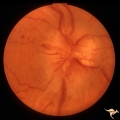
Segmental Atrophy - Temporal | Segmental Atrophy - Temporal - Nutritional Amblyopia (alcohol) Discs show bilateral temporal pallor with hyperemia of the remaining disc tissue - Pair with IIA2_02a. 1971. Anatomy: Optic disc. Pathology: Optic atrophy. Disease/Diagnosis: Toxic optic atrophy from alcohol. Clinical: Central visual lo... | Image |
| 29 |
 |
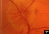
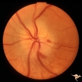
Multiple Sclerosis Slits and Thinning in Peripapillary (Retinal) Nerve Riber Layer | Multiple slit defect in the superior arcuate nerve fiber layer. Anatomy: Peripapillary nerve fiber layer. Pathology: Slit-like atrophy. Disease/Diagnosis: Multiple sclerosis optic neuropathy. Clinical: No symptoms. | Image |
| 30 |
 |
Multiple Sclerosis Slits and Thinning in Peripapillary (Retinal) Nerve Riber Layer | Multiple slit defect in the superior arcuate nerve fiber layer. Anatomy: Peripapillary nerve fiber layer. Pathology: Slit-like atrophy. Disease/Diagnosis: Multiple sclerosis optic neuropathy. Clinical: No symptoms. | Image |
| 31 |
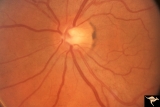 |
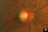
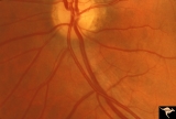
B115 Disc Swelling, Ischemic Papillopathies, AION | Normal eye in patient who later developed AION. Note generous optic cup. June 2, 1991. Same patient as B1_16b. Anatomy: Optic disc. Pathology: Normal. Clinical: Asymptomatic. | Image |
| 32 |
 |
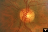
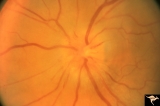
B114 Disc Swelling, Ischemic Papillopathies, AION | AION in a disc with an optic cup. Extraordinary exception with AION. Note ischemic vascular changes in disc surface. Anatomy: Optic disc. Pathology: Axoplasmic stasis due to ischemia. Disease/ Diagnosis: AION. Clinical: Visual loss. | Image |
| 33 |
 |
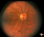
B116 Disc Swelling, Ischemic Papillopathies, AION | Typical AION in disc with optic cup. December 23, 2996. 5 years later in same patient as B1_15a. Anatomy: Optic disc. Pathology: Axoplasmic stasis due to ischemia. Disease/ Diagnosis: AION. Clinical: Visual loss. | Image |
| 34 |
 |
Multiple Sclerosis Slits and Thinning in Peripapillary (Retinal) Nerve Riber Layer | Multiple slit defect in the superior arcuate nerve fiber layer. Pair with IIB2_6b. Anatomy: Peripapillary nerve fiber layer. Pathology: Slit-like atrophy. Disease/Diagnosis: Multiple sclerosis optic neuropathy. Clinical: No symptoms. | Image |
| 35 |
 |
Multiple Sclerosis Slits and Thinning in Peripapillary (Retinal) Nerve Riber Layer | Multiple slit like defects in the inferior arcuate nerve fibers. Pair with IIB2_3b. Anatomy: Peripapillary nerve fiber layer. Pathology: Slit-like atrophy. Disease/Diagnosis: Multiple sclerosis optic neuropathy. Clinical: No symptoms. | Image |
| 36 |
 |
Multiple Sclerosis Slits and Thinning in Peripapillary (Retinal) Nerve Riber Layer | Multiple slit and wedge defects in the nerve fiber layer. Pair with IIB2_3a. Anatomy: Peripapillary nerve fiber layer. Pathology: Slit-like atrophy. Disease/Diagnosis: Multiple sclerosis optic neuropathy. Clinical: No symptoms. | Image |
| 37 |
 |
Multiple Sclerosis Slits and Thinning in Peripapillary (Retinal) Nerve Riber Layer | Multiple slit defect in the superior arcuate nerve fiber layer. Magnified. Pair with IIB2_6a. Anatomy: Peripapillary nerve fiber layer. Pathology: Slit-like atrophy. Disease/Diagnosis: Sclerosis optic neuropathy. Clinical: No symptoms. | Image |
| 38 |
 |
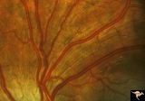
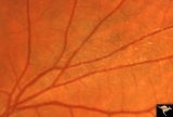
Multiple Sclerosis Slits and Thinning in Peripapillary (Retinal) Nerve Riber Layer | Left eye. Upper arcuate nerve fiber layer contains multiple low density slits. These indicate nerve fiber loss. Anatomy: Peripapillary nerve fiber layer. Pathology: Slit-like atrophy. Disease/Diagnosis: Multiple sclerosis optic neuropathy. Clinical: No symptoms. | Image |
| 39 |
 |
Multiple Sclerosis Slits and Thinning in Peripapillary (Retinal) Nerve Riber Layer | Multiple slit defect in the superior arcuate nerve fiber layer in a 13 year old boy. Right eye. Pair with IIB2_7a. Anatomy: Peripapillary nerve fiber layer. Pathology: Slit-like atrophy. Disease/Diagnosis: Multiple sclerosis optic neuropathy. Clinical: No symptoms. | Image |
| 40 |
 |
Multiple Sclerosis Slits and Thinning in Peripapillary (Retinal) Nerve Riber Layer | Multiple slit defect in the superior arcuate nerve fiber layer in a 13 year old boy. Pair with IIB2_7b. Anatomy: Peripapillary nerve fiber layer. Pathology: Slit-like atrophy. Disease/Diagnosis: Multiple sclerosis optic neuropathy. Clinical: No symptoms. | Image |
| 41 |
 |
Multiple Sclerosis Slits and Thinning in Peripapillary (Retinal) Nerve Riber Layer | Need magnification - Left eye - Peculiar punctate dotted surface of internal limiting membrane reflexes. Pairs with IIB2_01a & IIB2_02b. Anatomy: Peripapillary nerve fiber layer. Pathology: Slit-like atrophy. Disease/Diagnosis: Multiple sclerosis optic neuropathy. Clinical: No symptoms. | Image |
| 42 |
 |
Multiple Sclerosis Slits and Thinning in Peripapillary (Retinal) Nerve Riber Layer | Need magnification - Left eye - Inferior arcuate nerve fiber slits. Pairs with IIB2_01b & IIB2_01c. Anatomy: Peripapillary nerve fiber layer. Pathology: Slit-like atrophy. Disease/Diagnosis: Multiple sclerosis optic neuropathy. Clinical: No symptoms. | Image |
| 43 |
 |
Multiple Sclerosis Slits and Thinning in Peripapillary (Retinal) Nerve Riber Layer | Need magnification - Left eye - Inferior arcuate nerve fiber slits. Pairs with IIB2_01a & IIB2_01c. Anatomy: Peripapillary nerve fiber layer. Pathology: Slit-like atrophy. Disease/Diagnosis: Multiple sclerosis optic neuropathy. Clinical: No symptoms. | Image |
| 44 |
 |
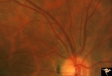
B104 Disc Swelling, Ischemic Papillopathies, AION | Ischemic swelling. 57 year old man. Anatomy: Optic disc. Pathology: Axoplasmic stasis due to ischemia. Disease/ Diagnosis: AION. Clinical: Visual loss. | Image |
| 45 |
 |
B107 Disc Swelling, Ischemic Papillopathies, AION | Pallid ischemic swelling. 41 year old man. Anatomy: Optic disc. Pathology: Axoplasmic stasis due to ischemia. Disease/ Diagnosis: AION. Clinical: Viusal loss. | Image |
| 46 |
 |
B111 Disc Swelling, Ischemic Papillopathies, AION | Acute AION. Anatomy: Optic disc. Pathology: Axoplasmic stasis due to ischemia. Disease/ Diagnosis: AION. Clinical: Visual loss. | Image |
| 47 |
 |
B113 Disc Swelling, Ischemic Papillopathies, AION | 57 year old woman with AION. Anatomy: Optic disc. Pathology: Axoplasmic stasis due to ischemia. Disease/ Diagnosis: AION. Clinical: Visual loss. | Image |
| 48 |
 |
B105 Disc Swelling, Ischemic Papillopathies, AION | Pallid ischemic swelling. 48 year old woman, flight attendant. Anatomy: Optic disc. Pathology: Axoplasmic stasis due to ischemia. Disease/ Diagnosis: AION. Clinical: Visual loss. | Image |
| 49 |
 |
B106 Disc Swelling, Ischemic Papillopathies, AION | Red ischemic swelling. 49 year old man. Anatomy: Optic disc. Pathology: Axoplasmic stasis due to ischemia. Disease/ Diagnosis: AION. Clinical: Visual loss. | Image |
| 50 |
 |
B108 Disc Swelling, Ischemic Papillopathies, AION | Pallid ischemic swelling. Woman with vasculitis. Anatomy: Optic disc. Pathology: Axoplasmic stasis due to ischemia. Disease/ Diagnosis: AION. Clinical: Visual loss. | Image |