Best known for his world-renowned neuro-ophthalmology unit based at the University of California, San Francisco, William Hoyt, MD collected here more than 850 of his best images covering a wide range of disorders.
William F. Hoyt, MD, Professor Emeritus of Ophthalmology, Neurology and Neurosurgery, Department of Ophthalmology, University of California, San Francisco.
NOVEL: https://novel.utah.edu/
TO
| Title | Description | Type | ||
|---|---|---|---|---|
| 226 |
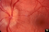 |
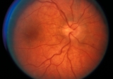
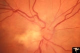
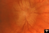
Drusen with Vertical Retinal Folds | PP36a & b:Both left eye: Buried drusen. Note vertical retinal folds. Anatomy: Optic disc. Pathology: Drusen of the optic disc. Disease/Diagnosis: Drusen of the optic disc. | Image |
| 227 |
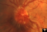 |
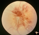
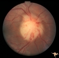
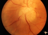
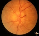
E01 Disc Swelling with Central Vein Occlusion | Left eye. Central retinal vein occlusion with disc swelling. Anatomyt: Optic disc; Retina. Pathology: Vasculitis. Disease/ Diagnosis: Disc swelling due to retinal vasculitis. | Image |
| 228 |
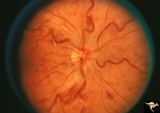 |
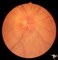
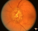
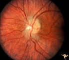
E03 Disc Swelling with Central Retinal Vein Occlusion | 36 year old woman with visual obscurations of right eye. Early CRVO, papillophlebitis. Steroid responsive. Anatomy: Optic disc; Retina. Pathology: Central retinal vein occlusion. Disease/ Diagnosis: Disc swelling due to central retinal vein occlusion. Clinical: Decreased vision in right eye. Acuity ... | Image |
| 229 |
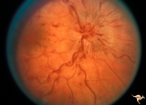 |
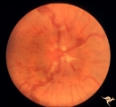
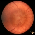
E04 Disc Swelling with Central Retinal Vein Occlusion | Acute CRVO, right eye with disc swelling. Male patient. Same patient as E05. Anatomy: Optic disc; Retina. Pathology: Central retinal vein occlusion. Disease/ Diagnosis: Disc swelling due to central retinal vein occlusion. Clinical: Visual blurring. | Image |
| 230 |
 |
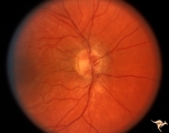
E05 Disc Swelling with Central Retinal Vein Occlusion | Resolving CVRO, right eye. Two months following slide E04. Male patient. Anatomy: Optic disc; Retina. Pathology: Central retinal vein occlusion. Disease/ Diagnosis: Resolved disc swelling after central retinal vein occlusion. Clinical: No symptoms. | Image |
| 231 |
 |
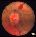
E06 Disc Swelling with Central Retinal Vein Occlusion | Acute disc swelling one week after onset of symptoms. Anatomy: Optic disc; Retina. Pathology: Central retinal vein occlusion. Disease/ Diagnosis: Disc swelling due to central retinal vein occlusion. Clinical: Visual blurring. | Image |
| 232 |
 |
E07 Disc Swelling with Central Vein Occlusion | 24 year old male. Papillophlebitis (CRVO) with optic disc edema. Right eye. Anatomy: Optic disc; Retina. Pathology: Central retinal vein occlusion. Disease/ Diagnosis: Disc swelling due to central retinal vein occlusion. Clinical: ??Branch retinal artery occlusion [sic]. | Image |
| 233 |
 |
E08 Disc Swelling with Central Vein Occlusion | Pituitary adenoma with right chronic CRVO with optociliary bypass vessels. Anatomy: Optic disc; Retina. Pathology: Central retinal vein occlusion. Disease/ Diagnosis: Disc swelling due to central retinal vein occlusion. | Image |
| 234 |
 |
E09 Disc Swelling with Central Vein Occlusion | Chronic disc swelling due to CRVO. Anatomy: Optic disc; Retina. Pathology: Central retinal vein occlusion. Disease/ Diagnosis: Disc swelling due to central retinal vein occlusion. | Image |
| 235 |
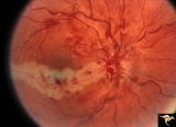 |
E10 Disc Swelling with Central Vein Occlusion | Cilioretinal artery infarction after a central retinal vein occlusion. Anatomy: Optic disc; Retina. Pathology: Central retinal vein occlusion. Disease/ Diagnosis: Disc swelling due to central retinal vein occlusion. | Image |
| 236 |
 |
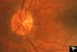
E11 Disc Swelling with Central Vein Occlusion | Old retinal vein occlusion with optociliary bypass vessel at 3:00. Right eye. Anatomy: Optic disc; Retina. Pathology: Central retinal vein occlusion. Disease/ Diagnosis: Disc swelling due to central retinal vein occlusion. | Image |
| 237 |
 |
E12 Disc Swelling with Central Vein Occlusion | 2nd attack of papillophlebitis. There is an optociliary bypass vessel at 4:00. Anatomy: Optic disc; Retina. Pathology: Central retinal vein occlusion. Disease/ Diagnosis: Disc swelling due to central retinal vein occlusion. | Image |
| 238 |
 |
Early Papilledema due to Brain Tumor - Resolving | Left eye. Same eye as P_34a. One month post op, papilledema resolving. Boy. Anatomy: Optic disc. Pathology: Papilledema. Disease/Diagnosis: Papilledema from posterior fossa hemangioblastoma. | Image |
| 239 |
 |
Early Papilledema due to Tumor | Left eye. Asymmetric Papilledema with posterior fossa hemangioblastoma. Left - moderate papilledema. Blurring of disc. Young man. Anatomy: Optic disc. Pathology: Papilledema. Disease/Diagnosis: Papilledema from posterior fossa hemangioblastoma. | Image |
| 240 |
 |
F101 Optic Disc Lymphosarcoma | Optic disc lymphosarcoma. This disc has been infiltrated by neoplastic cells. Anatomy: Optic disc. Pathology: Lymphosarcoma. Disease/ Diagnosis: Lymphosarcoma. | Image |
| 241 |
 |
F102 Myeloblastic Leukemia | Myeloblastic leukemia. Left eye. Pair with F1_03. Anatomy: Optic disc. Pathology: Neoplastic (metastatic) papillopathy. Disease/ Diagnosis: Myeloblastic leukemia. | Image |
| 242 |
 |
F103 Myeloblastic Leukemia | Myeloblastic leukemia. Right eye. Pair with F1_02. Anatomy: Optic disc. Pathology: Neoplastic (metastatic) papillopathy. Disease/ Diagnosis: Myeloblastic leukemia. | Image |
| 243 |
 |
F104 Esthesio Neuroblastoma | Esthesioneuroblastoma. Tumor cells infiltrating the optic disc. 20/200 vision. Anatomy: Optic disc. Pathology: Esthesioneuroblastoma. Disease/ Diagnosis: Esthesioneuroblastoma. | Image |
| 244 |
 |
F105 Histiocytosis Infiltrate of Disc | Histiocytosis infiltrate of right disc with simultaneous infiltration of the hypothalamus with skin lesions on eye lids and chest. Same patient as F1_06. Anatomy: Optic disc. Pathology: Histiocytosis infiltrate. Disease/ Diagnosis: Histiocytosis infiltrate. Clinical: Patient presented with skin lesi... | Image |
| 245 |
 |
F106 Histiocytosis Infiltrate of Disc | More fully developed and chronic histiocytosis infiltrate of right disc with simultaneous infiltration of the hypothalamus with skin lesions on eye lids and chest. Same patient as F1_05, one year later. Anatomy: Optic disc. Pathology: Histiocytosis infiltrate of disc. Disease/ Diagnosis: Histiocytos... | Image |
| 246 |
 |
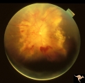
F107 Metastatic Breast Cancer to the Disc | Metastatic breast cancer to the disc. Notice mass on inferior portion of disc. Also notice tangled capillary dilation within the mass indicating infiltration. This disc tumor was radiated. It disappeared leaving a pale flat atrophic nerve. The patient died. Histologic study of the eye revealed metas... | Image |
| 247 |
 |
F108 Acute Disc Swelling | Chinese man with acute disc swelling. Blind in both eyes. Had large thalamic mass. (Lymphoma). Anatomy: Optic disc. Pathology: Lymphoma. Disease/ Diagnosis: Lymphoma. | Image |
| 248 |
 |
F109 T-Cell Leukemia Infiltrate | T-Cell leukemia infiltrate. 14 year old boy with T-Cell leukemia infiltrating the disc. Anatomy: Optic disc. Pathology: T-Cell leukemia. Disease/ Diagnosis: Neoplastic (metastatic) papillopathy | Image |
| 249 |
 |
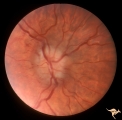
F201 Optic Nerve Sheath Meningioma | Right eye. Woman with ophthalmoplegia proptosis for 14 years. Visual field reduced due to optic nerve sheath meningioma. Notice large optociliary vessel temporally. Anatomy: Optic disc. Pathology: Chronic optic disc swelling caused by optic nerve sheath meningioma. Disease/ Diagnosis: Chronic optic ... | Image |
| 250 |
 |
F202 Optic Nerve Sheath Meningioma | Optic nerve sheath meningioma. Note optociliary vein at 3:00. The disc is atrophic. Anatomy: Optic disc. Pathology: Chronic optic disc swelling caused by optic nerve sheath meningioma. Disease/ Diagnosis: Chronic optic disc swelling caused by optic nerve sheath meningioma. | Image |