AAO-NANOS Neuro-Ophthalmology Clinical Collection: Derived from the AAO-NANOS Clinical Neuro-Ophthalmology collection produced on CD. The images are of selected cases from the NANOS teaching slide exchange, and the CD was produced under the direction of Larry Frohman, MD and Andrew Lee, MD.
The American Academy of Ophthalmology (AAO); The North American Neuro-Ophthalmology Association (NANOS).
NOVEL: https://novel.utah.edu/
TO
| Title | Creator | Description | ||
|---|---|---|---|---|
| 1 |
 |
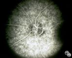
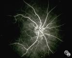
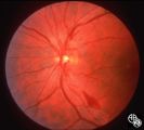
Isolated Congenital Optic Disc Anomalies | Rosa A. Tang, MD | This patient has optic disc drusen and evidence of a superimposed optic neuropathy, including loss of visual field, an ipsilateral afferent pupillary defect, and optic atrophy. Although optic disc drusen typically causes visual field loss without visual acuity loss superimposed, ischemic optic neuro... |
| 2 |
 |
Isolated Optic Neuritis/Neuropathy | John A. Charley, MD | Several Primary Mutations result in Leber's hereditary optic neuropathy, including mitochondrial deletions at positions 11778, 3460, and 14484. Although the 11778 is the most common mutation, the 11484 has the best prognosis for spontaneous recovery. This case exhibits the 3460 mutation. |
| 3 |
 |
Isolated Optic Neuritis/Neuropathy | John A. Charley, MD | Several Primary Mutations result in Leber's hereditary optic neuropathy, including mitochondrial deletions at positions 11778, 3460, and 14484. Although the 11778 is the most common mutation, the 11484 has the best prognosis for spontaneous recovery. This case exhibits the 3460 mutation. |
| 4 |
 |
Isolated Optic Neuritis/Neuropathy | John A. Charley, MD | Several Primary Mutations result in Leber's hereditary optic neuropathy, including mitochondrial deletions at positions 11778, 3460, and 14484. Although the 11778 is the most common mutation, the 11484 has the best prognosis for spontaneous recovery. This case exhibits the 3460 mutation. |
| 5 |
 |
Isolated Optic Neuritis/Neuropathy | John A. Charley, MD | Several Primary Mutations result in Leber's hereditary optic neuropathy, including mitochondrial deletions at positions 11778, 3460, and 14484. Although the 11778 is the most common mutation, the 11484 has the best prognosis for spontaneous recovery. This case exhibits the 3460 mutation. |
| 6 |
 |
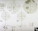
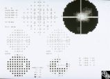
Isolated Optic Neuritis/Neuropathy | John A. Charley, MD | Several Primary Mutations result in Leber's hereditary optic neuropathy, including mitochondrial deletions at positions 11778, 3460, and 14484. Although the 11778 is the most common mutation, the 11484 has the best prognosis for spontaneous recovery. This case exhibits the 3460 mutation. |
| 7 |
 |
Isolated Hereditary Optic Neuritis/Neuropathy | Gregory S. Kosmorsky, MD | Leber's hereditary optic neuropathy is a mitochondrial hereditary optic neuropathy that usually affects young males but may occur at any age and in males or females. The clinical features are usually acute bilateral simultaneous or sequential visual loss with a central acuity and central visual fiel... |
| 8 |
 |
Isolated Hereditary Optic Neuritis/Neuropathy | Gregory S. Kosmorsky, MD | Leber's hereditary optic neuropathy is a mitochondrial hereditary optic neuropathy that usually affects young males but may occur at any age and in males or females. The clinical features are usually acute bilateral simultaneous or sequential visual loss with a central acuity and central visual fiel... |
| 9 |
 |
Isolated Hereditary Optic Neuritis/Neuropathy | Gregory S. Kosmorsky, MD | Leber's hereditary optic neuropathy is a mitochondrial hereditary optic neuropathy that usually affects young males but may occur at any age and in males or females. The clinical features are usually acute bilateral simultaneous or sequential visual loss with a central acuity and central visual fiel... |
| 10 |
 |
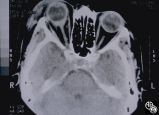
Neuro-Ophthalmic Imaging-CT Scan | Larry P. Frohman, MD | This patient was assaulted with a blunt object and suffered acute blindness due to traumatic optic neuropathy. Note how the lateral orbital wall has been fractured and displaced posteromedially into the region of the anterior optic canal. |
| 11 |
 |
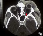
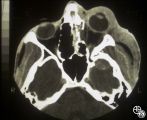
Neuro-Ophthalmic Imaging-CT Scan | Larry P. Frohman, MD | This 70-year-old woman sustained traumatic optic neuropathy in a motor vehicle accident. Note the funnel-shaped hemorrhage within the optic nerve sheath just posterior to the globe. |
| 12 |
 |
Neuro-Ophthalmic Imaging-CT Scan | Larry P. Frohman, MD | This patient was assaulted with a blunt object and suffered acute blindness due to traumatic optic neuropathy. Note how the lateral orbital wall has been fractured and displaced posteromedially into the region of the anterior optic canal. |
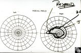
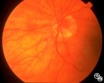
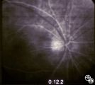
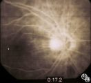
| 13 |
 |
Isolated Optic Neuritis/Neuropathy | Larry P. Frohman, MD | The patient is a 62-year-old female who presented in August 1996 with visual loss OD that she first noted as loss of her superior field in May 1996. She felt that it had been static since, and perhaps was even a little better in the week before she was seen. There was no pain, even with ocular rotat... |
| 14 |
 |
Isolated Optic Neuritis/Neuropathy | Larry P. Frohman, MD | The patient is a 62-year-old female who presented in August 1996 with visual loss OD that she first noted as loss of her superior field in May 1996. She felt that it had been static since, and perhaps was even a little better in the week before she was seen. There was no pain, even with ocular rotat... |
| 15 |
 |
Isolated Optic Neuritis/Neuropathy | Larry P. Frohman, MD | The patient is a 62-year-old female who presented in August 1996 with visual loss OD that she first noted as loss of her superior field in May 1996. She felt that it had been static since, and perhaps was even a little better in the week before she was seen. There was no pain, even with ocular rotat... |
| 16 |
 |
Isolated Optic Neuritis/Neuropathy | Larry P. Frohman, MD | The patient is a 62-year-old female who presented in August 1996 with visual loss OD that she first noted as loss of her superior field in May 1996. She felt that it had been static since, and perhaps was even a little better in the week before she was seen. There was no pain, even with ocular rotat... |
| 17 |
 |
Isolated Optic Neuritis/Neuropathy | Larry P. Frohman, MD | The patient is a 62-year-old female who presented in August 1996 with visual loss OD that she first noted as loss of her superior field in May 1996. She felt that it had been static since, and perhaps was even a little better in the week before she was seen. There was no pain, even with ocular rotat... |
| 18 |
 |
Isolated Optic Neuritis/Neuropathy | Larry P. Frohman, MD | The patient is a 62-year-old female who presented in August 1996 with visual loss OD that she first noted as loss of her superior field in May 1996. She felt that it had been static since, and perhaps was even a little better in the week before she was seen. There was no pain, even with ocular rotat... |
| 19 |
 |
Isolated Optic Neuritis/Neuropathy | Larry P. Frohman, MD | The patient is a 62-year-old female who presented in August 1996 with visual loss OD that she first noted as loss of her superior field in May 1996. She felt that it had been static since, and perhaps was even a little better in the week before she was seen. There was no pain, even with ocular rotat... |
| 20 |
 |
Isolated Optic Neuritis/Neuropathy | Larry P. Frohman, MD | The patient is a 62-year-old female who presented in August 1996 with visual loss OD that she first noted as loss of her superior field in May 1996. She felt that it had been static since, and perhaps was even a little better in the week before she was seen. There was no pain, even with ocular rotat... |
| 21 |
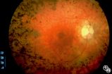 |
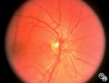
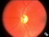
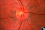
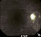
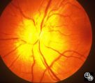
Optic Neuropathies | Rosa A. Tang, MD | Optic atrophy might be a false-localizing sign of optic nerve disease, as retinal disease may secondarily produce optic atrophy. In retinitis pigmentosa, for example, patients may exhibit a waxy disc palor. Fundus imaging. Anatomy: Retina. Disease/Diagnosis: Retinitis pigmentosa; Optic atrophy. |
| 22 |
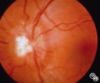 |
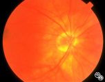
Systemic Disorders With Optic Nerve and Retinal Findings | Mark J. Kupersmith, MD | Sarcoidosis is an inflammatory multisystem granulomatous disease that may result in an inflammatory or infiltrative optic neuropathy, papilledema from increased intracranial pressure due to meningeal inflammation or intracranial granuloma, or may present with an optic disc granuloma. Pair with 91_32... |
| 23 |
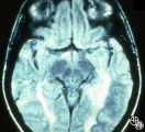 |
Magnetic Resonance Imaging in Detection of Extracranial Internal Carotid Artery Dissection | Marilyn C. Kay, MD | This 28-year-old woman presented with a 4-week history of bilateral visual loss. She had a known history of multiple sclerosis. Her vision was 20/60 OD and 20/40 OS, with an RAPD OS and optic pallor OU. Her fields and MRI are shown. Optic tract lesions usually result in an incongruous homonymous hem... |
| 24 |
 |
Neuro-Ophthalmic Case With Notable Field Changes | Marilyn C. Kay, MD | This 28-year-old woman presented with a 4-week history of bilateral visual loss. She had a known history of multiple sclerosis. Her vision was 20/60 OD and 20/40 OS, with an RAPD OS and optic pallor OU. Her fields and MRI are shown. Optic tract lesions usually result in an incongruous homonymous hem... |
| 25 |
 |
Systemic Disorders With Optic Nerve and Retinal Findings | Mark J. Kupersmith, MD | Sarcoidosis is an inflammatory multisystem granulomatous disease that may result in an inflammatory or infiltrative optic neuropathy, papilledema from increased intracranial pressure due to meningeal inflammation or intracranial granuloma, or may present with an optic disc granuloma. Pair with 91_31... |