Collection of materials relating to neuro-ophthalmology as part of the Neuro-Ophthalmology Virtual Education Library.
NOVEL: https://novel.utah.edu/
TO
1 - 25 of 16
| Title | Creator | Description | Subject | ||
|---|---|---|---|---|---|
| 1 |
 |
Corectopia | Meagan Seay, DO | These are photos of a patient with unilateral corectopia. This patient's corectopia is of unclear etiology and possibly related to birth trauma. | Corectopia; Unilateral; Photos |
| 2 |
 |
Carotid Cavernous Fistula | Adam Botwinick, MD; Rudrani Banik, MD | Power point of case presentation of 66-year-old female with chronic red eye OU x 2 months, misdiagnosed as conjunctivitis. Exam showed dilated, tortuous episcleral vessels OU with proptosis OU and elevated intraocular pressure. MRI showed suspicion of carotid cavernous fistula (CCF), confirmed by ... | Carotid Cavernous Fistula; Dural CCF; Chemosis; Corkscrew Vessels; Proptosis; Embolization; Neurointerventional Radiology |
| 3 |
 |
Modern Imaging of Optic Disc Drusen | Meagan Seay, DO | This is a short powerpoint describing imaging techniques (specifically OCT-EDI, fundus autofluorescence, and B-scan ultrasonography) for optic disc drusen. Examples of these techniques are included. | Optic Disc Drusen; Imaging; OCT-EDI; Fundus Autofluorescence; B-scan Ultrasonography |
| 4 |
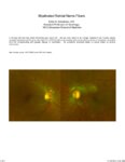 |
Myelinated Retinal Nerve Fibers | Scott N. Grossman, MD | A 33 year old man has noted chronically poor vision OS - left eye color noted to be 'orange' instead of red. fundus photos revealed myelinated retinal nerve fiber layer OU (OS>OD) with corresponding linear paracentral scotoma on Humphrey visual field 24-2 OS corresponding with greatest degree of my... | Myelinated Retinal Nerve Fibers |
| 5 |
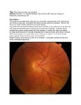 |
Situs Inversus Optic Disc Anomaly | Michael Hii, Medical Student; Ryan Walsh, MD | This patient was incidentally-noted to have anomalous appearance of the optic discs, right more so than left, consistent with situs inversus optic disc anomaly. She did not have any visual deficits related to this exam finding. ; The patient's fundus photos demonstrate situs inversus of the optic ... | Situs Inversus Optic Disc Anomaly |
| 6 |
 |
Straatsma Syndrome | Charles A. McBride, OD | Triad of ipsilateral retinal nerve fiber myelination, myopia and amblyopia. Includes fundus photos and OCT showing normal macular architecture OD and thickened and hyper reflective RNFL outside macular region. | Straatsma Syndrome; Myelinated Nerve Fibers; myelination; anisometropia; amblyopia |
| 7 |
 |
Tonometry | Ore-ofe Adesina, MD | Presentation covering the measurement of intraocular pressure, Tonometry. Covers indentation, applantation and electronic tonometry. | Tonometry; Intraocular Pressure |
| 8 |
 |
Fundus Fluorescein Angiography: What Is It and When Is It Useful for Neuro-Ophthalmology? | Clare L. Fraser, MBBS; Elisa E. Cornish, PhD | An introduction to the use of fluorescein angiography. | Fluorescein Angiography; Visual Exam |
| 9 |
 |
Dual Visual Field Defect (Quadrantanopia and Central Scotoma) Unmasks the Hidden Brain Lesion in a Patient with Non-arteritic Ischemic Optic Neuropathy | A. Mohan Kannam; B. Rajat Kapoor; C. Ramesh Kekunnaya, FRCS; Virender Sachdeva, MS, DNB | This submission is an interesting case that highlights the co-existence of two different visual field defects in the same patient presenting to us with clinical picture of non arteritic ischemic optic neuropathy. The correct interpretation of the visual field defects led to the appropriate localizat... | Hemianopia; Central Field Defect; Non-Arteritic Ischemic Optic Neuropathy; Ischemic Infarct |
| 10 |
 |
The History of the International Neuro-Ophthalmology Society | Klara Landau, MD, FEBO | This presentation provides an ovreview of hte hisotry of the International Neuro-ophthalmology Society (INOS), with maps and photos. | International Neuro-Ophthalmology Society: INOS |
| 11 |
 |
Acute Idiopathic Blind Spot Enlargement (AIBSE): Case Report | Andrew R. Osborn, MD; James C. O'Brien, MD | We present a single case of Acute Idiopathic Blind Spot Enlargement (AIBSE) in a young female patient, including her clinical course and relevant imaging studies. Her case description is followed by a brief discussion surrounding the current understanding of this rare entity. Associated Images: http... | Acute Idiopathic Blind Spot Enlargement (AIBSE); Outer Retinopathy |
| 12 |
 |
Orbit Evaluation | Allison Crum, MD | Presentation covering the evaluation of the orbit. This includes external examination of facial symmetry and skin. Also covered is the evaluation of the orbit. | Orbit |
| 13 |
 |
Giant Cell Arteritis | NANOS | Giant cell arteritis is a condition that can cause vision loss, new persistent headaches, scalp tenderness, and jaw pain with chewing. It is due to inflammation of blood vessels primarily of the head and neck. | Giant Cell Arteritis; Patient Brochure |
| 14 |
 |
Idiopathic Intracranial Hypertension | NANOS | Idiopathic intracranial hypertension (IIH), also called pseudotumor cerebri, is a condition in which there is high pressure in the fluid surrounding your brain, spinal cord, and optic nerves. This can cause headaches and problems with vision. | Idiopathic Intracranial Hypertension; Patient Brochure |
| 15 |
 |
Uveo-Meningeal Syndromes: Vogt-Koyanagi-Harada (VKH) Disease | Rachana Haliyur, MD, PhD; Emily Cole, MD, MPH; Therese Sassalos, MD; Sangeeta Khanna, MD | Ocular inflammatory symptoms with concurrent neuro-ophthalmologic manifestations can be diagnostically challenging. We provide a general overview of uveo-meningeal syndromes, which comprises a heterogeneous group of disorders that involve inflammation of the uveal tract, retina, and meninges. The pr... | VKH; Vogt-Koyanagi-Harada; Uveomeningeal Syndrome; Ocular Inflammation; Uveitis; CSF Pleocytosis; Serous Retinal Detachments |
| 16 |
 |
Giant Cell Arteritis: Diagnostic Prediction Models, Temporal Artery Biopsy and Epidemiology | Edsel Ing MD, PhD FRCSC MPH CPH MIAD MEd MBA, | Giant cell arteritis (GCA) is the most common primary vasculitis in the elderly and can cause irreversible blindness, aortitis, and stroke. Diagnostic confirmation of GCA usually entails temporal artery biopsy (TABx) - a time-consuming and invasive test, or ultrasound. The primary treatment of GCA i... | Giant Cell Arteritis; Diagnostic Prediction Model; Epidemiology; Temporal Artery Biopsy; Differential Diagnosis |
1 - 25 of 16