Collection of materials relating to neuro-ophthalmology as part of the Neuro-Ophthalmology Virtual Education Library.
NOVEL: https://novel.utah.edu/
TO
| Title | Creator | Description | Subject | ||
|---|---|---|---|---|---|
| 1 |
 | The Anterior Chamber Examination | Sovik De Sirkar, MSIII; Ore-ofe Adesina, MD | Description of the anterior chamber examination. | Anterior Chamber Examination |
| 2 |
 | The Anterior Vitreous Examination | Sovik De Sirkar, MSIII; Ore-ofe Adesina, MD | Description of the anterior vitreous examination. | Anterior Vitreous Examination |
| 3 |
 | Computed Tomography (CT) | Devin D. Mackay, MD | Explantation of computed tomography (CT) examinations. | Computed Tomography (CT) |
| 4 |
 | Conjunctival Examination | Sovik De Sirkar, MSIII; Ore-ofe Adesina, MD, | Description of the conjunctival examination. | Conjunctival Examination |
| 5 |
 | The Cornea and Scleral Examination | Sovik De Sirkar, MSIII; Ore-ofe Adesina, MD | Description of the cornea and scleral examination. | Cornea; Scleral Examination |
| 6 |
 | Corneal Staining | Sovik De Sirkar, MSIII; Ore-ofe Adesina, MD | Description of the corneal staining technique. | Corneal Staining |
| 7 |
 | CT Angiography (CTA) | Devin D. Mackay, MD | Explanation of using computed tomography angiography (CTA) in examinations. | CT Angiography (CTA) |
| 8 |
 | CT Venography (CTV) | Devin D. Mackay, MD | Explanation of using computed tomography venography (CTV). | CT Venography (CTV) |
| 9 |
 | Diffusion Tensor Imaging (DTI) | Devin D. Mackay, MD | Explanation of using diffusion tensor imaging (DTI) in examinations. | Diffusion Tensor Imaging (DTI) |
| 10 |
 | Digital Subtraction Angiography | Devin D. Mackay, MD | Explanation of using digital subtraction angiography in examinations. | Digital Subtraction Angiography |
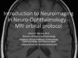
| 11 |
 | Doppler Ultrasonography | Devin D. Mackay, MD | Explanation of using doppler ultrasonography in examinations. | Doppler Ultrasonography |
| 12 |
 | Double Maddox Rod Test | Nagham Al-Zubidi, MD | A description of the double maddox rod test, the equipment used and the techniques for interpreting the exam. | Double Maddox Rod Test |
| 13 |
 | The Eyelid Examination | Sovik De Sirkar, MSIII; Ore-ofe Adesina, MD | Description of the eyelid examination. | Eyelid Examination |
| 14 |
 | Finger to Nose Perimetry | John Pula, MD | A description of the use of the finger to nose perimetry test is covered. | Non-organic Vision Loss |
| 15 |
 | Functional MRI | Devin D. Mackay, MD | Explanation of using functional MRI in examinations. | Functional MRI |
| 16 |
 | Fundus Photography | Devin D. Mackay, MD; Valérie Biousse, MD, | Explanation of using fundus photography in examinations. | Fundus Photography |
| 17 |
| Indirect Ophthalmoscope | Devin D. Mackay, MD; Valérie Biousse, MD | Explanation of using the indirect ophthalmoscope in examinations. | Indirect Ophthalmoscope |
| 18 |
 | Iris and Ciliary Body Examination | Sovik De Sirkar, MSIII; Ore-ofe Adesina, MD | Description of the iris and ciliary body examination. | Iris; Ciliary Body Examination |
| 19 |
 | The Lens Examination | Sovik De Sirkar, MSIII; Ore-ofe Adesina, MD | Description of the lens examination. | Lens Examination |
| 20 |
 | Magnetic Resonance Imaging (MRI) | Devin D. Mackay, MD | Explanation of using magnetic resonance imaging (MRI) in examinations. | Magnetic Resonance Imaging (MRI) |
| 21 |
 | Mirror Test for Malingering | Walsh and Hoyt Clinical Neuro-Ophthalmology, 6th Edition | Description of the mirror test. | Mirror Test; Malingering |
| 22 |
 | Monocular Hemianopia | Walsh and Hoyt Clinical Neuro-Ophthalmology, 6th Edition | Description of testing for a non-physiologic cause of a monocular hemianopia. | Monocular Hemianopia |
| 23 |
 | MR Angiography (MRA) | Devin D. Mackay, MD | Explanation of using MR angiography in examinations. | MR Angiography (MRA) |
| 24 |
 | MR Venography (MRV) | Devin D. Mackay, MD | Explanation of using MR venography (MRV) in examinations. | MR Venography (MRV) |
| 25 |
 | MRI Orbital Protocol | Devin D. Mackay, MD | Description of the MRI orbital protocol. | MRI Orbital Protocol |