|
|
Creator | Title | Description | Subject | Date |
| 1 |
 |
Huang, Lin Eric | HIF-1? mediates tumor hypoxia to confer a perpetual mesenchymal phenotype for malignant progression | Although tumor progression involves genetic and epigenetic alterations to normal cellular biology, the underlying mechanisms of these changes remain obscure. Numerous studies have shown that hypoxia-inducible factor 1? (HIF-1?) is overexpressed in many human cancers and up-regulates a host of hypoxi... | | |
| 2 |
 |
Couldwell, William T. | Intraoperative aneurysmal subarachnoid hemorrhage after rupture of a previously undiagnosed intracranial aneurysm during Chiari decompression: case report and literature review | Type 1 Chiari malformation occurs when caudal displacement of cerebellar tonsils below the level of foramen magnum obstructs the normal flow of cerebrospinal fluid (CSF) between the cranial and spinal spaces, a condition that often needs surgical decompression to restore normal CSF circulation. Abru... | | 2014-01-01 |
| 3 |
 |
Couldwell, William T. | Hemorrhagic and nonhemorrhagic Rathke cleft cysts mimicking pituitary apoplexy | Object. Rathke cleft cysts (RCCs) are infrequently symptomatic, and apoplexy is one of the most unusual presentations. Only a few cases of apoplexy associated with RCCs have been reported, and their clinical, imaging, surgical, and pathological features are poorly understood. In the cases that have ... | | 2008-01-01 |
| 4 |
 |
Gluf, Wayne M.; Gottfried, Oren N.; Schmidt, Meic H. | Cavernous hemangioma of the skull presenting with subdural hematoma | Cavernous hemangioma of the calvarium is a very rare disease, and patients usually present with headaches or a visible skull deformity. Few reports of patients presenting with intradiploic or epidural hemorrhages are found in the literature. No cases of an intradural hemorrhage from a cavernous hema... | Hemangioma, Cavernous; Skull; Hematoma, Subdural | 2004-07-15 |
| 5 |
 |
Schmidt, Meic H. | Cavernous hemangioma of the skull presenting with subdural hematoma | Cavernous hemangioma of the calvaria is a very rare disease, and patients usually present with headaches or a visible skull deformity. Few reports of patients presenting with intradiploic or epidural hemorrhages are found in the literature. No case of an intradural hemorrhage from a cavernous hemang... | Calvaria; Cavernous hemangioma | 2004 |
| 6 |
 |
Couldwell, William T. | Cranioplasty with the Medpor porous polyethylene Flexblock implant | The authors describe the use of a porous polyethylene Flexblock implant for cosmetic cranioplasty. The implant may be used to cover any small- or medium-sized (< 8 cm) cranial defect, offering similar cosmetic results to standard alloplast cranioplasty while decreasing operation time. The porous i... | Cranioplasty; Medpor; Alloplast; Polyethylene; Skull defect | 1994 |
| 7 |
 |
Schmidt, Meic H.; Apfelbaum, Ronald I. | Atlantoaxial transarticular screw fixation: a review of surgical indications, fusion rate, complications, and lessons learned in 191 adult patients | Object. In this, the first of two articles regarding C1-2 transarticular screw fixation, the authors assessed the rate of fusion, surgery-related complications, and lessons learned after C1-2 transarticular screw fixation in an adult patient series. Methods. The authors retrospectively reviewed 191 ... | Atlantoaxial junction; Craniocervical junction; Instability; Transarticular screw fixation; Fusion | 2005 |
| 8 |
 |
Couldwell, William T.; Gottfried, Oren N. | Distal ventriculoperitoneal shunt failure secondary to clostridium difficile colitis | Distal ventriculoperitoneal shunt obstruction is typically associated with cerebrospinal fluid (CSF) infection, fluid pseudocysts, bowel obstruction, bowel perforation, or improper shunt placement in the abdomen. We describe a unique etiology for distal shunt obstruction secondary to Clostridium di... | | 2005-01-01 |
| 9 |
 |
Couldwell, William T. | Distal ventriculoperitoneal shunt failure secondary to Clostridium Difficile Colitis | Distal ventriculoperitoneal shunt obstruction is typically associated with cerebrospinal fluid (CSF) infection, fluid pseudocysts, bowel obstruction, bowel perforation, or improper shunt placement in the abdomen. We describe a unique etiology for distal shunt obstruction secondary to Clostridium dif... | Shunt Failure | 2005-03 |
| 10 |
 |
Garber, Sarah | Neurosurgical complications of direct thrombin inhibitors - catastrophic hemorrhage after mild traumatic brain injury in a patient receiving dabigatran | Dabigatran etexilate is an oral anticoagulant that acts as a direct, competitive thrombin inhibitor. Large randomized clinical trials have shown higher doses of dabigatran (150 mg taken twice daily) to be superior to warfarin in terms of stroke and systemic embolism rates in patients with nonvalvula... | | 2012-01-01 |
| 11 |
 |
Dailey, Andrew T. | Guidelines for the performance of fusion procedures for degenerative disease of the lumbar spine. Part 8: lumbar fusion for disc herniation and radiculopathy | Standards. There is insufficient evidence to recommend a treatment standard. Guidelines. There is insufficient evidence to recommend a treatment guideline. Options. 1) Lumbar spinal fusion is not recommended as routine treatment following primary disc excision in patients with a herniated lumbar... | Fusion; Lumbar spine; Degenerative disease; Lumbar fusion; Disc herniation; Radiculopathy | 2005 |
| 12 |
 |
Couldwell, William T. | Radiographic features of tumefactive giant cavernous angiomas | Giant cavernous angiomas (GCAs) are very rare, and imaging features of GCAs can be very different from those of typical cavernous angiomas (CAs), making them a diagnostic challenge. The purpose of the study was to evaluate the radiographic features of GCAs, with an emphasis on the differentiating fe... | Cavernous angiomas; Cavernous malformations; Tumefactive | 2008 |
| 13 |
 |
Dailey, Andrew T. | Subaxial cervical spine trauma classification: the subaxial injury classification system and case examples | Object. The authors review a novel subaxial cervical trauma classification system and demonstrate its application through a series of cervical trauma cases. Methods. The Spine Trauma Study Group collaborated to create the Subaxial Injury Classification (SLIC) and Severity score. The SLIC system is... | Cervical spine trauma; Injury severity score; Spinal cord injury; Subaxial Injury Classification; SLIC; Discoligamentous integrity; Neurological status | 2008 |
| 14 |
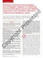 |
Dailey, Andrew T.; Brodke, Darrel S. | Differences between neurosurgeons and orthopaedic surgeons in classifying cervical dislocation injuries and making assessment and treatment decisions: a multicenter reliability study | Variability exists in the management of cervical spinal injuries. The goal of this study was to assess the effect of training specialty (orthopedic surgery vs neurosurgery) on management of cervical dislocations. | Cervical dislocation injuries; Classification; Orthopedic surgeons; Orthopaedic surgeons; Assessment decisions; Treatment decisions | 2008 |
| 15 |
 |
Digre, Kathleen B.; Couldwell, William T.; Amini, Amin | Photophobia in a blind patient: An alternate visual pathway. Case report. | Photophobia is a common neurological and ophthalmological symptom that has been associated with a growing number of neurosurgical conditions, especially compressive lesions. The exact signaling pathways and neurophysiological features of the disorder are not well understood; however, data from multi... | Photophobia, Blindness, Signaling Pathway, Pretectal Nuclei, Trigeminal Pathway | 2006-12-14 |
| 16 |
 |
Couldwell, William T. | Giant fusiform aneurysm in an adolescent with PHACES syndrome treated with a high-flow external carotid artery-M3 bypass | The acronym PHACES describes a rare neurocutaneous syndrome that comprises posterior fossa malformations, facial hemangiomas, arterial anomalies, coarctation of the aorta and cardiac defects, eye abnormalities, and sternal defects. Facial hemangiomas constitute the hallmark of this disorder. Giant ... | | 2007 |
| 17 |
 |
Couldwell, William T.; Schmidt, Richard H.; Salzman, Karen L.; Chin, Steven S. | Glioblastoma multiforme of the pineal region | Glioblastoma multiforme (GBMs) tumors are exceedingly rare tumors in the pineal region. We present three cases in which patients presented with a pineal/posterior third ventricular region mass and review all the previously reported cases in the literature. Pineal region GBM seems to be a very aggre... | Glioblastoma multiforme; Hydrocephalus; Leptomeningeal dissemination; Malignant glioma; Perinaud's syndrome; Pineal region; Spinal metastases | 2006 |
| 18 |
 |
Dailey, Andrew T. | Magnetic resonance neurography for cervical radiculopathy: a preliminary report | MAGNETIC RESONANCE NEUROGRAPHY was used to directly image cervical spinal nerves in patients with clinical and radiographic evidence of cervical radiculopathy. A magnetic resonance imaging phased-array coil system was used to obtain high-resolution coronal T1-weighted spin echo, coronal/axial T2-wei... | | 1996 |
| 19 |
 |
Couldwell, William T. | Medpor implant in cranioorbitomaxillary reconstruction: institutional experience and a review of the literature | Autologous materials remain the gold standard for complex skull base and craniofacial reconstruction, but they carry additional morbidity associated with the second harvest procedure and with prolonged operation time. These autologous materials also resorb in a way that is not predictable, rendering... | Medpor; Alloplastic material; Autogenous tissue; Craniofacial reconstruction; Skull base surgery; Methylmethacrylate; Silicone | 2008 |
| 20 |
 |
Schmidt, Meic H. | Benzoporphyrin derivative and light-emitting diode for use in photodynamic therapy: applications of space light-emitting diode technology | Photodynamic therapy (PDT) is a cancer treatment modality that recently has been applied as adjuvant therapy for rain tumors. PDT consists of intravenously injecting a photosensitizer, which preferentially accumulates in tumor ells, into a patient and then activating the photosensitizer with a light... | Benzoporphyrin; Photodynamic therapy; Brain tumors; Photofrin | 1998 |
| 21 |
 |
Couldwell, William T. | Efficacy of clip-wrapping in treatment of complex pediatric aneurysms | Purpose: Pediatric aneurysms (PAs) are distinct from their adult counterparts with respect to typical location, aneurysm type, and known predisposing risk factors. Many strategies have been employed to treat PAs, but, although it has been used frequently in adults, clip-wrapping in pediatric patient... | | 2012-01-01 |
| 22 |
 |
Couldwell, William T.; Osborne, Anne G. | Hypertrophic olivary degeneration after surgical removal of cavernous malformations of the brain stem: report of four cases and review of the literature | Background: Hypertrophic olivary degeneration (HOD) is a pathological phenomenon that occurs after injury to the dentato-olivary pathway. Its hallmarks include hypertrophy of the olive with increased T2 signal intensity on magnetic resonance imaging, and often manifests with palatal tremor and osci... | Hypertrophic olivary degeneration; Surgery; Cavernous malformation; Brainstem | 2008 |
| 23 |
 |
Patel, Bhupendra C. | Lateral orbital wall approach to the cavernous sinus: laboratory investigation | Object. Lesions of the cavernous sinus remain a technical challenge. The most common surgical approaches involve some variation of the standard frontotemporal craniotomy. Here, the authors describe a surgical approach to access the cavernous sinus that involves the removal of the lateral orbital wal... | | 2012-01-01 |
| 24 |
 |
Couldwell, William T. | Communication between malignant glioma cells and vascular endothelial cells through gap junctions | Object. Extensive invasion and angiogenesis are hallmark features of malignant gliomas. Communication between malignant glioma cells and surrounding astrocytes occurs, resulting in transformation of the astrocytic phenotype. In the present study, the authors examined whether malignant glioma cells a... | | 2003-01-01 |
| 25 |
 |
Kestle, John R. W. | Reduction of hemorrhage risk after stereotactic radiosurgery for cavernous malformations | The benefits of radiosurgery for cavernous malformations are difficult to assess because of the unclear natural history of this vascular lesion, the inability to image malformation vessels, and the lack of an imaging technique that defines "cure." The authors selected for radiosurgery 47 patients w... | Cavernous malformation; Gamma knife; Stereotactic radiosurgery | 1995 |