1 - 25 of 10
| Creator | Title | Description | Subject | Date | ||
|---|---|---|---|---|---|---|
| 1 |
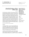 |
Gerig, Guido | Abnormal vessel tortuosity as a marker of treatment response of malignant gliomas: preliminary report | Despite multiple advances in medical imaging, noninvasive monitoring of therapeutic efficacy for malignant gliomas remains problematic. An underutilized observation is that malignancy induces characteristic abnormalities of vessel shape. These characteristic shape abnormalities affect both capillari... | 2004-01-01 | |
| 2 |
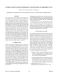 |
Gerig, Guido | Closed and open source neuroimage analysis tools and libraries at UNC | The emergence of open-source libraries and development tools in the last decade has changed the process of academic software development in many ways. In medical image processing and visualization this change is especially evident, also because open source projects are actively furthered by grant fu... | 2006-01-01 | |
| 3 |
 |
Gerig, Guido | Computational anatomy to assess longitudinal trajectory of brain growth | This paper addresses the challenging problem of statistics on images by describing average and variability. We describe computational anatomy tools for building 3-D and spatio-temporal 4-D atlases of volumetric image data. The method is based on the previously published concept of unbiased atlas bui... | 2006-01-01 | |
| 4 |
 |
Gerig, Guido | Group mean differences of voxel and surface objects via nonlinear averaging | Building of atlases representing average and variability of a population of images or of segmented objects is a key topic in application areas like brain mapping, deformable object segmentation and object classification. Recent developments in image averaging, i.e. constructing an image which is cen... | 2006-01-01 | |
| 5 |
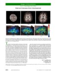 |
Gerig, Guido | Images in neuroscience: Fetal and Neonatal Brain Development | In the top row of longitudinal T1-weighted magnetic resonance images of the same child (and same scale), note the dramatic increase in total brain size as well as in white matter intensity over early development. In the bottom row of diffusion tensor images, white matter tractography of a neonate, o... | 2006-01-01 | |
| 6 |
 |
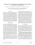
Gerig, Guido | Multiscale medial shape-based analysis of image objects | Medial representation of a three-dimensional (3-D) object or an ensemble of 3-D objects involves capturing the object interior as a locus of medial atoms, each atom being two vectors of equal length joined at the tail at the medial point. Medial representation has a variety of beneficial properties,... | 2003-01-01 | |
| 7 |
 |
Gerig, Guido | Reduced relationship to cortical white matter volume revealed by Tractography-based segmentation of the corpus callosum in young children with developmental delay | Objective: The corpus callosum is the primary anatomical substrate for inter-hemispheric communication, which is important for a range of adaptive and cognitive behaviors in early development. Previous studies that have measured the corpus callosum in developmental populations have been limited by t... | 2006-01-01 | |
| 8 |
 |
Gerig, Guido | Statistics of populations of images and its embedded objects: driving applications in neuroimaging | Work in progress towards modeling shape statistics of multi-object complexes is presented. Constraints defined by the set of objects such as a compact representation of object shape relationships and correlation of shape changes might have advantages for automatic segmentation and group discriminati... | 2006-01-01 | |
| 9 |
 |
Gerig, Guido | Tumor-induced structural radiometric asymmetry in brain images | This paper presents a general framework for analyzing structural and radiometric asymmetry in brain images. In a healthy brain, the left and right hemispheres are largely symmetric across the mid-sagittal plane. Brain tumors may belong to one or both of the following categories: mass-effect, in whic... | 2001-01-01 | |
| 10 |
 |
Gerig, Guido | Unbiased diffeomorphic atlas construction for computational anatomy | Construction of population atlases is a key issue in medical image analysis, and particularly in brain mapping. Large sets of images are mapped into a common coordinate system to study intra-population variability and inter-population differences, to provide voxel-wise mapping of functional sites, a... | 2004-01-01 |
1 - 25 of 10