Collection of materials relating to neuro-ophthalmology as part of the Neuro-Ophthalmology Virtual Education Library.
NOVEL: https://novel.utah.edu/
TO
| Title | Creator | Description | Subject | ||
|---|---|---|---|---|---|
| 1 |
 |
Cotton Wool Spots: The Basics | Arnav Gupta, BHSc; Rahul Sharma, MD, MPH | A presentation describing cotton wool spots, an abnormal finding on funduscopic exam of the retina of the eye. | Cotton Wool Spots; Retina |
| 2 |
 |
Disc Cupping: The Basics | Arnav Gupta, BHSc; Rahul Sharma, MD, MPH | A presentation describing optic disc cupping, due to damage of optic nerve fibres. | Disc Cupping; Optic Disc |
| 3 |
 |
Progressive Supranuclear Palsy (PSP) | Molly Cincotta, MD; Ali G. Hamedani, MD, MHS | Objectives:; To provide an overview of PSP and its pathophysiology;; To present typical clinical features of the disease with a focus on ocular findings;; To provide a template for work up, diagnosis and treatment; ; To demonstrate typical eye movement abnormalities seen in PSP | Progressive Supranuclear Palsy (PSP) |
| 4 |
 |
Computed Tomography (CT): Principles, Technique, and Neuro-ophthalmic Applications | Alex Fraser, MD | Presentation covering Computed Tomography principles, adverse effects, comparison vs. MRI, and assorted examples of neuro-ophthalmic interest. | Computed Tomography (CT) |
| 5 |
 |
Examining the Pediatric Patient for Non-Neuro-ophthalmologists | John Pula, MD | Ten need-to-know pearls for examining the pediatric patient, for non-neuro-ophthalmologists. | Pediatric Patient Exam |
| 6 |
 |
Examining the Comatose Patient for Non-Neuro-ophthalmologists | John Pula, MD | Seven need-to-know pearls for examining the pediatric patient, for non-neuro-ophthalmologists. | Comatose Patient Exam |
| 7 |
 |
Posterior Cortical Atrophy | Natali V. Baner, MD; Ali G. Hamedani, MD, MHS | PowerPoint providing an overview of the definition, clinical presentation and treatment of posterior cortical atrophy | Posterior Cortical Atrophy |
| 8 |
 |
Dementia: Overview and Classification | Molly Cincotta, MD; Whitley Aamodt, MD; Ali G. Hamedani, MD, MHS | PowerPoint providing a broad overview of dementia, including definition, clinical findings, work up, diagnosis, classification, and management. | Dementia |
| 9 |
 |
Multiple System Atrophy: Overview and Neuro-ophthalmologic Features | Pavan Vaswani, MD, PhD, Movement Disorders Fellow; Ali G. Hamedani, MD, MHS | Objectives: Know the key pathologic features of Multiple System Atrophy; Recognize the clinical presentation, including neuro-ophthalmologic features; Understand the symptomatic therapies and prognosis | Multiple System Atrophy |
| 10 |
 |
Dementia with Lewy Bodies: Overview and Neuro-ophthalmologic features | Pavan Vaswani, MD, PhD; Ali G. Hamedani, MD, MHS | Objectives: Recognize the difference between Dementia with Lewy Bodies and Parkinson disease dementia; Recognize the clinical presentation of DLB and differentiating features from Alzheimer disease dementia; Understand the symptomatic therapies and prognosis | Dementia; Lewy Bodies |
| 11 |
 |
Vascular Dementia | Whitley Aamodt, MD; Ali G. Hamedani, MD, MHS | PowerPoint providing an overview of vascular dementia, including the pathophysiology, clinical symptoms, diagnosis, and management. | Vascular Dementia |
| 12 |
 |
Amyotrophic Lateral Sclerosis (ALS) | Natali V. Baner, MD; Ali G. Hamedani, MD, MHS | PowerPoint providing an overview of the definition, clinical presentation and treatment of amyotrophic lateral sclerosis (ALS). | Amyotrophic Lateral Sclerosis (ALS) |
| 13 |
 |
Frontotemporal Dementia: Overview and Neuro-ophthalmologic Features | Pavan Vaswani, MD, PhD; Ali G. Hamedani, MD, MHS | Objectives: Understand the diagnostic criteria for the frontotemporal dementias; Differentiate behavioral variant FTD and the common variants of primary progressive aphasia; Recognize neuro-ophthalmologic and imaging features seen in FTD syndromes | Frontotemporal Dementia |
| 14 |
 |
Medicolegal and Ethical Considerations in Ophthalmology | M. Tariq Bhatti, MD | Slideshow describing topic. | Ethics; Legal |
| 15 |
 |
Diagnostic Error of Neuro-ophthalmologic Conditions: State of the Science | Leanne Stunkel, MD; David E. Newman-Toker, MD, PhD; Nancy J. Newman, MD; Valérie Biousse, MD | Diagnostic error is prevalent and costly, occurring in up to 15% of US medical encounters and affecting up to 5% of the US population. One-third of malpractice payments are related to diagnostic error. A complex and specialized diagnostic process makes neuro-ophthalmologic conditions particularly vu... | Diagnostic Errors |
| 16 |
 |
Protecting Human Subjects in Biomedical Research | Lisa R. Latchney, MS, CCRC | PowerPoint discussion of the history and development of ethics regulations in health research. | Ethical Issues in Research; Consent |
| 17 |
 |
Manuscripts: You Can Write These! | Elaine Smolock, PhD | Overview of writing techniques and parts of the manuscript, basic approach to writing results sections, what makes a good introduction, crafting a meaningful discussion, abstract and title suggestions, and how to get your editor's attention. | Writing Techniques |
| 18 |
 |
Acute Optic Neuritis | Neil R. Miller, MD, FACS | Overview of acute optic neuritis. | Optic Neuritis |
| 19 |
 |
Afferent Visual Pathway Disorders: Typical vs Atypical Optic Neuritis | Carmen Chan, FRCP, FRCOphth, FRCSEd(Ophth), FHKAM(Ophthalmology) | Discussion of typical vs atypical optic neuritis. | Optic Neuritis |
| 20 |
 |
Superior Segmental Optic Disc Hypoplasia (SSOH) "Topless Disc Syndrome" | Sparsh Jain, Medical Student; Ryan Walsh, MD | This is a case of superior segmental optic disc hypoplasia that was found incidentally after a screening visual field test revealed an asymptomatic inferior field defect in the left eye. The patient has a unilateral SSOH in the left eye. | Superior Segmental Optic Disc Hypoplasia (SSOH) |
| 21 |
 |
Bergmeister Papilla | Sumayya Almarzouqi, MD | A brief overview of Bergmeister papilla, a rare congenital disc anomaly. It arises from the center of the optic disc consists of a small tuft of fibrous tissue and represents a remnant of the fetal hyaloid artery. | Bergmeister Papilla |
| 22 |
 |
Modern Imaging of Optic Disc Drusen | Meagan Seay, DO | This is a short powerpoint describing imaging techniques (specifically OCT-EDI, fundus autofluorescence, and B-scan ultrasonography) for optic disc drusen. Examples of these techniques are included. | Optic Disc Drusen; Imaging; OCT-EDI; Fundus Autofluorescence; B-scan Ultrasonography |
| 23 |
 |
Suprasellar Meningioma | Sumayya Almarzouqi, MD | Description of a case of suprasellar or sellar mass causeing chiasmal compression. | Suprasellar Meningioma |
| 24 |
 |
Anaesthesia for Eye Surgery and Associated Complications Slides | Julie Smith, MBBS, FANZCA | Lecture covering commonly performed eye surgery and anaesthetic techniques. | Eye Surgery; Anesthesia |
| 25 |
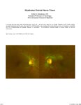 |
Myelinated Retinal Nerve Fibers | Scott N. Grossman, MD | A 33 year old man has noted chronically poor vision OS - left eye color noted to be 'orange' instead of red. fundus photos revealed myelinated retinal nerve fiber layer OU (OS>OD) with corresponding linear paracentral scotoma on Humphrey visual field 24-2 OS corresponding with greatest degree of my... | Myelinated Retinal Nerve Fibers |