A collection of videos relating to the diagnosis and treatment of eye movement disorders. This collection includes many demonstrations of examination techniques.
Dan Gold, D.O., Associate Professor of Neurology, Ophthalmology, Neurosurgery, Otolaryngology - Head & Neck Surgery, Emergency Medicine, and Medicine, The Johns Hopkins School of Medicine.
A collection of videos relating to the diagnosis and treatment of eye movement disorders.
NOVEL: https://novel.utah.edu/
TO
| Title | Description | Type | ||
|---|---|---|---|---|
| 26 |
 |
Leukemic Leptomeningeal Carcinomatosis Causing 4th and 6th Nerve Palsies | 𝗢𝗿𝗶𝗴𝗶𝗻𝗮𝗹 𝗗𝗲𝘀𝗰𝗿𝗶𝗽𝘁𝗶𝗼𝗻: This is a 55-yo-man with CML that recurred as AML. Diagonal diplopia developed, and on examination he was found to have a partial right 6th nerve palsy, in addition to a left hypertropia that increased in right gaze, down... | Image/MovingImage |
| 27 |
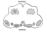 |
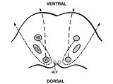
Medullary Structures Relevant to the Ocular Motor and Vestibular Consequences of Lateral Medullary (Wallenberg) Syndrome | This is an axial section of the medulla showing the structures that, when damaged, are responsible for the vestibular and ocular motor features of the lateral medullary or Wallenberg syndrome. The nucleus prepositus hypoglossi (NPH) and medial vestibular nucleus (MVN) complex is important for horizo... | Image |
| 28 |
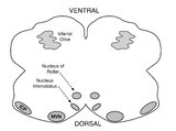 |
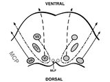
Medullary Structures Relevant to Upbeat Nystagmus | This is an axial section of the medulla, slightly more caudal as compared to (please refer to figure "medullary structures relevant to the ocular motor and vestibular consequences of the lateral medullary (Wallenberg) syndrome). Again seen are the inferior cerebellar peduncle (ICP) and caudal aspect... | Image |
| 29 |
 |
Mild 6th Nerve Palsy Due to Pontine Stroke | This is a 70-year-old woman with HTN and diabetes who presented with horizontal diplopia for several weeks, worse in right gaze. There was a very subtle abduction paresis OD with full motility elsewhere. With cover-uncover testing, there was a small esotropia in right gaze (esodeviation seen with al... | Image/MovingImage |
| 30 |
 |
Monocular Downbeat Nystagmus Due to a Posterior Fossa Cyst | This is a 40-yo-man who experienced months of imbalance and was found to have an epidermoid cyst (immediately posterior to the 4th ventricle), which was resected. Months after surgery, he experienced monocular vertical oscillopsia. On examination, there was subtle downbeat nystagmus (DBN) in the rig... | Image/MovingImage |
| 31 |
 |
Monocular Horizontal Pendular Nystagmus in MS | 𝗢𝗿𝗶𝗴𝗶𝗻𝗮𝗹 𝗗𝗲𝘀𝗰𝗿𝗶𝗽𝘁𝗶𝗼𝗻: Both of these patients have MS and monocular (OS) horizontal pendular nystagmus. The first patient seen in the video has normal afferent function and no evidence of optic nerve disease in either eye, while the second pati... | Image/MovingImage |
| 32 |
 |
Nystagmus Due to Paraneoplastic (Anti-Yo) Brainstem and Cerebellar Degeneration | 𝗢𝗿𝗶𝗴𝗶𝗻𝗮𝗹 𝗗𝗲𝘀𝗰𝗿𝗶𝗽𝘁𝗶𝗼𝗻: This is a 40-yo-woman with anti-Yo antibody associated with ovarian cancer. Initial symptoms 2.5 years prior (to this video) included imbalance and dysarthria. She complained of oscillopsia which was due to her upbeat nys... | Image/MovingImage |
| 33 |
 |
Ocular Bobbing Due to Hepatic Encephalopathy | 𝗢𝗿𝗶𝗴𝗶𝗻𝗮𝗹 𝗗𝗲𝘀𝗰𝗿𝗶𝗽𝘁𝗶𝗼𝗻: This is a 55-year-old man presented with hepatic encephalopathy, and found to have ocular bobbing. Head CT did not show any acute changes. Ocular bobbing almost always localizes to the pons, although cerebellar pathology ... | Image/MovingImage |
| 34 |
 |
Ocular Motor & Vestibular Features of the MLF Syndrome | 𝗢𝗿𝗶𝗴𝗶𝗻𝗮𝗹 𝗗𝗲𝘀𝗰𝗿𝗶𝗽𝘁𝗶𝗼𝗻: This 61-year-old woman with HTN and DM presented for evaluation of acute onset diagonal diplopia. Adduction OS was about 60% of normal while medialization OS improved with convergence. In right gaze, dissociated abducti... | Image/MovingImage |
| 35 |
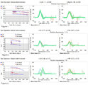 |
Ocular Motor & Vestibular Features of the MLF Syndrome (Figures 1, 2, and 3) | This 61-year-old woman with HTN and DM presented for evaluation of acute onset diagonal diplopia. Adduction OS was about 60% of normal while medialization OS improved with convergence. In right gaze, dissociated abducting nystagmus was present OD, and there was a clear adduction lag when asking he... | Image |
| 36 |
 |
Ocular Motor Signs in Progressive Supranuclear Palsy (PSP) | 𝗢𝗿𝗶𝗴𝗶𝗻𝗮𝗹 𝗗𝗲𝘀𝗰𝗿𝗶𝗽𝘁𝗶𝗼𝗻: This is a 65-yo-woman complaining of imbalance and double vision. She had significant convergence insufficiency (and would close her right eye with near viewing), providing an explanation for her diplopia. Convergence ins... | Image/MovingImage |
| 37 |
 |
Ocular Motor Signs in SCA 6 | 𝗢𝗿𝗶𝗴𝗶𝗻𝗮𝗹 𝗗𝗲𝘀𝗰𝗿𝗶𝗽𝘁𝗶𝗼𝗻: This is a 45-yo-man who was recently diagnosed with SCA 6. There was no clear spontaneous downbeat nystagmus (DBN) in primary gaze, although DBN could clearly be provoked by convergence. Other ocular motor features includ... | Image/MovingImage |
| 38 |
 |
Oculogyric Crisis | 𝗢𝗿𝗶𝗴𝗶𝗻𝗮𝗹 𝗗𝗲𝘀𝗰𝗿𝗶𝗽𝘁𝗶𝗼𝗻: This is a patient with neuroleptic-induced oculogyric crisis. 𝗡𝗲𝘂𝗿𝗼-𝗼𝗽𝗵𝘁𝗵𝗮𝗹𝗺𝗼𝗹𝗼𝗴𝘆 𝗮𝗻𝗱 𝗡𝗲𝘂𝗿𝗼-𝗼𝘁𝗼𝗹𝗼𝗴𝘆 𝗧𝗲𝘅𝘁𝗯�... | Image/MovingImage |
| 39 |
 |
One-and-a-Half Syndrome Due to Pontine Hemorrhage | This is a 50-year-old woman who, while exercising in the gym, suddenly experienced vertigo, nausea, vomiting, tingling in the left arm, and diplopia. MRI demonstrated a brainstem hemorrhage that involved the right greater than left pons. Examination demonstrated a right horizontal gaze palsy due to ... | Image/MovingImage |
| 40 |
 |
Opsoclonus Provoked by Convergence | 𝗢𝗿𝗶𝗴𝗶𝗻𝗮𝗹 𝗗𝗲𝘀𝗰𝗿𝗶𝗽𝘁𝗶𝗼𝗻: This is a 40-yo-man with post-infectious opsoclonus-myoclonus syndrome. Opsoclonus was intermittently evident in primary position, but was consistently provoked (and intensified) by convergence. Occasionally, opsoclonus (... | Image/MovingImage |
| 41 |
 |
Oscillopsia and Bilateral Vestibular Loss with Gentamicin Ototoxicity | Patients with bilateral vestibular loss commonly experience oscillopsia with head movements, or an inability to stabilize retinal images with subsequent bouncing or jumping of the environment due to loss of vestibular function. This causes significant blurring of vision and disorientation, dizziness... | Image/MovingImage |
| 42 |
 |
Oscillopsia: A Common Symptom of Bilateral Vestibular Loss | 𝗢𝗿𝗶𝗴𝗶𝗻𝗮𝗹 𝗗𝗲𝘀𝗰𝗿𝗶𝗽𝘁𝗶𝗼𝗻: This video is an example of what a patient with bilateral vestibular loss experiences while walking. Without a VOR, there is no mechanism to ensure retinal stability of the world with each head movement, and oscillopsia (... | Image/MovingImage |
| 43 |
 |
Paraneoplastic Downbeat Nystagmus and Cerebellar Ataxia Due to Small Cell Lung Carcinoma | 𝗢𝗿𝗶𝗴𝗶𝗻𝗮𝗹 𝗗𝗲𝘀𝗰𝗿𝗶𝗽𝘁𝗶𝗼𝗻: This is a 61-year-old woman (non-smoker) who developed a gait disorder, dizziness and oscillopsia that was progressive over 2 months. Exam demonstrated spontaneous downbeat nystagmus with side pocket nystagmus in lateral ... | Image/MovingImage |
| 44 |
 |
Pendular Nystagmus and Ocular Motor Signs in MS | 𝗢𝗿𝗶𝗴𝗶𝗻𝗮𝗹 𝗗𝗲𝘀𝗰𝗿𝗶𝗽𝘁𝗶𝗼𝗻: This is a 30-year-old man with a 15 year history of multiple sclerosis. For the last 12 months, he experienced horizontal oscillopsia. On examination, there were ocular motor abnormalities including gaze-evoked nystagmus,... | Image/MovingImage |
| 45 |
 |
Pendular Nystagmus and Vision Loss | Three patients are presented here, each with poor vision (counting fingers or worse) related to retinitis pigmentosa in one patient (Usher's syndrome) and optic neuropathy in two patients, each of whom developed pendular nystagmus after vision loss developed. Visually mediated movements normally pre... | Image/MovingImage |
| 46 |
 |
Periodic Alternating Nystagmus and Perverted Head-shaking Nystagmus in Cerebellar Degeneration | 𝗢𝗿𝗶𝗴𝗶𝗻𝗮𝗹 𝗗𝗲𝘀𝗰𝗿𝗶𝗽𝘁𝗶𝗼𝗻: This is a 60-yo-woman with several years of worsening imbalance, diplopia (hers was actually unrelated to cerebellar pathology [although she did have an esotropia greater at distance that was cerebellar in origin] and due... | Image/MovingImage |
| 47 |
 |
Pons: 6th and 7th Nerve Anatomy and the Central Segmental Tract | From this cross-section of the pons, the proximity of the 6th nucleus to the 7th nerve fascicles is apparent. This is the basis of the so-called facial colliculus syndrome, where an ipsilesional horizontal gaze palsy from a nuclear 6th lesion (usually related to stroke or demyelination) can be seen ... | Image |
| 48 |
 |
Pons: 6th, 7th, 8th, and Middle Cerebellar Peduncle Anatomy | From this cross-section of the pons, the proximity of the 7th and 8th fascicles can be appreciated, and a lateral inferior pontine syndrome (anterior inferior cerebellar artery territory), which could involve both of these fascicles, could cause acute prolonged vertigo accompanied by a + ipsilateral... | Image |
| 49 |
 |
Pontine Hemorrhage Causing Oculopalatal Tremor and Multiple Cranial Neuropathies | This is a 45-yo-woman who had a dorsal pontine cavernoma that bled 2 years prior to this video. Symptoms included diplopia and oscillopsia. On examination, she had left>right facial palsies (upper and lower face from involvement of the nucleus/fascicle - i.e., lower motor neuron palsies) and sixth n... | Image/MovingImage |
| 50 |
 |
Positional Downbeat Nystagmus Mimicking Anterior Canal BPPV | Although positional downbeat nystagmus (pDBN) can indicate the rare anterior canal variant of benign paroxysmal positional vertigo, central mimics are common causes of pDBN. pDBN may be seen in multiple system atrophy (MSA), or seen with posterior fossa lesions, with a common example being a stroke ... | Image/MovingImage |