Best known for his world-renowned neuro-ophthalmology unit based at the University of California, San Francisco, William Hoyt, MD collected here more than 850 of his best images covering a wide range of disorders.
William F. Hoyt, MD, Professor Emeritus of Ophthalmology, Neurology and Neurosurgery, Department of Ophthalmology, University of California, San Francisco.
NOVEL: https://novel.utah.edu/
TO
Filters: Date: "1977" Collection: "ehsl_novel_wfh"
1 - 25 of 16
| Title | Description | Type | ||
|---|---|---|---|---|
| 1 |
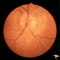 |
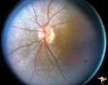
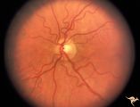
A201 Disc Swelling with Big Blind Spot Syndrome | Blind spot larger than could be explained by visible edema. Subretinal white dots probably indicate margin of blind spot. Anatomy: Optic disc; Retina. Pathology: Unknown. Disease/ Diagnosis: Big blind spot syndrome. Clinical: symptoms: photosias, blurred vision signs: Disc swelling; white spots in t... | Image |
| 2 |
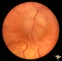 |
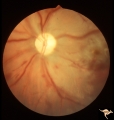
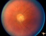
Bilateral Papilledema | Left eye. Bilateral Papilledema from vitamin A toxicity in young girl. Anatomy: Optic disc. Pathology: Bilateral papilledema. Disease/Diagnosis: Pseudotumor due to vitamin A toxicity in a young girl. Clinical notes: Headache. | Image |
| 3 |
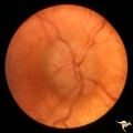 |
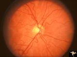
Bilateral Papilledema | Right eye. Bilateral Papilledema from vitamin A toxicity in young girl. Anatomy: Optic disc. Pathology: Bilateral papilledema. Disease/Diagnosis: Pseudotumor due to vitamin A toxicity in a young girl. Clinical notes: Headache. | Image |
| 4 |
 |
Cerebellar Macular Degenerative Disease | Cerebellar degeneration with granular maculae changes and bone spicules. Right eye. Anatomy: Retina. Pathology: Cerebellar macular degenerative disease. Disease/Diagnosis: Spinal Cerebellar Degeneration Type 7 (SCA-7). Clinical notes: Blindness and cerebellar degeneration. | Image |
| 5 |
 |
Cerebellar Macular Degenerative Disease | Cerebellar degeneration with granular maculae changes and bone spicules. Right eye. Anatomy: Retina. Pathology: Cerebellar macular degenerative disease. Disease/Diagnosis: Spinal Cerebellar Degeneration Type 7 (SCA-7). Clinical notes: Blindness and cerebellar degeneration. | Image |
| 6 |
 |
Cerebellar Macular Degenerative Disease | Cerebellar degeneration with granular maculae changes and bone spicules. Left eye. Anatomy: Retina. Pathology: Cerebellar macular degenerative disease. Disease/Diagnosis: Spinal Cerebellar Degeneration Type 7 (SCA-7). Clinical notes: Blindness and cerebellar degeneration. | Image |
| 7 |
 |
Cerebellar Macular Degenerative Disease | Cerebellar degeneration with granular maculae changes and bone spicules. Left eye. Anatomy: Retina. Pathology: Cerebellar macular degenerative disease. Disease/Diagnosis: Spinal Cerebellar Degeneration Type 7 (SCA-7). Clinical: Blindness and cerebellar degeneration. | Image |
| 8 |
 |
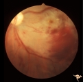
G206 Purtchers Traumatic Retinopathy | Left eye. After auto accident in which the patient's chest was squeezed. Same eye as G2_07. Anatomy: Optic disc. Pathology: Varied peripapillary ischemic retinopathy. Disease/ Diagnosis: Purtchers traumatic retinopathy. | Image |
| 9 |
 |
G207 Purtchers Traumatic Retinopathy | Left eye. Large pre-retinal hemorrhage. Same eye as G2_06. Anatomy: Optic disc. Pathology: Varied peripapillary ischemic retinopathy. Disease/ Diagnosis: Purtchers traumatic retinopathy. | Image |
| 10 |
 |
H73 Superior Segmental Optic Hypoplasia (SSOH) Topless Disc Syndrome | Bilateral SSOH. Left eye. Same patient as H_73. Anatomy: Optic disc. Pathology: Superior segmental optic hypoplasia (SSOH). Disease/ Diagnosis: Congenital anomaly. | Image |
| 11 |
 |
H74 Superior Segmental Optic Hypoplasia (SSOH) Topless Disc Syndrome | Bilateral SSOH. Right eye. Same patient as H_74. Anatomy: Optic disc. Pathology: Superior segmental optic hypoplasia (SSOH). Disease/ Diagnosis: Congenital anomaly. | Image |
| 12 |
 |
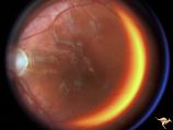
Paraneoplastic Retinopathy | Man with oat cell carcinoma of the lung with paraneoplastic retinopathy. Narrowed arterioles. Note absence of obvious retinal pigmentary degeneration. Cancer associated retinopathy syndrome (CAR Syndrome or Sawyer-Sellhorst Syndrome) (Ref: Sawyer, Sellhorst, Hoyt). Anatomy: Retina. Pathology: Oat c... | Image |
| 13 |
 |
Paraneoplastic Retinopathy | Man with oat cell carcinoma of the lung with paraneoplastic retinopathy. Narrowed arterioles. Note absence of obvious retinal pigmentary degeneration. Cancer associated retinopathy syndrome (CAR Syndrome or Sawyer-Sellhorst Syndrome) (Ref: Sawyer, Sellhorst, Hoyt). Anatomy: Retina. Pathology: Oat c... | Image |
| 14 |
 |
Retinal (Macular) Involvement in Subacute Sclerosing Pan Encephalopathy | Retinal (macular) involvement in Subacute Sclerosing Pan Encephalopathy (SSPE). Note optic disc pallor. Anatomy: Retina. Pathology: Cerebral and retinal degeneration. Disease/Diagnosis: Subacute Sclerosing Pan Encephalopathy (SSPE). Clinical: Progressive visual loss and progressive cerebral degenera... | Image |
| 15 |
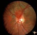 |
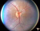
Von Hippel Lindau Disease (Hemangioblastoma of the Optic Disc) | Von Hippel Lindau Disease with a retinal hemangioblastoma on her optic disc. Anatomy: Optic disc. Pathology: Hemangioblastoma. Disease/Diagnosis: Von Hippel Lindau disease. Clinical: No visual symptoms. Patient had cerebellar ataxia. Imaging: R1_C1b is Arteriogram showing hemangioblastoma of the cer... | Image |
| 16 |
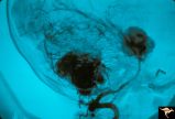 |
Von Hippel Lindau Disease (Hemangioblastoma of the Optic Disc) | Von Hippel Lindau Disease; Arteriogram showing hemangioblastoma of the cerebellum and midbrain. Anatomy: Brain. Pathology: Hemangioblastoma. Disease/Diagnosis: Von Hippel Lindau disease. Clinical: No visual symptoms. Patient had cerebellar ataxia. Imaging: Arteriogram showing hemangioblastoma of the... | Image |
1 - 25 of 16