Best known for his world-renowned neuro-ophthalmology unit based at the University of California, San Francisco, William Hoyt, MD collected here more than 850 of his best images covering a wide range of disorders.
William F. Hoyt, MD, Professor Emeritus of Ophthalmology, Neurology and Neurosurgery, Department of Ophthalmology, University of California, San Francisco.
NOVEL: https://novel.utah.edu/
TO
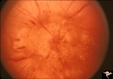
1 - 25 of 8
| Title | Description | Type | ||
|---|---|---|---|---|
| 1 |
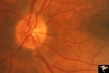 |
E12 Disc Swelling with Central Vein Occlusion | 2nd attack of papillophlebitis. There is an optociliary bypass vessel at 4:00. Anatomy: Optic disc; Retina. Pathology: Central retinal vein occlusion. Disease/ Diagnosis: Disc swelling due to central retinal vein occlusion. | Image |
| 2 |
 |
H38 Segmental Hypoplasia; Retinal; Tilted (Dysverted) Disc | Right eye. Man with tilted (dysverted) disc with inferior nasal crescent and high myopia. Same patient as H_39. Anatomy: Optic disc; Retina. Pathology: Hypoplasia secondary to retinal lesion. Disease/ Diagnosis: Segmental optic disc hypoplasia. Clinical: Man with bitemporal visual field defects. | Image |
| 3 |
 |
H39 Segmental Hypoplasia, Retinal, Tilted (Dysverted) Disc | Visual field of patient in H_38 showing upper temporal field depression caused by inferior nasal hypoplasia. Anatomy: Optic disc; Retina. Pathology: Hypoplasia secondary to retinal lesion. Disease/ Diagnosis: Segmental optic disc hypoplasia. Clinical: Man with bitemporal visual field defects. | Image |
| 4 |
 |
H78 Inferior Segmental Optic Hypoplasia (ISOH) | ISOH with inferior choroidal crescent. Patient had superior visual field defect. Anatomy: Optic disc. Pathology: Inferior segmental optic hypoplasia (ISOH). Disease/ Diagnosis: Congenital anomaly. | Image |
| 5 |
 |
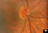
Resolution of Papilledema Following Optic Nerve Sheath Decompression (ONSD) | Left eye. 17 year old boy. Cryptococcal meningitis. Resolution of papilledema following optic nerve sheath decompression (ONSD) in November 1, 1974. Same eye as P_53a on December 1974. Atrophic. Note "high-water" marks. Anatomy: Optic disc. Pathology: Papilledema. Disease/Diagnosis: Resolving papill... | Image |
| 6 |
 |
Resolution of Papilledema Following Optic Nerve Sheath Decompression (ONSD) | Left eye. 17 year old boy. Cryptococcal meningitis. Module developed papilledema. June 1974. Anatomy: Optic disc. Pathology: Papilledema. Disease/Diagnosis: Resolving papilledema. | Image |
| 7 |
 |
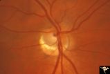
Resolution of Papilledema Following Optic Nerve Sheath Decompression (ONSD) | Left eye. 17 year old boy. Cryptococcal meningitis. Resolution of papilledema following optic nerve sheath fenestration (ONSF) on November 1, 1974. Same eye as P_53a on November 7, 1974, one week following ONSF. Anatomy: Optic disc. Pathology: Papilledema. Disease/Diagnosis: Resolving papilledema. | Image |
| 8 |
 |
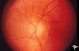
Resolution of Papilledema Following Optic Nerve Sheath Decompression (ONSD) | Left eye. 17 year old boy. Cryptococcal meningitis. Same eye as P_53a. Increased papilledema. August 1974. Anatomy: Optic disc. Pathology: Papilledema. Disease/Diagnosis: Resolving papilledema. | Image |
1 - 25 of 8