Best known for his world-renowned neuro-ophthalmology unit based at the University of California, San Francisco, William Hoyt, MD collected here more than 850 of his best images covering a wide range of disorders.
William F. Hoyt, MD, Professor Emeritus of Ophthalmology, Neurology and Neurosurgery, Department of Ophthalmology, University of California, San Francisco.
NOVEL: https://novel.utah.edu/
TO
Filters: Date: "1962" Collection: "ehsl_novel_wfh"
1 - 25 of 9
| Title | Description | Type | ||
|---|---|---|---|---|
| 1 |
 |
Buried Drusen | 5 year old boy. Bilateral buried drusen. Notice the lumpy nasal disc elevation. This patient had a twin brother whose optic disc drusen were exposed. Anatomy: Optic disc. Pathology: Drusen of the optic disc. Disease/Diagnosis: Drusen of the optic disc. Clinical notes: Normally functioning eye with ... | Image |
| 2 |
 |
Buried Drusen | 5 year old boy. Bilateral buried drusen. Notice the lumpy nasal disc elevation. This patient had a twin brother whose optic disc drusen were exposed. Anatomy: Optic disc. Pathology: Drusen of the optic disc. Disease/Diagnosis: Drusen of the optic disc. Clinical notes: Normally functioning eye with ... | Image |
| 3 |
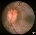 |
C301 Nodular Papillopathies (Sarcoid) | Disc swelling. Sarcoid papillopathy. Note infiltrative nodule at 9:00 on the disc.The patient had proven sarcoid. Perivenous inflammatory cuffing visible on image C3_02. Right eye. Pair with C3_02. Anatomy: Optic disc; Retina. Pathology: Axoplasmic stasis due to sarcoid infiltration. Disease/ Diagn... | Image |
| 4 |
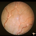 |
C302 Nodular Papillopathies (Sarcoid) | Perivenous Inflammatory Cuffing in a Patient with Proven Sarcoid. Left eye. Pair with C3_01. Anatomy: Retina. Pathology: Axoplasmic stasis due to sarcoid infiltration with retinal venous exudation? Disease/ Diagnosis: Sarcoid papillopathy with perivenous inflammatory disease. Clinical: Unknown? | Image |
| 5 |
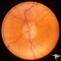 |
Chronic Papilledema with Pseudo Drusen | Left eye. Chronic papilledema with pseudo drusen due to cerebral pontine angle tumor. Anatomy: Optic disc. Pathology: Papilledema Disease/Diagnosis: Chronic papilledema with pseudo drusen. | Image |
| 6 |
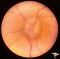 |
Chronic Papilledema with Pseudo Drusen | Right eye. Chronic papilledema with pseudo drusen due to cerebral pontine angle tumor. Anatomy: Optic disc Pathology: Papilledema Disease/Diagnosis: Chronic papilledema with pseudo drusen | Image |
| 7 |
 |
E09 Disc Swelling with Central Vein Occlusion | Chronic disc swelling due to CRVO. Anatomy: Optic disc; Retina. Pathology: Central retinal vein occlusion. Disease/ Diagnosis: Disc swelling due to central retinal vein occlusion. | Image |
| 8 |
 |
H25 Dysplasia with Hypoplasia (Elevated Dysplasia with Anomalous Vessels) | Right eye. Elevated dysplasia with anomalous blood vessel pattern and peri-papillary choroidal malformation. Same patient as H_26. Anatomy: Optic disc. Pathology: Dysplasia of the optic disc. Disease/ Diagnosis: Elevated dysplasia with hypoplasia. | Image |
| 9 |
 |
H26 Dysplasia with Hypoplasia (Elevated Dysplasia with Anomalous Vessels) | Left eye. Dysplasia with grossly anomalous vascular pattern. Elevated dysplasia. Same patient as H_25. Anatomy: Optic disc. Pathology: Dysplasia of the optic disc. Disease/ Diagnosis: Elevated dysplasia with hypoplasia. | Image |
1 - 25 of 9