Collection of materials relating to neuro-ophthalmology as part of the Neuro-Ophthalmology Virtual Education Library.
NOVEL: https://novel.utah.edu/
TO
- NOVEL971
Filters: Collection: "ehsl_novel_novel"
| Title | Creator | Description | Subject | ||
|---|---|---|---|---|---|
| 226 |
 |
Embryology of the Eye | Yesha Shah, BSA, BBA; Amanda Henderson, MD | Video lecture covering the embryology of the eye. | Embryology; Eye |
| 227 |
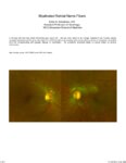 |
Myelinated Retinal Nerve Fibers | Scott N. Grossman, MD | A 33 year old man has noted chronically poor vision OS - left eye color noted to be 'orange' instead of red. fundus photos revealed myelinated retinal nerve fiber layer OU (OS>OD) with corresponding linear paracentral scotoma on Humphrey visual field 24-2 OS corresponding with greatest degree of my... | Myelinated Retinal Nerve Fibers |
| 228 |
 |
Cerebellar Anatomy on MRI | Joshua East, MD; Nicholas A. Koontz, MD; Devin D. Mackay, MD | Overview of structural anatomy of the cerebellum and surround structures on MRI images of the brain. | Cerebellar Anatomy; MRI |
| 229 |
 |
Conjunctiva Muller's Muscle Resection | Patrick Burchell, MD; Jeffrey Nerad, MD | Demonstration of conjunctiva Muller's muscle resection (CMMR). | Conjunctiva Muller's Muscle Resection; CMMR |
| 230 |
 |
Anterior Ptosis | Patrick Burchell, MD; Jeffrey Nerad, MD | Demonstration of anterior ptosis repair, levator advancement. | Anterior Ptosis |
| 231 |
 |
Pituitary Surgery | Jonathan Forbes | Operative video of endoscopic endonasal resection of pituitary macroadenoma. Describes intracapsular versus extracapsular techniques. | Pituitary Surgery |
| 232 |
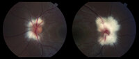 |
Peripapillary Myelinated Nerve Fibers | John J. Chen, MD, PhD | Fundus photographs of a 19-year old female with prominent peripapillary myelinated nerve fibers in both eyes that was incidentally found on routine eye examination. | Myelinated Nerve Fibers |
| 233 |
|
Myelinated Nerve Fibers | John J. Chen, MD, PhD | Fundus photographs of a 19-year old female with prominent peripapillary myelinated nerve fibers in both eyes that was incidentally found on routine eye examination. | Myelinated Nerve Fibers |
| 234 |
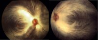 |
Myelinated Nerve Fibers | Carmen Chan,RN, PhD, FAAN | Fundus photos from a patient with extensive myelinated nerve fibers. The patient had normal visual functions. | Myelinated Nerve Fibers |
| 235 |
 |
Common Vitreo-Retinal Procedures and Surgeries | Luke B. Lindsell, OD, MD | Brief presentation on Common vitreo-retinal procedures and surgeries. | Vitreo-Retinal Procedures; Surgeries |
| 236 |
 |
Myotonic Dystrophy | Brian Villafuerte, MD, Ezequiel Piccione, MD | Presentation covering an overview of myotonic dystrophy. | Myotonic Dystrophy |
| 237 |
 |
Muscular Dystrophy Classification | Brian Villafuerte, MD, Ezequiel Piccione, MD | Presentation covering an overview of muscular dystrophy classification. | Muscular Dystrophy Classification |
| 238 |
 |
Congenital Hydrocephalus | Mays El-Dairi, MD | Presentation covering an overview of congenital hydrocephalus. | Congenital Hydrocephalus |
| 239 |
 |
Fibrous Dysplasia | Mays El-Dairi, MD | Presentation covering an overview of fibrous dysplasia. | Fibrous Dysplasia |
| 240 |
 |
Optic Neuropathy: A Recipe for Blindness | Karim Kozhaya, MD; Alaa Bou Ghannam, MD; Alfredo Sadun, MD, PhD | An epidemic of blindness and peripheral neuropathy struck Cuba in the early 90s. By the end of 1993, 7% of the population was affected. Most patients were men and presented with sub-acute, painless, bilateral loss of vision. The etiology of the disease pondered local and international scientists, es... | Cuban Epidemic Optic Neuropathy; Leber's Hereditary Optic Neuropathy; Mitochondrial Insufficiency; Nutritional Optic Neuropathy; Pale Optic Nerve |
| 241 |
 |
Heavy Eye Syndrome | Meagan D. Seay, DO; Bradley J. Katz, MD | A brief overview of heavy eye syndrome. | Heavy Eye Syndrome |
| 242 |
 |
Brown Syndrome | Meagan Seay, DO | A brief overview of Brown Syndrome. | Brown Syndrome |
| 243 |
 |
Intraocular Pressure (IOP) Measurement: A Simple Guide | Nandini Singh; Kirsty Sumerville Mcalester; Alicia Yap; Anne Lee | This video demonstrates the technique for measuring intraocular pressure (IOP) and the use of the tonopen. | Intraocular Pressure (IOP); Tonometry |
| 244 |
 |
Optic Tract Syndrome Secondary to Lacunar Infarction | Justin J. Grassmeyer, PhD; Kenan Xiao, MD; Jason T. Helvey, MD; Sachin Kedar, MD | Lesions of the optic tract produce a characteristic triad of clinical findings: contralateral homonymous hemianopia, contralateral relative afferent pupillary defect, and bilateral optic disc atrophy. This case describes clinical features, radiological findings, and optical imaging characteristics f... | Optic Tract Syndrome; Optic Tract; Lacunar Infarction; Homonymous Hemianopia |
| 245 |
 |
Brain Surface Anatomy | Arooj Ahmad, MD; Devin D. Mackay, MD | These images depict labeled structures of the surface anatomy of the different facies of the brain. | Neuroanatomy; Brain Surface Anatomy |
| 246 |
 |
The Anatomic Course of Cranial Nerve IV | Divya Chauhan, MD | Overview of the intracranial course of the trochlear nerve. | Cranial Nerve IV; Trochlear Nerve; Anatomy |
| 247 |
 |
The Anatomic Course of Cranial Nerve VI | Divya Chauhan, MD | Overview of the intracranial course of the abducens nerve. | Cranial Nerve VI; Abducens Nerve; Anatomy |
| 248 |
 |
CSF Composition | Divya Chauhan, MD | Overview of the composition of cerebrospinal fluid. | Cerebrospinal Fluid; CSF |
| 249 |
 |
The Internal Carotid Arteries and Branches | Katherine Hutchins, MD; Devin D. Mackay, MD | Illustrations, MRA, CTA, and cerebral angiography images of the internal carotid artery and its branches. | Vascular Anatomy; Internal Carotid Artery; Anterior Cerebral Artery; Middle Cerebral Artery; Anterior Circulation |
| 250 |
 |
The Vertebrobasilar System | Katherine Hutchins, MD; Devin D. Mackay, MD | Illustrations, MRA, and CTA images of the vertebrobasilar system and branches. | Vascular Anatomy; Basilar Artery; Vertebral Artery; AICA; PICA; Superior Cerebellar Artery; Posterior Cerebral Artery; Posterior Circulation |