Best known for his world-renowned neuro-ophthalmology unit based at the University of California, San Francisco, William Hoyt, MD collected here more than 850 of his best images covering a wide range of disorders.
William F. Hoyt, MD, Professor Emeritus of Ophthalmology, Neurology and Neurosurgery, Department of Ophthalmology, University of California, San Francisco.
NOVEL: https://novel.utah.edu/
TO
Filters: Collection: "ehsl_novel_wfh"
| Title | Description | Type | ||
|---|---|---|---|---|
| 201 |
 |
Cerebroretinal Microangiopathy (Susac Syndrome) | There is a plaque superior to the disc at 12:00. This plaque is not the result of embolism, but is the result of the microangioplastic process underlying the syndrome. There is a ghost vessel inferiorly at 5:00 off the disc. (NANOS 2001 by Egan, RA). Anatomy: Retina. Pathology: Microangiopathy invo... | Image |
| 202 |
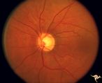 |
Cerebroretinal Microangiopathy (Susac Syndrome) | There is an occlusion of the superior nasal retinal arteriole visible as a white ghost vessel at 11:00. Note: Collateral filling of the distal branches in two places. (NANOS 2001 by Egan, RA). Anatomy: Retina. Pathology: Microangiopathy involving brain, auditory nerve and retina. Disease/Diagnosis:... | Image |
| 203 |
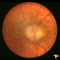 |
Chronic Atrophic Papilledema | Left eye. Left eye blind. Chronic Atrophic Papilledema. Obese woman (300 lbs) with large tentorial meningioma. "Pseudo Pseudotumor". Anatomy: Optic disc. Pathology: Papilledema. Disease/Diagnosis: Papilledema from large tentorial meningioma. | Image |
| 204 |
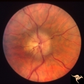 |
Chronic Atrophic Papilledema | Right eye. Chronic Atrophic Papilledema. Obese woman (300 lbs) with large tentorial meningioma. "Pseudo Pseudotumor" Anatomy: Optic disc. Pathology: Papilledema. Disease/Diagnosis: Papilledema from large tentorial meningioma. | Image |
| 205 |
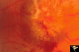 |
Chronic Papilledema due to Brain Tumor | Right eye. Chronic papilledema wth white centrally located exudates in a man with hemispheric glioma. Anatomy: Optic disc. Pathology: Papilledema. Disease/Diagnosis: Chronic papilledema. | Image |
| 206 |
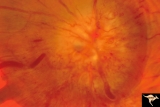 |
Chronic Papilledema due to Brain Tumor | Left eye. Chronic papilledema with white centrally located exudates in a man with hemispheric glioma. Anatomy: Optic disc. Pathology: Papilledema. Disease/Diagnosis: Chronic papilledema. | Image |
| 207 |
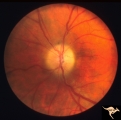 |
Chronic Papilledema due to Brain Tumor - Resolved | Left eye - same as P_40b - follow up after 4 months. Chronic papilledema resolved after treatment showing residual atrophy. | Image |
| 208 |
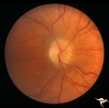 |
Chronic Papilledema due to Brain Tumor - Resolved | Right eye - same as P_40a - follow up after 4 months. Chronic papilledema resolved after treatment showing residual atrophy. Anatomy: Optic disc. Pathology: Papilledema. Disease/Diagnosis: Chronic papilledema. | Image |
| 209 |
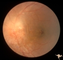 |
Chronic Papilledema in Resolution. Sequence | Left eye 2 weeks after presentation. Chronic papilledema in resolution. Note first evidence of a vertical choroidal fold. Anatomy: Optic disc. Pathology: Papilledema. Disease/Diagnosis: Papilledema. | Image |
| 210 |
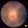 |
Chronic Papilledema in Resolution. Sequence | Left eye 4 weeks after presentation. Chronic papilledema in resolution. Notice more extensive vertical choroidal fold temporally ("high-water" marks) | Image |
| 211 |
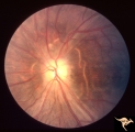 |
Chronic Papilledema in Resolution. Sequence | Left eye 7 weeks after presentation. Chronic papilledema in resolution. Note the profound optic atrophy with blurred disc margins and circumferential receptor layer folds ("high-water" marks) | Image |
| 212 |
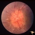 |
Chronic Papilledema in Resolution. Sequence | Left eye at presentation. Chronic papilledema. Anatomy: Optic disc Pathology: Papilledema Disease/Diagnosis: Papilledema | Image |
| 213 |
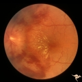 |
Chronic Papilledema with Hemorrhagic and Exudative Complications | Left eye one month after presentation. Resolving hemorrhage. Chronic papilledema with hemorrhagic and exudative complications due to Pseudotumor cerebri. Anatomy: Optic disc. Pathology: Papilledema. Disease/Diagnosis: Chronic papilledema with hemorrhagic and exudative complications | Image |
| 214 |
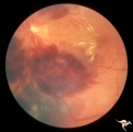 |
Chronic Papilledema with Hemorrhagic and Exudative Complications | Left eye at presesntation. Chronic papilledema with hemorrhagic and exudative complications due to Pseudotumor cerebri. Anatomy: Optic disc Pathology: Papilledema Disease/Diagnosis: Chronic papilledema with hemorrhagic and exudative complications. | Image |
| 215 |
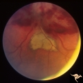 |
Chronic Papilledema with Hemorrhagic and Exudative Complications | Left eye one month after presentation. View below of resolving subretinal hemorrhage. Chronic papilledema with hemorrhagic and exudative complications due to Pseudotumor cerebri. | Image |
| 216 |
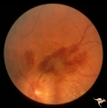 |
Chronic Papilledema with Hemorrhagic and Exudative Complications | Left eye one month after presentation. View above of resolving preretinal hemorrhage. Chronic papilledema with hemorrhagic and exudative complications due to Pseudotumor cerebri. | Image |
| 217 |
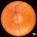 |
Chronic Papilledema with Pseudo Drusen | Left eye. Chronic papilledema with pseudo drusen due to cerebral pontine angle tumor. Anatomy: Optic disc. Pathology: Papilledema Disease/Diagnosis: Chronic papilledema with pseudo drusen. | Image |
| 218 |
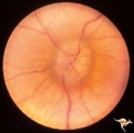 |
Chronic Papilledema with Pseudo Drusen | Right eye. Chronic papilledema with pseudo drusen due to cerebral pontine angle tumor. Anatomy: Optic disc Pathology: Papilledema Disease/Diagnosis: Chronic papilledema with pseudo drusen | Image |
| 219 |
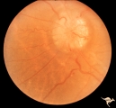 |
Chronic Papilledema with Pseudo Drusen | Right eye. Meningioma. Pseudo drusen from chronic papilledema. Woman. Anatomy: Optic disc Pathology: Papilledema Disease/Diagnosis: Chronic papilledema with pseudo drusen | Image |
| 220 |
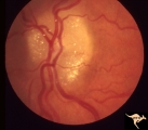 |
Chronic Papilledema with Pseudo Drusen | Left eye of 51 year old, 220 pound black woman. Pseudotumor cerebri, pseudo drusen, exudates. Anatomy: Optic disc. Pathology: Papilledema Disease/Diagnosis: Chronic papilledema with pseudo drusen | Image |
| 221 |
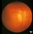 |
Chronic Papilledema with Pseudo Drusen | Chronic papilledema with pseudo drusen. Residual choroidal folds. Pseudo drusen. Anatomy: Optic disc. Pathology: Papilledema. Disease/Diagnosis: Chronic papilledema with pseudo drusen. | Image |
| 222 |
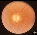 |
Chronic Papilledema with Pseudo Drusen | Left eye. Meningioma. Pseudo drusen from chronic papilledema. The patient's meningioma had blinded her left eye and caused chronic elevated intracranial pressure. Woman. Anatomy: Optic disc Pathology: Papilledema Disease/Diagnosis: Chronic papilledema with pseudo drusen | Image |
| 223 |
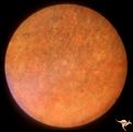 |
Congenital Retinal Cerebellar Degeneration | Congenital retinal blindness due to cerebellar degeneration syndrome. Granular retinal pigmentary degeneration. Pair with R2_B1_1a. Anatomy: Retina. Pathology: Optic atrophy. Disease/Diagnosis: Congenital retinal cerebellar degeneration. Clinical: Severe mental retardation and blindness. | Image |
| 224 |
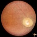 |
Congenital Retinal Cerebellar Degeneration | Congenital retinal blindness due to cerebellar degeneration syndrome. Optic disc pallor with arteriolar attenuation. Pair with R2_B1_1b. Anatomy: Retina. Pathology: Optic atrophy. Disease/ Diagnosis: Congenital retinal cerebellar degeneration. Clinical: Severe mental retardation and blindness. | Image |
| 225 |
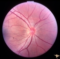 |
Congenitally Crowded Disc - Little Red Disc | Right eye: "little red disc". Congenitally blurred disc. 26 year old man. Anatomy: Optic disc Pathology: Normal variation of the optic disc Disease/Diagnosis: Normal variation of the optic disc. Congenital blurred disc. Little red disc. | Image |
