Best known for his world-renowned neuro-ophthalmology unit based at the University of California, San Francisco, William Hoyt, MD collected here more than 850 of his best images covering a wide range of disorders.
William F. Hoyt, MD, Professor Emeritus of Ophthalmology, Neurology and Neurosurgery, Department of Ophthalmology, University of California, San Francisco.
NOVEL: https://novel.utah.edu/
TO
1 - 25 of 14
| Title | Description | Type | ||
|---|---|---|---|---|
| 1 |
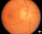 |
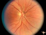
C308 Nodular Papillopathies (Sarcoid) | Nodular infiltrative papillopathy in a patient with sarcoid. Woman. Anatomy: Optic disc. Pathology: Axoplasmic stasis due to sarcoid infiltration. Disease/ Diagnosis: Sarcoid papillopahty. Clinical: Unknown? | Image |
| 2 |
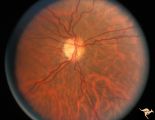 |
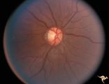
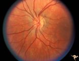
H67 Superior Segmental Optic Hypoplasia (SSOH) Topless Disc Syndrome | Bilateral SSOH. Right eye. Same patient as H_68 and H_69. Anatomy: Optic disc. Pathology: Superior segmental optic hypoplasia (SSOH). Disease/ Diagnosis: Congenital anomaly. | Image |
| 3 |
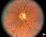 |
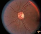
H68 Superior Segmental Optic Hypoplasia (SSOH) Topless Disc Syndrome | Bilateral SSOH. Left eye. Entire nerve looks hypoplastic. Same patient as H_67 and H_69. Anatomy: Optic disc. Pathology: Superior segmental optic hypoplasia (SSOH). Disease/ Diagnosis: Congenital anomaly. | Image |
| 4 |
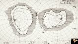 |
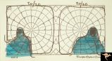
H69 Superior Segmental Optic Hypoplasia (SSOH) Topless Disc Syndrome | Visual field. Entire visual field is constricted. Patient's mother Type 1 diabetic. Same patient as H_68 and H_69. Anatomy: Optic disc. Pathology: Superior segmental optic hypoplasia (SSOH). Disease/ Diagnosis: Congenital anomaly. | Image |
| 5 |
 |
H70 Superior Segmental Optic Hypoplasia (SSOH) Topless Disc Syndrome | Bilateral SSOH. Right eye. Same patient as H_71 and H_72. Anatomy: Optic disc. Pathology: Superior segmental optic hypoplasia (SSOH). Disease/ Diagnosis: Congenital anomaly. | Image |
| 6 |
 |
H71 Superior Segmental Optic Hypoplasia (SSOH) Topless Disc Syndrome | Bilateral SSOH. Left eye. Same patient as H_70 and H_72. Anatomy: Optic disc. Pathology: Superior segmental optic hypoplasia (SSOH). Disease/ Diagnosis: Congenital anomaly. | Image |
| 7 |
 |
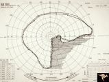
H72 Superior Segmental Optic Hypoplasia (SSOH) Topless Disc Syndrome | Visual field. Entire visual field is constricted. Patient's mother Type 1 diabetic. Same patient as H_70 and H_71. Anatomy: Optic disc. Pathology: Superior segmental optic hypoplasia (SSOH). Disease/ Diagnosis: Congenital anomaly. | Image |
| 8 |
 |
H93 Occipital Hemianoptic Hypoplasia | Visual field. Left eye. Right inferior homonymous. Same patient as H_94, H_95, H_96, H_97. Anatomy: Optic disc. Pathology: Occipital hemianoptic hypoplasia. Disease/ Diagnosis: Congenital defect of the occipital lobe. Imaging: MRI scan - See slide H97. | Image |
| 9 |
 |
H94 Occipital Hemianoptic Hypoplasia | Visual field. Right eye. Quatrantanopia. Same patient as H_93, H_95, H_96, H_97. Anatomy: Optic disc. Pathology: Occipital hemianoptic hypoplasia. Disease/ Diagnosis: Congenital defect of the occipital lobe. Imaging: MRI scan - See slide H97. | Image |
| 10 |
 |
H95 Occipital Hemianoptic Hypoplasia | Right eye with temporal field defect shows trans-synaptic band atrophy. Absence of nasal nerve fibers. Same patient as H_93, H_94, H_96, H_97. Anatomy: Optic disc. Pathology: Occipital hemianoptic hypoplasia. Disease/ Diagnosis: Congenital defect of the occipital lobe. Imaging: MRI scan - See slide ... | Image |
| 11 |
 |
H96 Occipital Hemianoptic Hypoplasia | Left eye has nasal quadrantic field defect. Same patient as H_93, H_94, H_95, H_97. Anatomy: Optic disc. Pathology: Occipital hemianoptic hypoplasia. Disease/ Diagnosis: Congenital defect of the occipital lobe. Imaging: MRI scan - See slide H97. | Image |
| 12 |
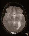 |
H97 Occipital Hemianoptic Hypoplasia | MRI scan shows left occipital lobe periventricular leuko-melacia. Same patient as H_93, H_94, H_95, H_96. Anatomy: Optic disc. Pathology: Occipital hemianoptic hypoplasia. DIsease/ Diagnosis: Congenital defect of the occipital lobe. Imaging: MRI scan. | Image |
| 13 |
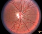 |
Neurofibromatosis-1 | Optic atrophy and hypoplasia of the optic disc associated with chiasmal glioma in a patient with NF-1. Anatomy: Optic disc. Pathology: Chiasmal glioma; Optic atrophy; Hypoplasia. Disease/Diagnosis: Neurofibromatosis type 1. Clinical: Proptosis; Blindness. | Image |
| 14 |
 |
P50 Chronic Papilledema with Subretinal Neo-Vascular Network | Chronic papilledema with subretinal neo-vascular network. Pseudotumor. Anatomy: Optic disc. Pathology: Papilledema. Disease/ Diagnosis: Chronic papilledema with sub-retinal neovascular network. | Image |
1 - 25 of 14