Best known for his world-renowned neuro-ophthalmology unit based at the University of California, San Francisco, William Hoyt, MD collected here more than 850 of his best images covering a wide range of disorders.
William F. Hoyt, MD, Professor Emeritus of Ophthalmology, Neurology and Neurosurgery, Department of Ophthalmology, University of California, San Francisco.
NOVEL: https://novel.utah.edu/
TO
| Title | Description | Type | ||
|---|---|---|---|---|
| 26 |
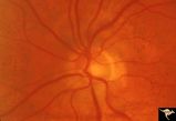 |
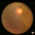
H79 Inferior Segmental Optic Hypoplasia (ISOH) | ISOH. Anatomy: Optic disc. Pathology: Inferior segmental optic hypoplasia (ISOH). Disease/ Diagnosis: Congenital anomaly. | Image |
| 27 |
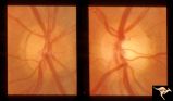 |
H90 Occipital Hemianoptic Hypoplasia | Note left disc (right side of image) is the eye with temporal field defect. Shows band atrophy. Anatomy: Optic disc. Pathology: Occipital hemianoptic hypoplasia. Congenital defect of the occipital lobe. | Image |
| 28 |
 |
Macular Cherry Red Spots in Niemann-Pick disease | Close up view of macular cherry red spots in Niemann-Pick disease. Same patient as R2A2a. Anatomy: Retina. Pathology: Retinal ganglion cell accumulation of lipid. Disease/Diagnosis: Niemann-Pick disease. Clinical: Severe mental retardation and blindness. Fatal. | Image |
| 29 |
 |
Macular Cherry Red Spots in Niemann-Pick disease | Macular cherry red spots in Niemann-Pick disease. Same patient as R2A2b. Anatomy: Retina. Pathology: Retinal ganglion cell accumulation of lipid. Disease/Diagnosis: Niemann-Pick disease. Clinical: Severe mental retardation and blindness. Fatal. | Image |
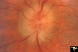
| 30 |
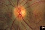 |
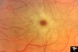
Post Papilledema Disc Blurring | Left eye. 8 year old boy. Post papilledema due to brain tumor. Note the entire peripapillary nerve fiber is blurred but the optic discs are barely elevated. Anatomy: Optic disc. Pathology: Brain tumor. Disease/Diagnosis: Papilledema. Clinical: Post papilledema due to brain tumor. | Image |
| 31 |
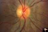 |
Post Papilledema Disc Blurring | Right eye. 8 year old boy. Post papilledema due to brain tumor. Note the entire peripapillary nerve fiber is blurred but the optic discs are barely elevated. Anatomy: Optic disc. Pathology: Post papilledema. Disease/Diagnosis: Post papilledema due to brain tumor. | Image |
| 32 |
 |
R3C6 Calcific Retinal Emboli | Calcific retinal emboli. There is a large calcium embolus totally occluding the superior branches of the central retinal artery. Note: calcium emboli of this type usually derive from the aortic valve. Close view of R3_C7. Anatomy: Retina. Pathology: Calcific aortic stenosis. Disease/ Diagnosis: Calc... | Image |
| 33 |
 |
R3C7 Calcific Retinal Emboli | Calcific retinal emboli. This woman had aortic stenosis and a heart murmur. There is a large calcium embolus totally occluding the superior branches of the central retinal artery. Note: calcium emboli of this type usually derive from the aortic valve disease. Note superior retinal infarction. Far v... | Image |
| 34 |
 |
Slow Flow (Chronic Hypoxic) Retinopathy | Examples of Slow flow (chronic hypoxic) retinopathy showing produced by a carotid-cavernous sinus fistula. Arteriole pressure was low in the retina and venous pressure was elevated. Note the characteristic dot and blot hemorrhages in the black and white photo (R3B2b). Anatomy: Retina. Pathology: Car... | Image |
| 35 |
 |
Slow Flow (Chronic Hypoxic) Retinopathy | Examples of Slow flow (chronic hypoxic) retinopathy produced by a carotid-cavernous sinus fistula. Arteriole pressure was low in the retina and venous pressure was elevated. Note the characteristic dot and blot hemorrhages in this black and white photo. Anatomy: Retina. Pathology: carotid-cavernous ... | Image |
| 36 |
 |
Slow Flow (Chronic Hypoxic) Retinopathy | Slow flow (chronic hypoxic) retinopathy. Optic disc change in left eye (b) secondary to reduced carotid artery perfusion. Patient was an elderly man with a innominant artery occlusion. Note the reduced arteriole caliber in the left disc (b) compared to the right (a). Central retinal artery pressure ... | Image |
| 37 |
 |
Slow Flow (Chronic Hypoxic) Retinopathy | Slow flow (chronic hypoxic) retinopathy. Optic disc change in left eye (b) secondary to reduced carotid artery perfusion. Patient was an elderly man with a innominant artery occlusion. Note the reduced arteriole caliber in the left disc (b) compared to the right (a). Central retinal artery pressure ... | Image |
| 38 |
 |
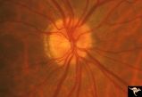
Slow Flow (Chronic Hypoxic) Retinopathy | Slow flow (chronic hypoxic) retinopathy (right eye) in a man with polycythaemia rubra vera. Hematological disease. Anatomy: Retina. Pathology: Hematological disease. Disease/Diagnosis: Slow flow (chronic hypoxic) retinopathy secondary to polycythaemia. Clinical: No visual symptoms. | Image |
| 39 |
 |
Slow Flow (Chronic Hypoxic) Retinopathy | Slow flow (chronic hypoxic) retinopathy (right eye) in a man with polycythaemia rubra vera. Hematological disease. Anatomy: Retina. Pathology: Hematological disease. Disease/Diagnosis: Slow flow (chronic hypoxic) retinopathy secondary to polycythaemia. Clinical: No visual symptoms. | Image |
| 40 |
 |
Tuberous Sclerosis | Retinal lesion in tuberous sclerosis in the form of a translucent disc in the superior temporal area of the retina. Note the dense calcification in the center of the lesion. Note how the lesion obscures the details of the arterioles which pass through it. Anatomy: Retina. Pathology: Astrocytic hamar... | Image |
| 41 |
 |
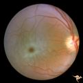
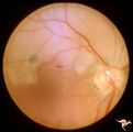
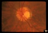
Unilateral Papilledema | Right eye. Has no optic cup. Disc is flat. Anatomy: Optic disc. Pathology: Unilateral papilledema. Disease/Diagnosis: Idiopathic intracranial hypertension (pseudotumor cerebri). Clinical: Transient monocular blindness (transient visual obscurations, amaurosis fugax); headache, sixth nerve palsy, ele... | Image |
| 42 |
 |
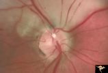
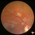
Unilateral Papilledema | Left eye. This eye has papilledema. Anatomy: Optic disc. Pathology: Unilateral papilledema. Disease/Diagnosis: Idiopathic intracranial hypertension (pseudotumor cerebri). Clinical: Transient monocular blindness (transient visual obscurations, amaurosis fugax); headache, sixth nerve palsy, elevated i... | Image |
| 43 |
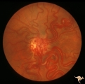 |
Vascular Disc Anomalies - Retinal Arteriovenous Malformations | Retinal arteriovenous malformations (Racemous angioma). Found in a 13 year old girl who had extension of this arteriovenous malformation up her right optic nerve into her thalamus and into her midbrain. Patient had large intra-cerebral AVM (Wyburn-Mason Syndrome). Patient died 10 years later of mass... | Image |
| 44 |
 |
Visible Drusen | PP21a: Right eye. Drusen barely visible. Note disc margin drusen at 1:00 and 2:30.; PP21b: Left eye shows multiple exposed drusen. Girl. Anatomy: Optic disc. Pathology: Drusen of the optic disc. Disease/Diagnosis: Drusen of the optic disc. Clinical: Normally functioning eye with drusen. | Image |